Skin Diagram — Explained by Medical Evidence, Not Myths
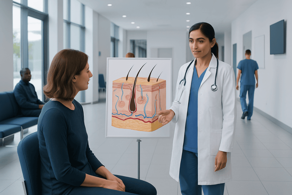
A skin diagram usually shows three main layers: epidermis, dermis, and subcutaneous tissue. Important structures include hair follicles, sweat glands, oil glands, blood vessels, nerves, and collagen.
Key Takeaways
- A skin diagram usually shows three main layers: epidermis, dermis, and subcutaneous tissue.
- Important structures include hair follicles, sweat glands, oil glands, blood vessels, nerves, and collagen.
- Different symptoms often come from different layers, which is why location matters in diagnosis.
- Skin diagrams are useful educational tools, but they do not replace a medical examination.
- Changes such as non-healing sores, rapidly changing moles, or severe infection signs need medical review.
A skin diagram is a visual map of the skin’s layers and structures, including the epidermis, dermis, and subcutaneous tissue. Understanding this anatomy helps explain how the skin protects the body, heals, senses touch, and develops common symptoms such as dryness, rash, pain, or swelling.
What a skin diagram shows
A skin diagram is a medical illustration that shows how the skin is built from the outside inward. In most diagrams, the skin is divided into three main layers: the epidermis on top, the dermis underneath, and the subcutaneous layer below them. These images also often label structures such as pores, hair follicles, sweat glands, oil glands, blood vessels, nerves, and connective tissue.
For many people, the value of a skin diagram is practical rather than academic. It helps explain why some problems stay on the surface, such as mild dryness, while others involve deeper inflammation, pain, swelling, cysts, or scarring. A clear understanding of where a symptom begins can make discussions with a clinician more meaningful and less confusing.
Medical evidence supports using anatomy-based explanations to understand skin health because structure and function are closely linked. The skin is not just a covering. It is an active organ that acts as a barrier, supports immune defense, regulates body temperature, stores water, helps produce vitamin D, and sends sensory information to the brain.
The three main layers of the skin
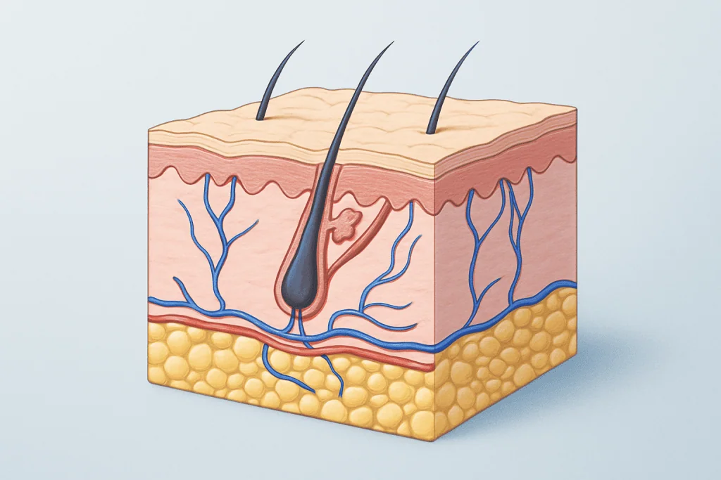
The epidermis is the outermost layer. It forms the body’s first barrier against the environment and is mainly made of cells called keratinocytes. The epidermis also contains melanocytes, which produce pigment, and specialized immune cells that help detect threats. In many skin diagrams, the epidermis is shown as several thin sublayers, although these may not all be labeled in simplified versions.
Beneath it lies the dermis, which gives the skin strength, flexibility, and much of its function. This layer contains collagen and elastin fibers, along with blood vessels, lymphatic vessels, nerves, sweat glands, sebaceous glands, and hair follicles. Because the dermis is richly supplied with nerves and vessels, conditions involving this layer may feel tender, swollen, itchy, or warm.
The deepest layer shown on a skin diagram is the subcutaneous tissue, also called the hypodermis or subcutis. This layer contains fat and connective tissue that cushion the body, help conserve heat, and support nearby structures. Injections, deeper infections, bruising, and some inflammatory conditions may involve this level rather than the surface alone.
- Epidermis: barrier, pigment, water retention
- Dermis: strength, sensation, circulation, glands
- Subcutaneous tissue: padding, insulation, energy storage
Key parts commonly labeled on a skin diagram
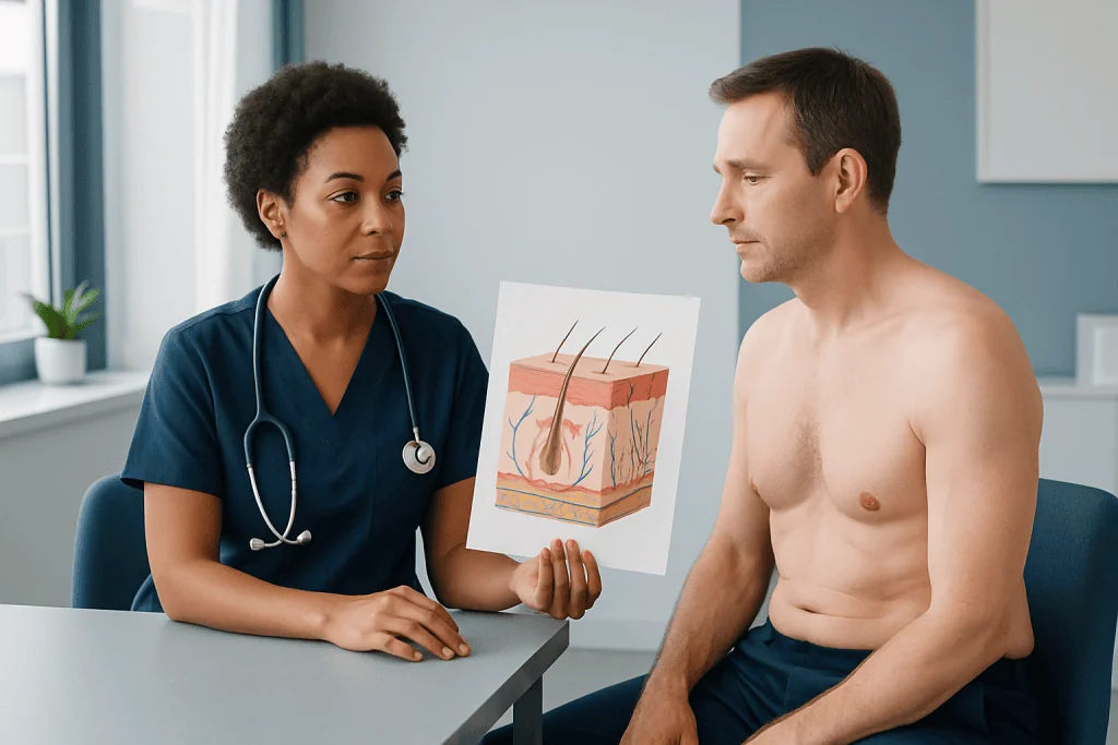
Many diagrams go beyond the three layers and label smaller structures that explain everyday skin functions. Hair follicles are tiny organs anchored in the dermis, often connected to sebaceous glands that produce oil, or sebum. This oil helps lubricate the skin and hair, but when pores become blocked, acne and inflammation can develop. A useful related overview may be acne.
Sweat glands are another common feature. Eccrine glands are found over much of the body and help regulate temperature by releasing sweat onto the skin surface. Apocrine glands, found mainly in areas such as the underarms and groin, become active around puberty and are associated with body odor after sweat is broken down by bacteria. Their ducts and openings may be shown in more detailed diagrams.
Blood vessels and nerves are especially important in understanding symptoms. Blood vessels support healing and temperature control, while nerves detect touch, pressure, pain, and temperature. A skin diagram may also label collagen and elastin fibers, which help explain why aging, sun exposure, inflammation, or injury can change skin texture and elasticity over time.
How skin structure relates to symptoms and common conditions
A skin diagram becomes most helpful when it is used to interpret symptoms. Surface scaling, roughness, and mild irritation often begin in the epidermis, where the barrier may be dry or disrupted. Redness, itching, and oozing can involve both the epidermis and dermis, as happens in inflammatory conditions such as eczema. Deeper lumps, painful nodules, or abscesses usually point to involvement below the surface.
Layer-based thinking also helps explain why some conditions heal cleanly while others leave marks or scars. If a problem remains superficial, healing may occur without lasting change. When inflammation extends into the dermis, the skin may repair itself with altered pigment, thickening, or scar formation. This is one reason clinicians pay close attention to depth, duration, and whether a lesion is changing over time.
Infections, allergic reactions, autoimmune disorders, friction injury, burns, and vascular problems can all affect different structures on a skin diagram. Hair follicle inflammation may cause folliculitis; blocked glands may form cysts; damage to deeper tissue may produce more swelling and pain than a surface rash alone. For suspicious growths or changing pigmented spots, assessment for skin cancer may be necessary.
How doctors use skin anatomy in diagnosis
Doctors do not diagnose a skin problem from a diagram alone, but they use the same anatomical principles that diagrams illustrate. During an examination, they look at the lesion’s color, shape, border, distribution, depth, texture, and whether it blanches, crusts, scales, or drains. They also ask about itch, pain, timing, sun exposure, new products, medications, travel, and family history.
Sometimes a close visual exam is enough to guide diagnosis and treatment. In other cases, additional tests may be needed. These may include skin swabs, allergy testing, blood tests, dermoscopy, imaging for deeper masses, or a biopsy. A biopsy allows a sample of skin to be examined under a microscope, which can clarify whether inflammation, infection, or abnormal cell growth is present.
If a lesion is large, unusual, or difficult to classify, referral to a dermatologist or another specialist may be appropriate. In selected situations, diagnostic evaluation can be followed by procedures such as skin biopsy or removal of a concerning lesion through skin cancer surgery. The exact approach depends on symptoms, examination findings, and overall health.
Treatment options depend on the layer and cause
There is no single treatment based only on a skin diagram, because treatment depends on the underlying cause. Surface dryness may improve with gentle cleansers, moisturizers, and avoiding irritants. Inflammatory rashes may require medicated creams or other therapies prescribed by a doctor. Bacterial, fungal, or viral infections need cause-specific treatment rather than trial-and-error home remedies.
Deeper or structural problems may need more targeted care. Cysts, abscesses, suspicious growths, or chronic wounds often require in-person assessment. Some conditions need procedures rather than topical therapy, especially when the problem lies in the deeper layers or involves uncontrolled tissue growth. When symptoms persist or recur, treatment may also include reviewing triggers such as friction, humidity, allergens, or underlying disease.
For skin cancers or precancerous lesions, treatment may involve local procedures or surgery depending on the diagnosis and extent. In broader cancer care planning, some patients may also need consultation about medical oncology. Near the end of the care pathway, multidisciplinary specialists at Acibadem International and its JCI-accredited hospitals diagnose and treat skin conditions for international patients when advanced evaluation is needed.
Skin care, prevention, and healthy habits
A good understanding of a skin diagram can improve everyday skin care by showing why the barrier matters. Gentle cleansing, regular moisturizing, and sun protection help support the epidermis and reduce unnecessary irritation. Products that are too harsh can disturb the barrier, increase dryness, and make existing conditions more noticeable.
Prevention also includes avoiding excessive sun exposure, using broad-spectrum sunscreen, seeking shade, and wearing protective clothing when practical. Sunlight affects not only the surface but also deeper collagen and elastic tissue, contributing to premature aging and increasing the risk of skin cancer over time. Self-checking the skin from head to toe can help a person notice new or changing lesions earlier.
Healthy routines such as staying hydrated, managing chronic conditions, avoiding smoking, and not picking at lesions can also support skin repair. When trying new products, patch testing on a small area may reduce the chance of widespread irritation. If a rash or sore worsens despite basic care, professional advice is safer than prolonged self-treatment.
When to seek medical care
Medical care is important when a skin problem is severe, spreading, or not improving. A person should arrange a clinical review if they have a rash that lasts, repeated infections, deep pain, significant swelling, a lump under the skin, or a sore that does not heal. Rapid changes in a mole or pigmented spot, including changes in size, border, color, or bleeding, also deserve prompt attention.
Urgent assessment is sensible if there are signs of possible infection such as fever, increasing redness, warmth, pus, or red streaks in the skin. Widespread blistering, facial swelling, trouble breathing, or skin symptoms after a new medicine need immediate medical help. Infants, older adults, and people with weakened immune systems may need earlier evaluation because skin problems can progress differently in these groups.
A skin diagram can guide understanding, but it cannot confirm what a lesion is or how deep it extends. If there is uncertainty, especially around persistent, painful, changing, or suspicious areas, consultation with a qualified doctor is the safest next step.
Frequently asked questions
What are the three layers shown on a skin diagram?
Most skin diagrams show the epidermis, dermis, and subcutaneous tissue. These layers work together to provide protection, sensation, temperature regulation, and support for healing.
Why is a skin diagram useful?
A skin diagram helps explain where a symptom may be coming from and why it feels or looks a certain way. It is especially helpful for understanding the difference between surface irritation, deeper inflammation, and under-the-skin lumps.
Can a skin diagram help diagnose a rash?
A skin diagram can improve understanding, but it cannot diagnose a rash by itself. Diagnosis depends on a medical history, examination, and sometimes tests such as a swab, allergy assessment, or biopsy.
What structures are usually labeled besides the skin layers?
Common labels include hair follicles, sweat glands, sebaceous glands, pores, nerves, blood vessels, collagen, and fat. These structures explain how the skin senses, heals, cools the body, and develops certain conditions.
Do all skin problems start in the same layer?
No, different skin problems affect different layers and structures. Dryness may mainly affect the outer barrier, while cysts, infections, or scarring often involve deeper tissue.
When should someone worry about a skin change?
A person should seek medical advice for a sore that does not heal, a rapidly changing mole, severe pain, spreading redness, or signs of infection. New symptoms after a medication, or skin changes with fever or breathing trouble, need prompt medical attention.
This article is for general information only and is not a substitute for professional medical advice. Please consult a qualified doctor about your individual situation.
Explore treatments in Turkey — costs, top hospitals & a free quote
JCI-accredited · board-certified surgeons · reply within 24h
Add us as a Preferred Source to see more of our trusted health content across Google Search, AI Overviews and Discover.

More from the Health Library

Brachiocephalic Vein — Explained by Medical Evidence, Not Myths

Sour Taste in Mouth: A Complete Medical Overview

Dental Sedation: A Complete Medical Overview

Libido — Explained by Medical Evidence, Not Myths

Concierge Medicine: An Evidence-Based Guide for Patients