Brachiocephalic Vein — Explained by Medical Evidence, Not Myths

The brachiocephalic veins are two large veins, one on each side, that join to form the superior vena cava. They return blood from the head, neck, upper chest, and arms to the heart.
Key Takeaways
- The brachiocephalic veins are two large veins, one on each side, that join to form the superior vena cava.
- They return blood from the head, neck, upper chest, and arms to the heart.
- Most brachiocephalic vein issues are related to clotting, compression, or medical devices such as central venous catheters.
- Symptoms can include arm, neck, or facial swelling, visible chest veins, discomfort, or no symptoms at all.
- Ultrasound, CT, MRI, and venography may be used to diagnose problems affecting this vein.
- Treatment depends on the cause and may include monitoring, medicines, catheter-based procedures, or surgery.
The brachiocephalic vein is a major vein in the upper chest that carries oxygen-poor blood from the head, neck, and arms back to the heart. Most people never notice it, but understanding its location and role can help explain certain scans, procedures, and symptoms such as swelling or prominent veins.
What is the brachiocephalic vein?
The brachiocephalic vein is a large vein in the upper chest that helps return blood to the heart. There are actually two brachiocephalic veins: a right brachiocephalic vein and a left brachiocephalic vein. Each forms where the internal jugular vein from the neck meets the subclavian vein from the arm, and together they join to create the superior vena cava, which empties into the right side of the heart.
In simple terms, the brachiocephalic veins are part of the body’s main drainage pathway for the upper half of the body. They carry oxygen-poor blood from the head, neck, upper limbs, and part of the chest back toward the heart so it can then be sent to the lungs for oxygen.
This vein is often discussed in anatomy reports, imaging results, and hospital procedures because it sits near important structures such as the aorta, lungs, trachea, and major arteries. Even so, most healthy people do not feel or notice the brachiocephalic vein in everyday life unless there is swelling, a clot, compression, or a device such as a catheter nearby.
Where it is located and why it matters
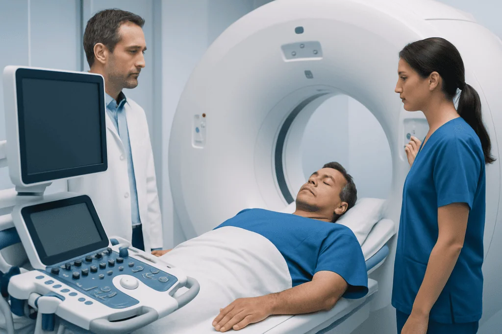
The right brachiocephalic vein is relatively short and runs downward to meet the left brachiocephalic vein. The left brachiocephalic vein is longer because it crosses the upper chest from left to right behind the breastbone area. This difference in length and course can matter during imaging, line placement, and when doctors evaluate possible compression or obstruction.
Because these veins lie deep in the chest, they are not usually examined directly by touch. Instead, they are seen on imaging tests such as ultrasound, computed tomography, magnetic resonance imaging, or contrast venography. Their location also explains why conditions in nearby organs, such as an enlarged thyroid, a chest mass, or swollen lymph nodes, can occasionally affect blood flow through them.
The brachiocephalic vein also matters in practical medical care. It may be involved when doctors place long-term intravenous access, evaluate unexplained swelling of the face or arm, or investigate suspected deep vein thrombosis in the upper body. Its anatomy helps guide safe planning for procedures and interpretation of chest scans.
What the brachiocephalic vein does
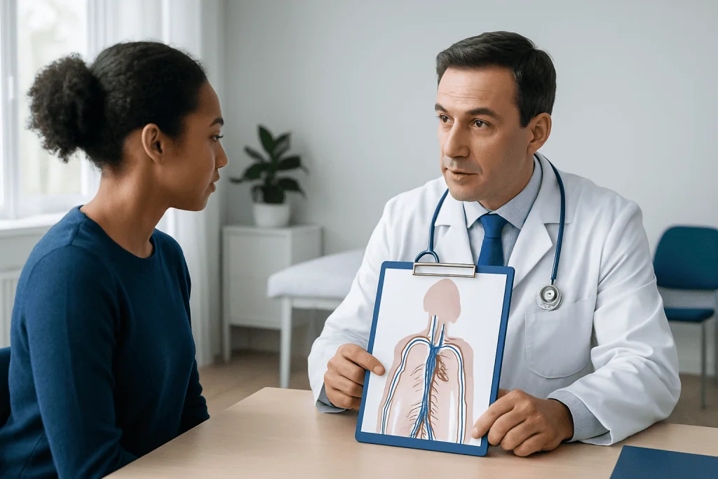
The main job of the brachiocephalic vein is to carry deoxygenated blood from the upper body back to the heart. It collects blood that has already delivered oxygen to tissues in the head, neck, shoulders, arms, and parts of the chest wall. This is a normal and essential part of circulation.
The brachiocephalic veins also help maintain balanced blood return from both sides of the upper body. Since the left vein crosses toward the right to join the right vein, this system allows blood from both arms and both sides of the head and neck to flow efficiently into the superior vena cava.
Although the vein itself does not pump blood, it works closely with breathing, body position, surrounding muscles, and the pressure changes created by the heartbeat. These factors all help venous blood move steadily toward the heart. If the vein becomes narrowed or blocked, that return flow can slow down and lead to congestion or swelling upstream.
Common problems that can affect the brachiocephalic vein
Most people will never develop a brachiocephalic vein disorder. When problems do occur, they usually involve a blood clot, external compression, narrowing after repeated catheter use, or less commonly, congenital differences in anatomy. Doctors may refer to a clot in this area as upper-extremity or central venous thrombosis.
A clot can develop because of reduced blood flow, irritation of the vein wall, or a tendency for blood to clot more easily. Risk may rise with central venous catheters, pacemaker wires, cancer, recent surgery, limited movement, inherited clotting conditions, or serious illness. The same principles that apply to venous thrombosis in other areas can apply here as well.
Compression is another possible issue. A nearby tumor, enlarged lymph node, thyroid enlargement, or other mass in the chest can press on the vein. In some cases, scar tissue or changes from prior treatment can narrow it. Rarely, people have an anatomical variant that is found incidentally on a scan and causes no symptoms.
Doctors may also pay close attention to this vein during placement of a central venous catheterization or other vascular access device. Most such procedures are performed safely, but because the vein is large and centrally located, correct placement and monitoring are important.
Possible symptoms and signs
Symptoms depend on whether blood flow is partly blocked, fully blocked, or only mildly affected. Some people have no symptoms at all, and the issue is found on imaging done for another reason. Others may notice swelling in one arm, the neck, or the face, especially on the side of the problem.
Other possible signs include a feeling of heaviness in the arm, discomfort in the shoulder or upper chest, visible enlarged veins across the chest or neck, skin color changes, or fullness around the collarbone area. If the narrowing develops slowly, the body may form alternate veins, which can make symptoms milder or more gradual.
Severe symptoms are less common but can occur if central venous flow is significantly impaired. Worsening swelling of the face, shortness of breath, dizziness, or symptoms suggesting broader obstruction of the superior vena cava need prompt medical assessment. These symptoms can overlap with other conditions, so a clinician usually needs imaging to confirm the cause.
- Arm, neck, or facial swelling
- Visible veins on the chest or neck
- Upper chest or shoulder discomfort
- Fullness, pressure, or heaviness in the arm
- No symptoms, with incidental finding on imaging
How doctors diagnose brachiocephalic vein problems
Diagnosis starts with a medical history and physical examination. A doctor asks about recent surgery, cancer treatment, intravenous lines, pacemaker leads, injuries, clotting disorders, and symptoms such as swelling, pain, or shortness of breath. The pattern of symptoms can suggest whether blood flow from the upper body may be reduced.
Imaging is usually needed because the brachiocephalic veins are deep in the chest. Ultrasound may help assess related veins in the neck and arm, but it can be limited for viewing the full central chest veins. CT venography or MRI can provide a clearer picture of the vein’s size, position, narrowing, compression, or clot. In some settings, contrast venography remains the most direct way to define the blockage.
Blood tests may be ordered depending on the situation, especially if clotting is suspected or if there may be an underlying inflammatory, cancer-related, or inherited tendency to form clots. If a device-related problem is suspected, the care team may also review line position or evaluate whether a catheter is still needed.
When a structural problem is seen, diagnosis focuses not only on the vein itself but also on the reason for the problem. That may include assessment for a chest mass, enlarged thyroid, prior radiation changes, or compression by neighboring structures. This step is important because treatment is guided by the cause, not only by the location.
Treatment options and what recovery may involve
Treatment for a brachiocephalic vein problem depends on the cause, the degree of blockage, and whether symptoms are mild or significant. If the finding is an anatomical variant or a mild narrowing without symptoms, careful observation may be enough. If there is a clot, treatment often includes anticoagulant medicines to reduce the risk of extension and help the body gradually clear the clot.
When a central line or another device has contributed to the problem, doctors may decide whether it should be repositioned or removed. In selected situations, interventional procedures may be considered, such as catheter-directed treatment, angioplasty, or stenting to improve blood flow. These decisions are individualized and usually involve vascular, interventional radiology, cardiology, or thoracic specialists.
If compression is caused by another condition, treatment focuses on the source of the pressure. For example, this may involve managing a chest mass, inflammatory process, or another nearby structural problem. Related evaluation can overlap with vascular surgery or other specialty care when the vein needs direct repair or reconstruction.
Recovery varies widely. Some people improve with medication and follow-up imaging, while others need a more procedural approach. Toward the end of the care pathway, a multidisciplinary team may coordinate imaging, blood-thinner management, and symptom monitoring. Acibadem International’s multidisciplinary specialists and JCI-accredited hospitals diagnose and treat vascular conditions for international patients when more advanced evaluation is needed.
Self-care, prevention, and when to seek medical care
There is no guaranteed way to prevent all brachiocephalic vein problems, but risk can sometimes be reduced by staying mobile after surgery or illness, following advice about catheter care, attending follow-up appointments, and managing conditions that increase clot risk. People with a history of thrombosis should discuss future procedures, travel, and medication plans with their doctor. General measures that support vascular health, such as not smoking and staying physically active, may also help overall circulation.
Self-care should not replace medical evaluation if swelling or prominent veins appear in the upper body. It is reasonable to note when symptoms began, whether one side is more affected, and whether there has been recent line placement, injury, infection, or cancer treatment. That information can help doctors decide which tests are most useful.
Medical care should be sought promptly for new swelling of the arm, neck, or face; unexplained chest discomfort; visible enlarged veins across the upper chest; or symptoms after a central line or pacemaker procedure. Emergency care is appropriate if symptoms are accompanied by shortness of breath, severe chest pain, fainting, or rapidly worsening swelling. These symptoms do not always mean a brachiocephalic vein problem, but they should be assessed without delay.
People who already know they have a venous condition may also be advised to follow specialist care and, if prescribed, continue monitoring while receiving therapies such as anticoagulation treatment. The safest plan is individualized and should come from a qualified clinician who understands the person’s full medical history.
Frequently asked questions
Is the brachiocephalic vein the same as the innominate vein?
Yes. The term innominate vein is an older name often used interchangeably with brachiocephalic vein. Most modern medical reports prefer brachiocephalic vein, but both terms usually refer to the same structure.
How many brachiocephalic veins are there?
There are two: a right brachiocephalic vein and a left brachiocephalic vein. They join together in the upper chest to form the superior vena cava, which carries blood into the heart.
Can a brachiocephalic vein problem cause swelling in the face or arm?
Yes. If blood flow through the vein is narrowed or blocked, blood can back up and cause swelling in the arm, neck, or face. The exact pattern depends on which side is affected and how severe the blockage is.
What causes a clot in the brachiocephalic vein?
Common causes include central venous catheters, pacemaker leads, cancer, recent surgery, reduced movement, and conditions that make blood clot more easily. Sometimes more than one risk factor is present at the same time.
Which test is best for seeing the brachiocephalic vein?
The best test depends on the clinical question. CT venography or MRI often shows the deep chest veins more clearly than standard ultrasound, while venography may be used in selected cases for a more direct view.
Are brachiocephalic vein anatomical variants dangerous?
Not usually. Many anatomical variants are incidental findings and do not cause symptoms or require treatment. A doctor can explain whether a specific variant is simply a normal difference or something that needs follow-up.
This article is for general information only and is not a substitute for professional medical advice. Please consult a qualified doctor about your individual situation.
Explore treatments in Turkey — costs, top hospitals & a free quote
JCI-accredited · board-certified surgeons · reply within 24h
Add us as a Preferred Source to see more of our trusted health content across Google Search, AI Overviews and Discover.

More from the Health Library

Preventive Dentistry: An Evidence-Based Guide for Patients

Ringworm Armpit — Explained by Medical Evidence, Not Myths

Outie Belly Button — Explained by Medical Evidence, Not Myths

Menquadfi: What Patients Need to Know

Orthotics — Explained by Medical Evidence, Not Myths