Spinal Stenosis: Leg Pain While Walking and Modern Treatment Options

Spinal stenosis most often affects the lower back and may cause leg pain, tingling, weakness, or heaviness during walking. Symptoms often improve when sitting or leaning forward, a pattern called neurogenic claudication.
Key Takeaways
- Spinal stenosis most often affects the lower back and may cause leg pain, tingling, weakness, or heaviness during walking.
- Symptoms often improve when sitting or leaning forward, a pattern called neurogenic claudication.
- Diagnosis usually includes a physical examination and imaging such as MRI, with other tests used when needed.
- Treatment may include activity changes, physiotherapy, pain-relieving medicines, spinal injections, or decompression surgery.
- Urgent medical attention is needed for new bladder or bowel problems, severe weakness, or numbness in the groin area.
Spinal stenosis is a narrowing of the spinal canal that can irritate or compress nerves, often causing leg pain, heaviness, or numbness while walking. Many people improve with careful diagnosis, physical therapy, medication, injections, or, when needed, modern surgical treatments.
Overview
Spinal stenosis means that the spaces within the spine have become narrowed. When this narrowing occurs, the spinal cord or the nerve roots that travel to the legs can become irritated or compressed. The condition is most common in the lower back, called lumbar spinal stenosis, but it can also affect the neck, known as cervical spinal stenosis.
A common and important symptom of lumbar spinal stenosis is leg pain while walking. Some people describe this as aching, cramping, heaviness, tingling, or weakness in one or both legs. The discomfort may start after walking a certain distance and improve when the person sits down, bends forward, or leans on a shopping cart. This pattern is often called neurogenic claudication.
Spinal stenosis usually develops gradually with age-related changes in the spine, such as arthritis, thickened ligaments, disc bulging, or bony overgrowth. Although the symptoms can affect daily life, many people are helped by non-surgical treatments. When symptoms are severe or persistent, modern surgical options can relieve pressure on the nerves and help restore function.
Symptoms of Spinal Stenosis
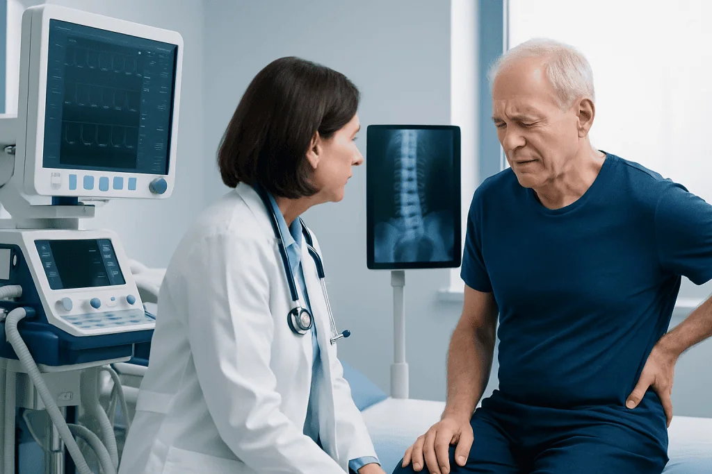
Symptoms vary depending on which part of the spine is affected and how much pressure is placed on the nerves. In lumbar spinal stenosis, symptoms most often involve the buttocks, thighs, calves, or feet. Pain may be accompanied by numbness, pins and needles, burning sensations, or a feeling that the legs are tired or weak.
A typical feature is that symptoms worsen with standing or walking and improve with sitting or bending forward. This happens because leaning forward can slightly open the spinal canal and reduce pressure on the nerves. Some people find they can walk farther when pushing a trolley or cycling than when walking upright.
Symptoms may include:
- Leg pain, cramping, or heaviness during walking
- Lower back pain, which may or may not be the main complaint
- Numbness, tingling, or burning in the legs or feet
- Weakness or reduced balance
- Difficulty standing for long periods
- In neck stenosis, hand clumsiness, arm symptoms, or walking imbalance
It is important to distinguish neurogenic claudication from vascular claudication, which is leg pain caused by reduced blood flow. Both can cause walking-related leg pain, but their treatments are different. A doctor may assess pulses, circulation, nerve function, and imaging findings to clarify the cause.
Causes and Risk Factors
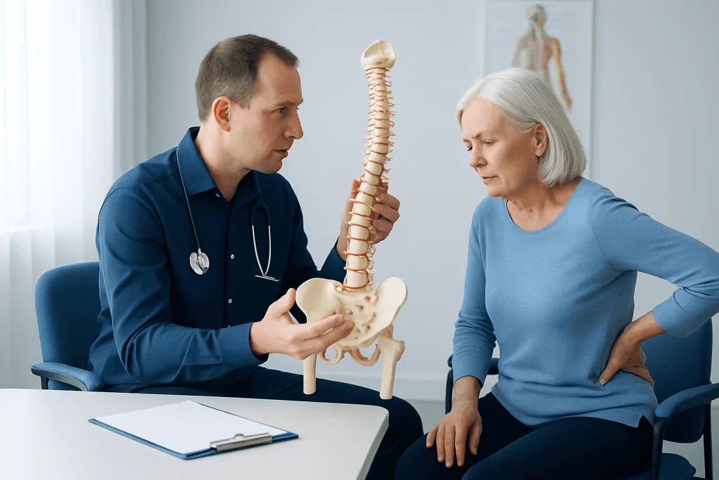
The most common cause of spinal stenosis is age-related wear and tear, also called degenerative change. Over time, the cushioning discs between the vertebrae may lose height and bulge. The small joints at the back of the spine can develop arthritis and enlarge. Ligaments may thicken, and bone spurs may form. Together, these changes can narrow the spinal canal or the openings where nerves exit.
Some people are born with a naturally narrow spinal canal, which may make symptoms appear earlier in life. Others develop stenosis after a spine injury, previous surgery, a slipped vertebra called spondylolisthesis, spinal curvature, or certain inflammatory or bone conditions. Disc herniation can also contribute to nerve compression, especially when combined with existing narrowing.
Risk factors include older age, a history of physically demanding work, previous spine injury, obesity, smoking, and conditions that affect posture or spinal alignment. Genetics can also influence disc health, arthritis, and canal size. However, imaging changes are not always the same as symptoms; some people have narrowing on MRI but little or no pain.
Because spinal stenosis develops in different ways, treatment should be individualized. The goal is not simply to treat an MRI image, but to match symptoms, examination findings, lifestyle needs, and imaging results to the most appropriate care plan.
Diagnosis
Diagnosis begins with a careful medical history. The doctor asks where the pain occurs, what triggers it, how far the person can walk, what relieves symptoms, and whether there is numbness, weakness, balance difficulty, or bladder and bowel change. Details about previous treatments, medical conditions, and medications are also important.
A physical examination usually includes checking posture, walking pattern, spinal movement, reflexes, muscle strength, sensation, and straight-leg testing. The doctor may also examine hip and knee joints because arthritis in these areas can mimic spine-related leg pain. Circulation in the legs may be assessed to help distinguish nerve-related walking pain from blood-flow problems.
Imaging is commonly used when symptoms persist, affect function, or suggest nerve compression. MRI is often the preferred test because it shows discs, nerves, ligaments, and the degree of canal narrowing. X-rays can show alignment, arthritis, instability, or spondylolisthesis. CT scans may be useful when MRI is not suitable or when detailed bone anatomy is needed.
In selected cases, additional tests may be recommended. Electromyography and nerve conduction studies can help evaluate nerve function and rule out other nerve disorders. Diagnostic injections may sometimes help identify which spinal level is causing pain. A complete diagnosis guides whether conservative care, injections, or surgery is most appropriate.
Treatment Options
Treatment depends on symptom severity, the degree of nerve compression, general health, and personal goals. Many people start with non-surgical care, especially if symptoms are mild to moderate and there is no progressive weakness. Conservative treatment aims to reduce pain, improve walking tolerance, strengthen supporting muscles, and help the person stay active safely.
Non-surgical options may include physiotherapy, posture training, stretching, core and hip strengthening, and low-impact exercise such as cycling or swimming. A physiotherapist may teach flexion-based exercises, walking strategies, and ways to pace activity. Medicines such as simple pain relievers, anti-inflammatory drugs, or nerve-pain medications may be considered, depending on the person’s health and medical history. All medication should be discussed with a qualified doctor, especially in older adults or people with kidney, stomach, heart, or blood pressure conditions.
Spinal injections may be used for some patients when pain limits rehabilitation or daily activity. Epidural steroid injections or targeted nerve root injections aim to reduce inflammation around irritated nerves. They do not correct the narrowing itself, but they may provide temporary relief and can be part of a broader treatment plan. The expected benefit varies, and risks and alternatives should be reviewed before the procedure.
Surgery may be considered when symptoms remain disabling despite appropriate non-surgical treatment, or when there is significant weakness, loss of function, or certain structural problems. The most common operation is decompression, such as laminectomy, where bone and thickened tissue are removed to create more space for the nerves. In some cases, fusion is added if there is spinal instability, deformity, or significant vertebral slipping. Minimally invasive techniques may be suitable for selected patients, using smaller incisions and targeted decompression, but the best approach depends on anatomy and the surgeon’s assessment.
Prevention and Self-Care
Not all cases of spinal stenosis can be prevented, especially when age-related changes or inherited anatomy play a role. However, healthy habits can reduce stress on the spine and help maintain mobility. The aim is to support the spine, protect nerve function, and prevent symptoms from becoming more limiting.
Regular low-impact activity is often helpful. Walking in shorter intervals, stationary cycling, water exercise, and guided strengthening can improve endurance without overloading the back. Exercises that build core, hip, and leg strength may improve posture and balance. Stretching the hip flexors, hamstrings, and lower back may also help some people, but exercises should be tailored to the individual.
Self-care strategies may include:
- Using a slightly forward-leaning posture when symptoms occur
- Taking planned sitting breaks during longer walks
- Maintaining a healthy weight to reduce mechanical load
- Avoiding smoking, which can affect disc and bone health
- Using supportive footwear and fall-prevention measures
- Following safe lifting techniques and avoiding sudden heavy strain
People with spinal stenosis should avoid prolonged bed rest unless advised for a specific short-term reason. Staying active within comfortable limits is usually better for strength, circulation, mood, and independence. A healthcare professional can help design a safe plan, especially when there are other conditions such as osteoporosis, diabetes, heart disease, or joint arthritis.
When to See a Doctor
A medical evaluation is recommended when leg pain while walking is persistent, recurrent, or limits daily activities. It is also important to seek advice if numbness, tingling, weakness, balance problems, or back pain are worsening. Early assessment can help identify the cause and start appropriate treatment before activity levels decline.
Urgent medical attention is needed if there is new loss of bladder or bowel control, numbness in the groin or saddle area, rapidly worsening leg weakness, fever with severe back pain, unexplained weight loss, or pain after a significant injury. These symptoms may indicate a condition that requires prompt evaluation.
Patients who are considering surgery should have a detailed discussion about expected benefits, possible risks, recovery time, and alternatives. A second opinion may be helpful when symptoms are complex or imaging findings involve several spinal levels. Decisions are usually best made through shared discussion between the patient, spine specialist, and rehabilitation team.
Acibadem International’s multidisciplinary specialists and JCI-accredited hospitals diagnose and treat spinal stenosis for international patients, including assessment, imaging, rehabilitation planning, interventional pain procedures, and surgical care when appropriate. Patients should always consult a qualified physician to determine the safest and most suitable plan for their individual condition.
Frequently asked questions
Why does spinal stenosis cause leg pain while walking?
Walking upright can reduce the available space around the nerves in the lower spine. In spinal stenosis, this may increase nerve irritation and cause pain, heaviness, numbness, or weakness in the legs. Sitting or leaning forward often relieves symptoms because it can slightly widen the spinal canal.
Is spinal stenosis the same as a slipped disc?
No. A slipped or herniated disc is one possible cause of nerve compression, while spinal stenosis refers to narrowing of the spinal canal or nerve openings. Many patients with stenosis have a combination of disc bulging, arthritis, ligament thickening, and bone overgrowth.
Can spinal stenosis improve without surgery?
Yes, many people manage symptoms with physiotherapy, activity modification, medication, and sometimes spinal injections. These treatments do not always reverse the narrowing, but they may reduce pain and improve walking tolerance. Surgery is usually considered when symptoms remain disabling or neurological problems progress.
What exercises are usually helpful for lumbar spinal stenosis?
Low-impact exercise such as stationary cycling, swimming, and guided walking intervals may be useful. Many people benefit from supervised core, hip, and leg strengthening, along with flexibility work. Exercises should be personalized, especially if there is osteoporosis, balance difficulty, or other joint problems.
When is surgery recommended for spinal stenosis?
Surgery may be recommended when pain, numbness, or walking limitation remains significant despite appropriate non-surgical care. It may also be advised sooner if there is progressive weakness or certain structural problems such as instability. The decision depends on symptoms, examination findings, imaging, general health, and patient goals.
Is minimally invasive spine surgery suitable for everyone?
Minimally invasive techniques can be helpful for selected patients, but they are not the best choice for every type of stenosis. Suitability depends on the location and severity of narrowing, spinal alignment, instability, previous surgery, and overall health. A spine specialist can explain which approach is safest and most appropriate.
References
- American Academy of Orthopaedic Surgeons
- North American Spine Society
- National Institute of Arthritis and Musculoskeletal and Skin Diseases
- Mayo Clinic
- National Institute for Health and Care Excellence
This article is for general information only and is not a substitute for professional medical advice. Please consult a qualified doctor about your individual situation.
Joint and spine care in Turkey — expert assessment & treatment
JCI-accredited · board-certified surgeons · reply within 24h
Add us as a Preferred Source to see more of our trusted health content across Google Search, AI Overviews and Discover.

Orthopedics & Spine
Care for bones, joints and the musculoskeletal system, including joint replacement, sports injuries and trauma.
125 specialists in this unitMore from the Health Library



Related Specialists

Dr. İmam Bakir Batı
Liver Transplant Center
Prof. Dr. Fuat Bilgen
Cardiovascular Surgery
Assoc. Prof. Dr. Mehmet Altiparmak
Aesthetic Plastic & Reconstructive Surgery
Prof. Dr. Mert Akan
Anesthesiology