Stage 1 Bladder Cancer CT Scan: Preparation, Procedure and Results
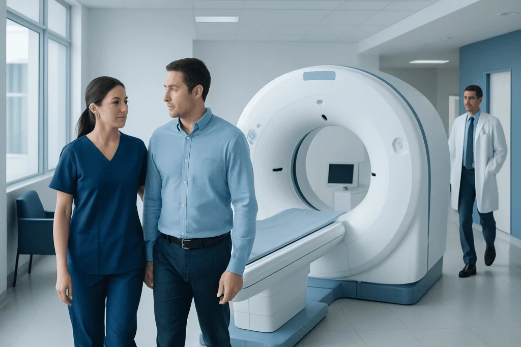
CT scans help evaluate the urinary tract and look for enlarged lymph nodes or possible spread outside the bladder. A CT scan cannot reliably determine the exact depth of bladder-wall invasion on its own; cystoscopy and biopsy provide confirmation.
Key Takeaways
- CT scans help evaluate the urinary tract and look for enlarged lymph nodes or possible spread outside the bladder.
- A CT scan cannot reliably determine the exact depth of bladder-wall invasion on its own; cystoscopy and biopsy provide confirmation.
- The scan itself usually takes only minutes, although the full appointment may take longer when contrast imaging is used.
- Most people can return to usual activities shortly after a CT scan, unless sedation or another procedure is also planned.
- Contrast dye is generally safe but should be discussed in advance for people with kidney disease, diabetes, thyroid conditions or prior contrast reactions.
A stage 1 bladder cancer CT scan is commonly used to examine the kidneys, ureters, bladder and nearby tissues for signs that cancer may extend beyond the bladder. It supports staging and treatment planning, but cystoscopy with tissue sampling remains essential for confirming the diagnosis and cancer stage.
Overview: the role of CT scanning in stage 1 bladder cancer
A stage 1 bladder cancer CT scan is an imaging examination used to assess the urinary tract and look for signs that cancer may have grown outside the bladder or reached lymph nodes or other organs. It is useful for the wider picture of staging, but it does not replace cystoscopy and biopsy, which are needed to identify the tumor type and confirm how deeply it has grown into the bladder wall.
Stage 1 bladder cancer generally means that cancer has grown into the connective tissue beneath the bladder lining but has not reached the bladder muscle. This is often called non-muscle-invasive bladder cancer, although accurate staging depends on tissue obtained during a procedure such as transurethral resection of a bladder tumor (TURBT).
CT imaging may be performed before treatment, after an initial diagnosis, or when symptoms and clinical findings suggest a need to examine the kidneys, ureters and surrounding structures. The care team interprets CT findings alongside pathology results, cystoscopy findings, urine tests and a person’s medical history.
How a CT scan works and who may need one
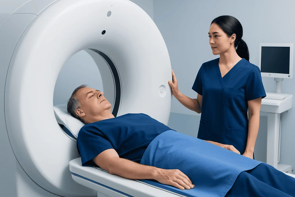
Computed tomography, or CT, uses X-rays and computer processing to create detailed cross-sectional images of the body. For bladder cancer assessment, clinicians may request a CT scan of the abdomen and pelvis. A CT urogram is a specialized approach that uses intravenous contrast material and timed images to show how contrast passes through the kidneys, ureters and bladder.
CT may be particularly helpful for people with blood in the urine, a newly diagnosed bladder tumor, higher-risk pathology features, urinary tract obstruction, unexplained flank pain, or concern for disease beyond the bladder. It can also identify some conditions that may coexist with or resemble bladder cancer, such as kidney stones, masses in the upper urinary tract, or swollen kidneys caused by blocked urine flow.
Not every person with an early bladder tumor needs the same imaging schedule. The decision is individualized according to tumor grade, size, number of tumors, pathology findings, symptoms, kidney function and previous imaging. A urologist can explain whether CT, ultrasound, MRI or another test is most appropriate.
- CT is especially useful for evaluating the kidneys and ureters.
- It can assess nearby organs, lymph nodes and distant sites when clinically indicated.
- It is less precise than tissue sampling for determining superficial versus deeper bladder-wall invasion.
Preparation and step-by-step procedure
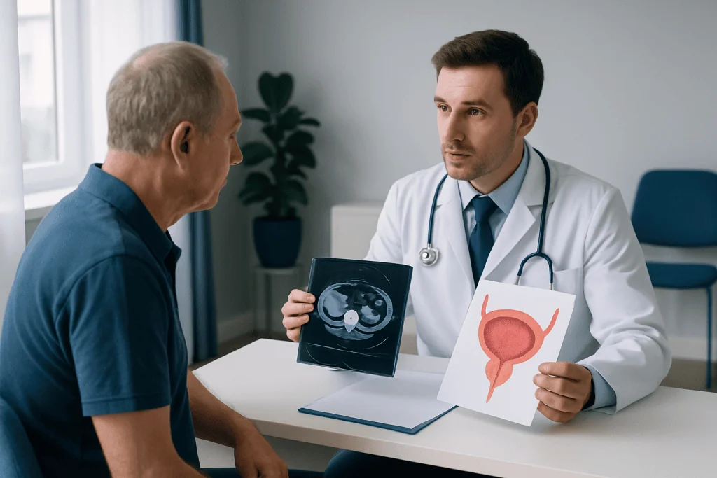
The imaging center provides specific instructions before the appointment. Some people may be asked not to eat for several hours before a contrast-enhanced CT scan, while clear fluids may still be allowed. Instructions differ by facility and scan type, so patients should follow the directions given by their radiology team.
Before the scan, staff usually ask about kidney disease, diabetes, thyroid conditions, asthma, pregnancy, medications and previous reactions to iodinated contrast. A recent blood test may be needed to check kidney function when contrast is planned. Patients should report a history of serious contrast reaction; the team can consider precautions or an alternative imaging approach.
During the examination, the person lies on a padded table that moves through the CT scanner. If contrast is used, it is injected through a small intravenous line. A temporary warm sensation, metallic taste or feeling of needing to urinate can occur and usually passes quickly. The radiographer may ask the person to hold their breath briefly while images are taken.
After imaging, the radiologist reviews the scan and sends a report to the referring clinician. The urology team then discusses the findings in the context of cystoscopy, pathology and any planned treatment. Drinking fluids afterward is often advised unless a clinician has recommended fluid restriction for another health reason.
What does a CT scan indicate about Stage 1 bladder cancer?
A CT scan can show a bladder mass in some cases and may reveal features that raise or lower concern for disease outside the bladder. It can help identify enlarged lymph nodes, urinary tract blockage, changes in the kidneys or ureters, and findings in the abdomen, pelvis or chest that require further assessment. A normal CT scan is reassuring regarding obvious spread, but it cannot rule out microscopic cancer cells.
Importantly, CT cannot consistently distinguish a tumor confined to the inner layers of the bladder from one that has invaded more deeply into the muscle. Small, flat or superficial tumors may not be visible on CT at all. For this reason, staging relies on cystoscopy and pathology from tumor tissue, usually obtained during TURBT.
If cancer is confirmed as stage 1, management commonly focuses on complete endoscopic tumor removal, assessment of recurrence and progression risk, and carefully planned cystoscopic surveillance. Depending on risk features, treatment may include medication delivered into the bladder. The exact plan should be guided by a urologist and the pathology report.
How long is a bladder CT scan?
The image acquisition portion of a bladder CT scan generally takes only a few minutes. However, a complete appointment may last around 30 to 60 minutes because it includes registration, safety screening, placement of an intravenous line when needed, possible contrast timing and post-scan instructions.
A CT urogram can take longer than a basic CT because images may be obtained at more than one time point. Delayed images allow the radiology team to see contrast as it travels through the urinary tract. The exact duration depends on the scan protocol and whether additional areas, such as the chest, also need imaging.
CT scanning is painless and does not usually require sedation. Remaining still is important for clear images. People with anxiety about enclosed spaces should mention this before the appointment; CT scanners are generally more open than MRI scanners, and staff can explain what to expect.
Which is better, a cystoscopy or a CT scan?
Cystoscopy and CT scanning serve different, complementary purposes, so one is not simply better than the other. Cystoscopy allows a urologist to look directly inside the bladder with a thin camera. It is the main test for detecting many bladder tumors, including small or flat abnormalities that CT may miss.
During cystoscopy, a suspicious area can be biopsied or removed. This tissue is examined under a microscope to determine the cancer type, grade and depth of invasion. These findings are central to confirming stage 1 disease and selecting treatment.
CT provides information that cystoscopy cannot provide as fully, including the appearance of the kidneys and ureters, the tissues around the bladder, lymph nodes and other organs. In a well-planned bladder cancer evaluation, the two tests are often used together rather than as alternatives.
For a confirmed or suspected tumor, a urologist may recommend bladder cancer treatment based on pathology, imaging and individual risk factors. Follow-up typically includes repeat cystoscopy because early bladder cancer can recur inside the bladder even after successful initial treatment.
Benefits, possible risks and recovery after CT imaging
The principal benefit of CT is that it provides rapid, detailed imaging of the urinary tract and surrounding anatomy. This can help clinicians identify issues that affect staging, select further tests and plan treatment safely. CT is widely available and is particularly useful when a prompt assessment is needed.
CT uses ionizing radiation, so clinicians aim to use it only when the expected diagnostic benefit outweighs the risk. Modern protocols are designed to keep radiation exposure as low as reasonably achievable. The amount of radiation varies with the type and number of scans performed.
Iodinated contrast can occasionally cause nausea, itching, hives or other allergic-like reactions. Severe reactions are uncommon, and imaging staff are trained to respond. Contrast may also require additional caution in people with significantly reduced kidney function. Patients should share all relevant health information so the team can make the examination as safe as possible.
Most people feel well immediately after the scan and can resume normal routines the same day. Those who receive contrast may be advised to drink fluids if medically appropriate. A clinician should be contacted promptly if symptoms such as trouble breathing, swelling, widespread rash or persistent vomiting occur after leaving the imaging center.
How fast does stage 1 bladder cancer spread?
Stage 1 bladder cancer does not have one predictable rate of spread. Many stage 1 tumors remain confined to the bladder for a long time, particularly when they are low grade and are completely removed. However, some tumors have a higher chance of returning or progressing, especially when they are high grade, large, multiple, recurrent, associated with carcinoma in situ, or have certain microscopic features.
Because progression risk varies, prompt evaluation and regular follow-up are important. A urologist uses the pathology report and tumor characteristics to classify risk and recommend an appropriate surveillance schedule. Follow-up may include cystoscopy, urine testing and imaging when indicated.
People should avoid interpreting a CT result alone as a definitive prediction of behavior. The pathology findings after tumor removal offer more meaningful information about likely risk. Discussing results with the treating urologist can clarify what the individual diagnosis means and what monitoring is needed.
When to seek medical care
Visible blood in the urine, even when painless or temporary, should be assessed by a healthcare professional. Other symptoms that warrant medical review include persistent burning during urination, frequent urination, new urgency, difficulty passing urine, recurrent urinary infections or unexplained pain in the side or lower back. These symptoms are common and can have non-cancer causes, but they should not be ignored.
Urgent medical care is appropriate for heavy bleeding with clots, inability to urinate, severe pain, fever with urinary symptoms, fainting, chest pain or signs of a severe allergic reaction after contrast. People already diagnosed with bladder cancer should contact their care team if new urinary symptoms develop between scheduled visits.
Bladder cancer care may involve urologists, radiologists, pathologists, medical oncologists and radiation oncologists, depending on the diagnosis and treatment needs. Acibadem International’s multidisciplinary specialists and JCI-accredited hospitals support diagnosis and treatment planning for international patients.
Frequently asked questions
Does a CT scan confirm stage 1 bladder cancer?
No. A CT scan can support staging by assessing the urinary tract, lymph nodes and nearby or distant organs, but it cannot reliably confirm the depth of invasion within the bladder wall. Cystoscopy and tissue examination after biopsy or tumor removal are required to establish the stage.
Is contrast always used for a bladder cancer CT scan?
Contrast is often used for CT urography because it helps show the kidneys, ureters and bladder in more detail. However, the imaging protocol depends on the clinical question and a person’s kidney function, allergy history and other health factors. The radiology and urology teams decide whether contrast is suitable.
Can a CT scan miss bladder cancer?
Yes. Small, flat and superficial bladder tumors may not be seen on CT imaging. Cystoscopy is more sensitive for examining the inner bladder lining, which is why it remains a key part of diagnosis and follow-up.
Do people need to fast before a bladder CT scan?
Some contrast-enhanced CT examinations require a short fasting period, while others do not. The imaging center will provide instructions based on the scan protocol. Patients should ask in advance if they are unsure, especially if they take regular medication or have diabetes.
What happens if the CT scan shows enlarged lymph nodes?
Enlarged lymph nodes can occur for several reasons, including inflammation or infection, and do not by themselves prove cancer spread. The treating team may recommend additional imaging, repeat assessment or other tests depending on the full clinical picture. Pathology and cystoscopy findings remain important when interpreting the result.
How soon are CT scan results available?
A radiologist usually reviews the images and prepares a report for the referring clinician, often within a few days, although timing varies by facility and urgency. The urologist should explain what the findings mean alongside pathology and cystoscopy results. Patients can ask when and how their results will be communicated.
References
- European Association of Urology
- American Urological Association
- National Cancer Institute
- Radiological Society of North America
- American Cancer Society
This article is for general information only and is not a substitute for professional medical advice. Please consult a qualified doctor about your individual situation.
Bladder Cancer in Turkey — costs, top hospitals & a free quote
JCI-accredited · board-certified surgeons · reply within 24h
Add us as a Preferred Source to see more of our trusted health content across Google Search, AI Overviews and Discover.

Medical Oncology Department
Medical treatment of cancer with chemotherapy, immunotherapy and targeted therapies under a multidisciplinary tumor board.
60 specialists in this unit