Stereotactic Laser Ablation: Procedure, Recovery and Results

Stereotactic laser ablation uses MRI guidance and a thin laser probe to heat and destroy selected abnormal brain tissue. It is considered surgery, although it usually involves a small skull opening rather than a large craniotomy.
Key Takeaways
- Stereotactic laser ablation uses MRI guidance and a thin laser probe to heat and destroy selected abnormal brain tissue.
- It is considered surgery, although it usually involves a small skull opening rather than a large craniotomy.
- Suitability depends on the diagnosis, lesion location and size, symptoms, prior treatment, and overall health.
- Many people leave hospital within one to a few days, but full recovery and treatment response vary substantially.
- Potential risks include swelling, bleeding, infection, seizures, and new or worsening neurological symptoms.
Stereotactic laser ablation is an image-guided, minimally invasive brain procedure that delivers controlled laser heat to a precisely selected area of abnormal tissue. It may be considered for certain brain tumors, radiation-related tissue injury, epilepsy-related targets, or other carefully evaluated lesions when open surgery is not the preferred option.
Overview: What Is Stereotactic Laser Ablation?
Stereotactic laser ablation, sometimes called stereotactic laser ablation (SLA), laser interstitial thermal therapy, or stereotactic laser surgery, is a minimally invasive neurosurgical procedure. A neurosurgeon places a very thin laser fiber into a planned area of the brain and uses real-time MRI temperature monitoring to deliver controlled heat. The heat damages targeted tissue while helping the team protect nearby healthy structures.
Stereotactic laser ablation brain surgery may be used for selected tumors, recurrent tumors, radiation necrosis (tissue injury after radiation therapy), and certain forms of epilepsy. It is not appropriate for every brain lesion or every person. The decision is made by a multidisciplinary team after reviewing imaging, symptoms, pathology when available, previous therapies, and the person’s individual goals of care.
Unlike open brain surgery, this procedure generally uses a small incision and a small opening in the skull. However, it remains a significant medical procedure requiring anesthesia, advanced imaging, and specialist follow-up. The aim may be to reduce or control a lesion, obtain tissue for diagnosis in some cases, improve symptoms, or reduce seizure activity, depending on the underlying condition.
How Stereotactic Laser Ablation Works
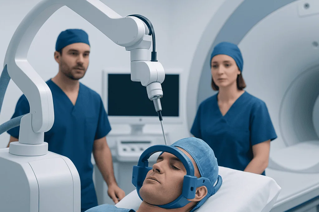
The word “stereotactic” refers to the use of three-dimensional imaging and navigation to reach a precise target in the brain. Before treatment, the surgical team studies MRI and sometimes CT images to map the safest route to the lesion. In some centers, stereotactic robotic assisted laser ablation may support accurate positioning of the laser probe, alongside the surgeon’s planning and clinical judgment.
Once the fiber is in place, the person is moved into an MRI scanner or treated with MRI-based monitoring. The laser creates heat at the tip of the probe, and MRI thermometry provides a live temperature map. This enables the team to watch the treatment zone and stop or adjust energy delivery when the planned boundary is reached.
Following treatment, the ablated tissue does not disappear immediately. The body gradually clears and remodels the treated area over time. Early MRI scans can show temporary enlargement from inflammation or swelling, so specialists interpret follow-up images alongside symptoms, neurological examinations, and the original diagnosis.
Who May Be a Candidate?

Candidacy is highly individualized. Stereotactic laser ablation may be considered when a lesion is relatively small and can be reached through a safe pathway, particularly when it is deep within the brain or located in an area where open surgery may carry added risk. It can also be an option when prior surgery or radiation has already been performed, although previous treatments must be carefully reviewed.
For people with brain tumors, the procedure may be discussed for a newly diagnosed lesion, a recurrent lesion, or a lesion suspected to represent radiation necrosis rather than active tumor. It may also be considered for carefully selected people with drug-resistant focal epilepsy when the seizure source has been identified. Related neurological assessment may include review of epilepsy history, seizure recordings, neuropsychological testing, and detailed brain imaging.
Lesion size, shape, number, and proximity to areas controlling movement, language, vision, memory, or sensation all affect the decision. A person’s general health, medications, blood-thinning treatment, ability to undergo anesthesia, and expectations for recovery are also important. A consultation with a neurosurgeon does not automatically mean the procedure is recommended; it is an opportunity to compare appropriate options.
Step by Step: What Happens During the Procedure?
Preparation usually begins with preoperative imaging, blood tests, anesthesia assessment, and a discussion of medicines that may need adjustment. The care team explains the planned target, possible benefits and limitations, alternatives, and potential complications. Depending on the indication, a biopsy may be performed during the same procedure or separately to clarify whether tissue is tumor, radiation injury, or another process.
On the day of surgery, the person receives anesthesia. The head is positioned securely, and the surgical team uses a navigation system, frame, or robotic guidance to identify the planned entry point. After making a small incision and a small opening in the skull, the surgeon advances the laser catheter along the preplanned path to the target.
The team then uses MRI guidance to monitor treatment temperatures and the developing ablation zone. After the desired treatment area has been covered, the laser fiber is removed and the incision is closed. The person is monitored in a recovery area and may spend time in a neurological observation unit or intensive care setting, depending on the diagnosis, location of treatment, and individual needs.
For patients considering this approach, laser ablation treatment should be discussed in the context of the underlying condition and all reasonable surgical, radiation, medical, and observation options.
Recovery Timeline and What Recovery Feels Like
How long does it take to recover from laser ablation surgery? Many people are able to leave hospital within one to a few days after an uncomplicated procedure, but the overall recovery period is different for each person. Returning to light daily activity may take days to a few weeks, while recovery of neurological symptoms, stamina, and readiness for work or travel can take longer. The diagnosis, treated brain region, extent of ablation, and any pre-existing symptoms are important factors.
What is the recovery like after stereotactic surgery? It is often less physically demanding than recovery after a large craniotomy because the incision is small and there is less disruption of the scalp and skull. Still, fatigue, headache, scalp tenderness, nausea, temporary changes in balance or thinking, and emotional adjustment can occur. Brain swelling can develop after treatment, so clinicians may prescribe medications such as corticosteroids or anti-seizure medicines when appropriate.
Follow-up commonly includes a wound check, neurological review, and repeat MRI scans. Family members or caregivers may be asked to watch for changes in speech, strength, vision, alertness, behavior, or seizures. Patients should follow individualized advice about driving, exercise, bathing, work, alcohol, medications, and travel, particularly if they have had seizures or new neurological symptoms.
Benefits, Limitations, Risks and Pain
Potential benefits of stereotactic laser ablation include a smaller incision, shorter hospital stay for many patients, and access to lesions that may be difficult to reach through open surgery. It may also offer an option for people who have already received radiation or surgery. However, its benefits depend on the disease being treated, and it does not replace open surgery, radiation therapy, systemic treatment, or observation in every situation.
Is laser ablation considered surgery? Yes. Although it is minimally invasive, it is a form of neurosurgery because it involves anesthesia, surgical placement of a probe through the skull, and treatment of brain tissue. It should be approached with the same careful discussion of risks, consent, recovery needs, and follow-up as other surgical procedures.
How painful is laser ablation? The procedure itself is performed under anesthesia, so patients do not feel the laser treatment while it is happening. Afterwards, pain is often related to the small scalp incision or headache and can frequently be managed with prescribed or over-the-counter pain relief recommended by the care team. Pain experience varies, and severe, increasing, or unusual headache should be reported promptly.
Possible complications include bleeding, infection, swelling, seizure, stroke-like neurological deficits, confusion, weakness, speech or vision changes, and complications related to anesthesia. Some risks are temporary, while others can be lasting. The treating neurosurgeon can explain the risks most relevant to the lesion’s location and the person’s health.
When to Seek Medical Care
After stereotactic laser surgery, patients should contact their surgical team promptly for worsening headache, fever, wound redness or drainage, repeated vomiting, new or worsening seizures, increasing sleepiness, confusion, or a meaningful change in balance, speech, vision, sensation, or strength. These symptoms do not always indicate a serious complication, but they require timely medical assessment.
Emergency care is needed for sudden weakness or facial drooping, difficulty speaking or understanding speech, loss of consciousness, a prolonged seizure, severe breathing difficulty, or the worst headache of a person’s life. Caregivers should follow the emergency plan provided by the clinical team and should not drive a person who is having an acute neurological emergency unless emergency services are unavailable.
Long-term follow-up is essential because symptoms and imaging changes can occur after the initial recovery period. Acibadem International’s multidisciplinary specialists and JCI-accredited hospitals evaluate and treat neurological conditions for international patients, coordinating neurosurgical care with neurology, oncology, radiology, rehabilitation, and supportive services when needed.
Frequently asked questions
What conditions can stereotactic laser ablation treat?
Stereotactic laser ablation may be used for selected brain tumors, recurrent lesions, radiation necrosis, and certain focal epilepsy targets. The appropriate use depends on the diagnosis, lesion characteristics, location, prior treatment, and the person’s overall health. A multidisciplinary review is often needed to determine whether it is suitable.
Is stereotactic laser ablation safer than open brain surgery?
It may reduce some risks associated with a larger incision and craniotomy, but it is not automatically safer for every lesion or patient. It has its own risks, including brain swelling, bleeding, seizures, and neurological changes. The safest option depends on the target area and the goals of treatment.
How long does stereotactic laser ablation take?
The total time varies because it includes anesthesia, positioning, image guidance, placement of the laser probe, MRI monitoring, and recovery from anesthesia. The ablation portion itself may be relatively brief, but patients should expect the overall process to take several hours. The surgical team can provide a more specific estimate after planning.
Will the treated brain lesion disappear immediately after laser ablation?
No. The treated tissue changes gradually after ablation, and early scans may show swelling or temporary enlargement of the treatment area. Follow-up MRI imaging helps the clinical team distinguish expected post-treatment changes from progression or other concerns. The timing and meaning of imaging changes depend on the underlying condition.
Can seizures occur after stereotactic laser ablation?
Yes. Seizures can occur after a brain procedure, especially when treatment involves an area associated with seizure activity or when there is post-procedure swelling. Some patients are prescribed anti-seizure medication before or after treatment. New, prolonged, or repeated seizures require urgent medical advice.
When can someone return to work after laser ablation surgery?
The timing depends on the type of work, neurological symptoms, fatigue, imaging findings, and whether further treatment is needed. Some people return to light activities within a few weeks, while others need a longer recovery period. The treating team should give individualized clearance, especially for driving, physically demanding work, or safety-sensitive roles.
References
- National Institute of Neurological Disorders and Stroke
- American Association of Neurological Surgeons
- National Cancer Institute
- Epilepsy Foundation
- Radiological Society of North America
This article is for general information only and is not a substitute for professional medical advice. Please consult a qualified doctor about your individual situation.
Explore treatments in Turkey — costs, top hospitals & a free quote
JCI-accredited · board-certified surgeons · reply within 24h
Add us as a Preferred Source to see more of our trusted health content across Google Search, AI Overviews and Discover.