Thalamus Brain — Explained by Medical Evidence, Not Myths
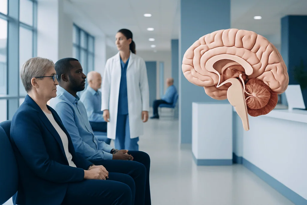
The thalamus is located deep in the brain, with one thalamus on each side near the third ventricle. It filters and relays most sensory information to the cerebral cortex; smell is the main sensory exception.
Key Takeaways
- The thalamus is located deep in the brain, with one thalamus on each side near the third ventricle.
- It filters and relays most sensory information to the cerebral cortex; smell is the main sensory exception.
- Different thalamic regions support different functions, including movement, alertness, memory and emotional processing.
- Stroke, bleeding, inflammation, tumors, infection and injury can affect the thalamus and cause symptoms that vary by location.
- Sudden weakness, numbness, speech difficulty, vision changes, severe imbalance or altered consciousness needs urgent medical assessment.
The thalamus is a small but important structure deep within the brain that helps organize and relay information between the body, brainstem and cerebral cortex. It contributes to sensation, movement, attention, consciousness, sleep and aspects of memory and emotion, but it is not a single “control center” for the brain.
Thalamus brain: what it is and why it matters
The thalamus brain refers to the thalamus, a pair of oval-shaped structures positioned deep in the center of the brain. It sits above the brainstem and below the cerebral cortex, the folded outer layer involved in conscious thought, voluntary movement, language and perception. Although relatively small, the thalamus has extensive connections with many brain regions.
Its best-known role is as a relay and processing hub. Much of the sensory information arriving from the body passes through the thalamus before reaching the cortex, where it can be recognized and interpreted. The thalamus does not simply forward every signal unchanged; it helps prioritize, regulate and coordinate information according to a person’s state of attention, sleep or wakefulness.
The thalamus is made up of several groups of nerve-cell nuclei, each with specialized connections and tasks. This organization helps explain why a problem affecting one part of the thalamus can cause a different pattern of symptoms than a problem affecting another part. Effects may involve sensation, movement, vision, memory, alertness or pain, depending on the area involved.
Where is the thalamus located?
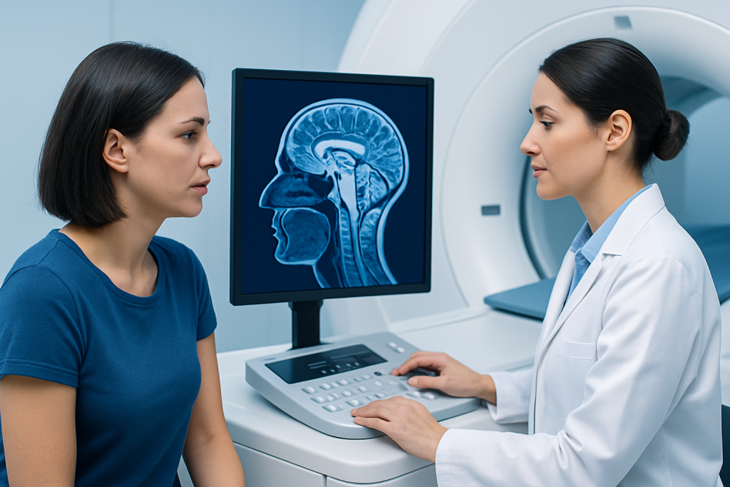
There are two thalami: one on the left and one on the right side of the brain. They lie within an area called the diencephalon, close to the center of the head. The two structures sit on either side of the third ventricle, a narrow fluid-filled space that is part of the brain’s ventricular system.
The thalamus is near several other important structures. Below it is the hypothalamus, which has key roles in hormone regulation, temperature, appetite and the autonomic nervous system. Behind it are areas involved in visual processing, while below and behind it are parts of the midbrain. Its central location allows the thalamus to communicate efficiently with the cortex, brainstem, spinal cord and deeper brain networks.
Because it is deep inside the brain, the thalamus cannot be assessed by looking at the outside of the head or through a routine physical examination alone. When a clinician suspects a disorder in this area, brain imaging such as computed tomography (CT) or magnetic resonance imaging (MRI) may be used alongside a detailed neurological examination.
What does the thalamus do?
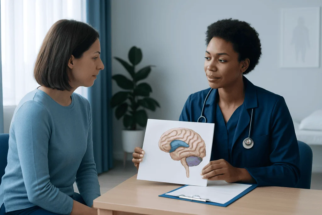
The thalamus helps process sensory information from the body. Signals related to touch, pressure, pain, temperature, hearing and vision generally pass through thalamic pathways before reaching the appropriate areas of the cerebral cortex. Smell is the main exception: olfactory signals initially reach cortical areas without the same primary thalamic relay.
The thalamus also participates in movement control through its connections with the basal ganglia, cerebellum and motor cortex. These networks help plan, start and refine voluntary movement. In addition, thalamic-cortical circuits are important for maintaining alertness, directing attention and supporting normal transitions between wakefulness and sleep.
Some thalamic regions are involved in memory, learning and emotional processing through their connections with the hippocampus and limbic system. This does not mean the thalamus stores every memory or creates every emotion. Rather, it contributes to broader networks in which several brain areas work together. Modern neuroscience describes the thalamus as an active coordinator, not merely a passive switchboard.
- Sensation: relays and helps regulate sensory input.
- Attention and consciousness: supports alertness and awareness.
- Movement: communicates with movement-control networks.
- Sleep: helps shape sleep rhythms and sensory responsiveness.
- Memory and emotion: contributes to connected brain circuits.
Common myths about the thalamus
A common oversimplification is that the thalamus is only a sensory relay station. Sensory processing is an important role, but the thalamus also has two-way communication with the cortex and interacts with networks involved in movement, attention, sleep, memory and behavior. Information moves through these circuits continuously rather than along one simple route.
Another myth is that a thalamic condition always causes loss of consciousness. Major disturbances in alertness can occur when certain thalamic regions or connected structures are affected, especially on both sides. However, many people with a small, one-sided thalamic lesion remain awake and may instead experience numbness, weakness, coordination difficulty, visual changes or cognitive symptoms.
It is also inaccurate to assume that all pain or sensory changes after a thalamic injury will be the same. The thalamus contains distinct nuclei with different connections. A person’s symptoms, recovery and care plan depend on the underlying cause, the precise location and size of the affected area, other medical conditions and the timing of treatment.
What can affect the thalamus?
Stroke is one of the more important causes of sudden thalamic dysfunction. A blockage in a small blood vessel can reduce blood flow to part of the thalamus, while a hemorrhagic stroke involves bleeding into brain tissue. Symptoms can begin suddenly and may include altered sensation on one side of the body, weakness, unsteadiness, speech or vision changes, confusion, or severe sleepiness. A suspected stroke is an emergency.
Other possible causes include head injury, brain tumors, infections, inflammatory or autoimmune disorders, and conditions that affect blood vessels. Less commonly, metabolic disturbances or lack of oxygen can injure deep brain structures, including the thalamus. Some neurological conditions affecting movement or memory involve thalamic networks, even when the thalamus is not the only area involved.
Damage to thalamic pathways may sometimes lead to persistent neuropathic pain, often called central post-stroke pain when it occurs after a stroke. Pain may be burning, aching, sharp or triggered by light touch or temperature changes. This type of pain is real and can be challenging, but a neurologist or pain specialist can help identify suitable management options.
How thalamic disorders are diagnosed and treated
Diagnosis begins with a medical history and neurological examination. A clinician may assess strength, sensation, reflexes, coordination, speech, eye movements, walking, attention and memory. The pattern and timing of symptoms can help identify whether the thalamus or another part of the nervous system may be involved.
Emergency CT imaging is often used when stroke or bleeding is suspected, while MRI can provide more detailed images of the thalamus and surrounding structures. Depending on the situation, clinicians may also request blood tests, blood-vessel imaging, heart assessment, electroencephalography (EEG), lumbar puncture or other targeted investigations. These tests are selected according to the symptoms and likely cause.
Treatment focuses on the cause rather than the thalamus alone. Stroke care may include urgent therapies to restore blood flow when appropriate, treatment for bleeding, and measures to reduce future vascular risk. Infections, inflammation, tumors and metabolic conditions each require condition-specific care. Rehabilitation may include physiotherapy, occupational therapy, speech and language therapy, cognitive rehabilitation or pain management to support function and independence.
Acibadem International’s multidisciplinary specialists and JCI-accredited hospitals evaluate and treat neurological conditions affecting international patients. Individual care decisions should always be made with a qualified clinician who can review imaging, symptoms and the person’s overall health.
When to seek medical care
Emergency medical care is needed for sudden neurological symptoms, even if they improve quickly. Warning signs include new weakness or numbness of the face, arm or leg; difficulty speaking or understanding speech; sudden loss of balance; unexplained confusion; vision changes; a severe sudden headache; seizures; fainting; or unusual drowsiness. These symptoms may be caused by stroke or another urgent neurological condition.
It is also sensible to arrange a non-urgent medical appointment for persistent or recurring numbness, unexplained burning pain, new tremor, changes in coordination, memory concerns, disrupted sleep with other neurological symptoms, or changes in daily functioning. These symptoms do not necessarily mean there is a thalamic problem, but they deserve appropriate evaluation.
There is no reliable home test for thalamus function, and online information cannot diagnose brain symptoms. Prompt assessment is especially important for people with high blood pressure, diabetes, high cholesterol, an irregular heartbeat, smoking history, previous stroke or transient ischemic attack, or a recent head injury.
Frequently asked questions
Is the thalamus part of the brain?
Yes. The thalamus is a paired structure deep within the brain, located near its center. It is part of the diencephalon and has widespread connections with the cerebral cortex, brainstem and other brain regions.
What is the main function of the thalamus?
The thalamus helps process and relay much of the sensory information that reaches the cerebral cortex. It also contributes to movement, attention, alertness, sleep, memory and emotional networks.
Does all sensory information pass through the thalamus?
Most sensory pathways, including those for touch, pain, vision and hearing, have important thalamic relays. Smell is the principal exception because olfactory signals initially reach cortical areas through a different route.
What happens if the thalamus is damaged?
Effects depend on the exact area and cause of damage. Possible symptoms include numbness, altered pain perception, weakness, poor coordination, vision or speech changes, memory difficulties, confusion, sleepiness or changes in alertness.
Can a person recover from a thalamic stroke?
Many people improve after a thalamic stroke, although recovery varies widely. Early medical care, rehabilitation, management of stroke risk factors and support for specific symptoms can all play important roles.
Can stress damage the thalamus?
Everyday stress is not known to directly cause a focal thalamic injury such as a stroke or bleeding. Long-term stress can affect sleep, mood and cardiovascular health, so managing it may still support overall brain and body health.
References
- National Institute of Neurological Disorders and Stroke
- American Stroke Association
- National Library of Medicine
- Merck Manual Consumer Version
This article is for general information only and is not a substitute for professional medical advice. Please consult a qualified doctor about your individual situation.
Explore treatments in Turkey — costs, top hospitals & a free quote
JCI-accredited · board-certified surgeons · reply within 24h
Add us as a Preferred Source to see more of our trusted health content across Google Search, AI Overviews and Discover.