Tibial Fracture Xray: Preparation, Procedure and Results

A tibial fracture xray usually includes front and side views of the tibia and nearby joints. No special preparation is usually needed, but patients should tell staff about pregnancy or a possible pregnancy.
Key Takeaways
- A tibial fracture xray usually includes front and side views of the tibia and nearby joints.
- No special preparation is usually needed, but patients should tell staff about pregnancy or a possible pregnancy.
- X-rays show most fractures well, while CT or MRI may be needed for complex injuries or suspected soft-tissue damage.
- Walking and healing timelines vary widely depending on the fracture location, stability, treatment and rehabilitation progress.
- Urgent assessment is important after significant leg trauma, worsening pain, numbness, a cold foot or an open wound.
A tibial fracture xray is a fast, commonly used imaging test that helps clinicians confirm a shinbone fracture, assess alignment and plan treatment. It is also used during follow-up to check whether the bone is healing in a stable position.
Overview: what a tibial fracture xray shows
A tibial fracture xray is an imaging examination of the tibia, the larger weight-bearing bone in the lower leg. It is commonly requested after a fall, sporting injury, road traffic collision or direct blow to the shin. The images can show whether a break is present, its location, whether the bone fragments have moved, and whether the knee or ankle may also be involved.
Most examinations use at least two views taken from different directions, commonly an anteroposterior (front-to-back) view and a lateral (side) view. Images usually include the knee and ankle where appropriate, because the full length of the tibia and its alignment matter when planning care. Tibial fracture x rays may be repeated after a cast, brace or surgery has been provided to confirm that the fracture remains well positioned.
An x-ray is often the first imaging test, but it does not answer every question. If a fracture extends into the knee joint, a clinician may request CT to define the joint surface in more detail. MRI can be useful when an x-ray is normal but symptoms still suggest a stress fracture, or when ligaments, cartilage or other soft tissues may be injured.
How the examination works and who may need it
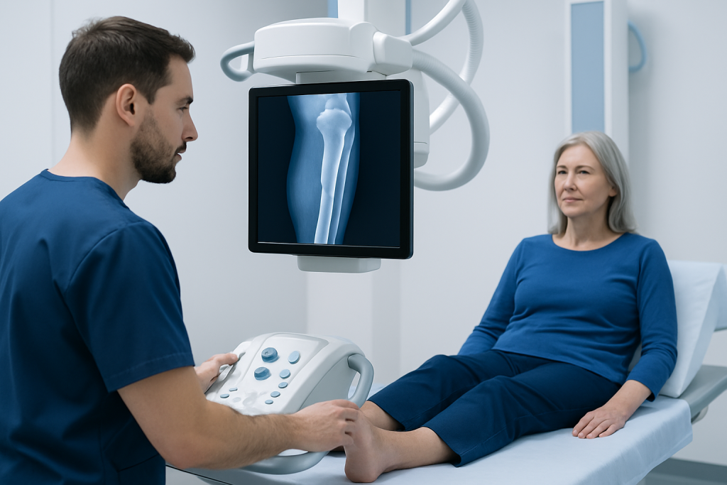
X-rays use a small, controlled amount of ionising radiation to create images of dense structures such as bone. A radiographer positions the lower leg between the x-ray detector and machine, then takes images from selected angles. The test is brief and does not require needles, contrast dye or sedation.
A tibial fracture xray may be appropriate for a person with pain, swelling, bruising, tenderness over the shin, difficulty bearing weight, an obvious change in leg shape or reduced movement after an injury. It may also be used to investigate persistent lower-leg pain in runners or other active people when a stress injury is suspected.
People who are pregnant or think they may be pregnant should inform the imaging team before the examination. X-rays of the leg generally expose the reproductive organs to very little scattered radiation, and staff can use appropriate measures to keep exposure as low as reasonably achievable. In an emergency, needed imaging should not be delayed, but the team will tailor the approach to the individual situation.
Preparation and step-by-step procedure
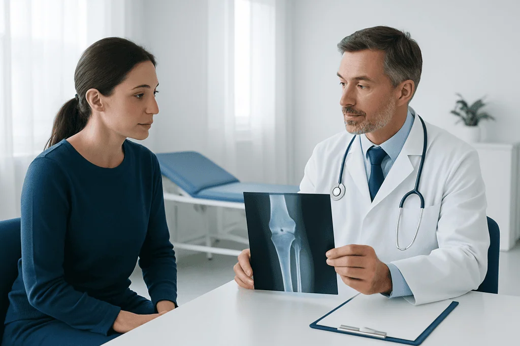
Preparation for a tibial fracture xray is usually simple. Patients may be asked to remove shoes, jewellery, metal objects or clothing with metal fasteners around the leg. Comfortable clothing that can be rolled above the knee and ankle can be helpful. If pain is significant, the team will work carefully around a splint or temporary immobilisation rather than asking the person to move beyond what is safe.
During the examination, the radiographer explains each position and supports the injured leg as needed. The person may lie on an x-ray table or sit, depending on comfort and the injury. For standard views, the leg is placed straight for a front image and gently turned or supported for a side image. It is important to remain still for a few seconds while each image is taken.
Tibial plateau fracture x ray projections require particular care because the tibial plateau forms the lower surface of the knee joint. Standard knee views may be supplemented by angled views when appropriate. Tibial plateau fracture x ray positioning is adapted to the person’s comfort and injury stability; the team should not force painful bending, rotation or weight-bearing positions.
The images are reviewed by a radiologist and the treating clinician in the context of the examination findings. In urgent cases, an initial interpretation may guide immediate treatment, with a formal radiology report following. The entire visit may take longer if pain control, splinting or additional imaging is needed.
Understanding results and the protocol for a fractured tibia
What is the protocol for a fractured tibia? The immediate protocol is to assess the person’s overall condition, check circulation and nerve function below the injury, control pain, protect the leg from movement and obtain appropriate imaging. If there is a suspected fracture, the leg is usually immobilised with a splint or brace until an orthopaedic assessment determines the safest definitive treatment.
The x-ray report may describe the fracture by location, such as the upper tibia near the knee, the shaft of the tibia, or the lower tibia near the ankle. It may also describe the fracture pattern, including whether it is displaced, angulated, shortened, comminuted (broken into several pieces), open, or extending into a joint. These features help determine whether non-surgical care is suitable or whether surgery is more likely to be recommended.
Stable fractures can sometimes heal with a cast, functional brace and scheduled follow-up x-rays. Fractures that are displaced, unstable, open, associated with significant soft-tissue injury, or involving an important joint surface may require operative stabilisation. The approach can include orthopedic surgery using internal fixation or, in selected complex injuries, temporary or definitive external fixation.
Follow-up images are compared over time. Clinicians look for maintained alignment and signs of new bone formation, often called callus, although healing appearances differ by fracture type and treatment method. An x-ray result is interpreted alongside pain, swelling, ability to move safely and physical examination findings rather than in isolation.
What are the 5 R's of fracture management?
What are the 5 R’s of fracture management? This is a commonly taught framework, though the wording can vary between clinical settings. It generally refers to recognition, resuscitation, reduction, retention and rehabilitation. The purpose is to ensure that urgent threats are addressed first and that the bone heals in the best achievable position and function is restored gradually.
Recognition means identifying a possible fracture and assessing the injury mechanism, pain, skin condition, deformity and neurovascular status. Resuscitation refers to stabilising the patient when needed, particularly after high-energy trauma, while treating pain and other injuries. An open fracture, severe bleeding, altered sensation or poor blood flow to the foot requires urgent emergency care.
Reduction means realigning bone fragments when they are displaced. It may be performed without surgery in some cases or achieved during an operation in others. Retention means holding the corrected position with a cast, brace, plate, screws, intramedullary nail or external fixator. Rehabilitation includes guided movement, strengthening, gradual weight-bearing and follow-up, aiming to restore safe mobility while protecting healing bone.
Not every tibia fracture follows exactly the same pathway. Decisions are individualised according to the fracture pattern, skin and muscle injury, health conditions, smoking status, activity needs and the person’s progress on repeat examinations and imaging.
How long does recovery take, and what happens after 8 weeks?
How many days will it take to walk normally after a tibia fracture? There is no single number of days that applies to everyone. Some people with stable injuries may begin carefully increasing weight-bearing earlier under medical guidance, while complex fractures can require a longer period of restricted weight-bearing. Walking normally often takes months rather than days because bone healing, muscle strength, balance, joint movement and confidence all need time to recover.
Clinicians commonly schedule reviews during the first weeks after injury to check the skin, swelling, comfort, alignment and x-ray appearance. The care team advises when to begin or progress weight-bearing. Starting too soon can move an unstable fracture or interfere with healing, while prolonged unnecessary inactivity can contribute to stiffness and weakness. A physiotherapist can provide an individual rehabilitation plan.
What happens to a tibia fracture after 8 weeks? At about eight weeks, many fractures show early or progressing healing on x-ray, but this does not automatically mean the bone is ready for unrestricted walking, sport or heavy work. Some tibial fractures heal more slowly, especially after high-energy injury, when blood supply has been affected, or when there are risk factors such as smoking, diabetes, infection or poor nutrition.
The clinician compares current tibial fracture x rays with earlier images and considers symptoms and examination findings. If healing is progressing, activity may be advanced gradually. If pain persists, alignment changes or healing appears delayed, further imaging or a change in management may be considered. Patients should not remove immobilisation or alter weight-bearing restrictions without advice from their treating team.
Benefits, limitations and possible risks
The main benefit of a tibial fracture xray is that it is quick, widely available and effective for identifying many bone fractures and checking their alignment. It helps clinicians make timely decisions about immobilisation, referral, surgery and follow-up. Repeat x-rays can provide a practical record of healing over time.
The radiation dose from a standard lower-leg x-ray is low. Nevertheless, imaging is requested when the expected clinical benefit outweighs the small radiation risk. Radiology teams use the lowest exposure that can produce diagnostic images and avoid unnecessary repeat studies.
There are also limitations. Very early stress fractures, subtle non-displaced fractures and cartilage, ligament or meniscal injuries may not be clearly visible on plain x-rays. CT or MRI may therefore be recommended when symptoms, examination findings or the fracture’s location raise further questions. Positioning can be uncomfortable after an acute injury, but staff can modify the examination and support the limb to reduce discomfort.
When to seek medical care
Medical assessment is needed promptly after a significant lower-leg injury, particularly when there is severe pain, inability to bear weight, marked swelling, deformity, a cracking sensation at the time of injury or tenderness directly over the tibia. A clinician can determine whether a tibial fracture xray or other imaging is appropriate and arrange safe immobilisation.
Emergency care is particularly important if bone is visible, there is an open wound near the injury, bleeding does not stop, the foot becomes pale, blue, cold or numb, pain rapidly worsens, or the leg feels unusually tight or swollen. These signs can indicate complications that need urgent treatment.
People already being treated for a tibia fracture should contact their clinical team if their cast or brace becomes too tight or damaged, pain and swelling worsen rather than improve, fever or wound drainage develops after surgery, or new numbness and weakness occurs. Acibadem International’s multidisciplinary specialists and JCI-accredited hospitals provide assessment and treatment for international patients with traumatic bone injuries.
Frequently asked questions
Does a tibial fracture always show on an x-ray?
Most acute tibial fractures can be seen on standard x-rays. However, very small, non-displaced or early stress fractures may not be obvious initially. If clinical suspicion remains high, a clinician may arrange repeat x-rays, CT or MRI.
Do I need to prepare for a tibial fracture xray?
Usually, no special preparation is needed. The patient may be asked to remove metal items, shoes or clothing that covers the area. It is important to tell the radiographer about pregnancy or a possible pregnancy and about severe pain or difficulty moving the leg.
Will a tibial fracture xray be painful?
The x-ray itself is painless. Positioning an injured leg can be uncomfortable, especially soon after trauma, but the radiographer will support the limb and avoid unnecessary movement. Pain relief and splinting may be arranged before imaging when needed.
Why might CT be needed after a tibial plateau x-ray?
CT provides detailed cross-sectional images and can show the shape and extent of a fracture involving the knee joint surface. It can help an orthopaedic surgeon assess depression, splitting or multiple fragments that may not be fully defined on plain x-rays. This information can guide treatment planning.
Can I walk on a tibia fracture while waiting for results?
A person should avoid putting weight on a suspected tibia fracture unless a clinician has specifically said it is safe. Weight-bearing on an unstable injury may worsen displacement or pain. Using a splint, crutches or other support as advised helps protect the leg until assessment is complete.
How do doctors know that a tibia fracture is healing?
Doctors assess healing using symptoms, physical examination and follow-up imaging. Improving pain and function, stable alignment and evidence of new bone formation on x-ray can all support healing. The timing varies, so activity should progress according to the treating clinician’s advice.
References
- American Academy of Orthopaedic Surgeons
- Radiological Society of North America
- National Institute for Health and Care Excellence
- American College of Radiology
This article is for general information only and is not a substitute for professional medical advice. Please consult a qualified doctor about your individual situation.
Joint and spine care in Turkey — expert assessment & treatment
JCI-accredited · board-certified surgeons · reply within 24h
Add us as a Preferred Source to see more of our trusted health content across Google Search, AI Overviews and Discover.