Tomosynthesis — Explained by Medical Evidence, Not Myths
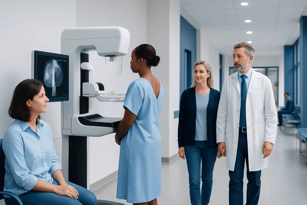
Tomosynthesis is a type of 3D mammogram used mainly for breast cancer screening and diagnostic imaging. It can make breast tissue easier to evaluate by reducing the effect of tissue overlap on images.
Key Takeaways
- Tomosynthesis is a type of 3D mammogram used mainly for breast cancer screening and diagnostic imaging.
- It can make breast tissue easier to evaluate by reducing the effect of tissue overlap on images.
- Tomosynthesis does not replace a medical exam or biopsy when a suspicious finding needs further evaluation.
- The test is quick, uses low-dose X-rays, and feels similar to a standard mammogram.
- Whether tomosynthesis is appropriate depends on age, symptoms, breast density, risk factors, and local screening guidance.
Tomosynthesis is an advanced form of mammography that creates thin, layered images of the breast, helping radiologists examine tissue more clearly than with standard 2D mammography alone. It is widely used in breast cancer screening and follow-up imaging because it can improve detection in some patients and reduce recalls caused by overlapping breast tissue.
What tomosynthesis is and why it is used
Tomosynthesis, also called digital breast tomosynthesis or a 3D mammogram, is a breast imaging technique that uses low-dose X-rays taken from multiple angles. A computer reconstructs these images into thin slices of the breast, allowing the radiologist to review one layer at a time rather than relying only on a single flat image.
In practical terms, tomosynthesis is used to look for signs of breast cancer and other changes in breast tissue. It is most often performed as part of routine screening in people without symptoms, but it may also be used when there is a breast lump, focal pain, nipple changes, or an abnormality found on a standard mammogram.
The main advantage of tomosynthesis is clarity. Breast tissue can overlap on conventional 2D mammograms, sometimes hiding a small cancer or creating an area that only looks suspicious because tissues are stacked on top of one another. By separating the breast into layers, tomosynthesis can help the radiologist distinguish real findings from overlap.
This test does not diagnose cancer by itself. Instead, it improves imaging assessment and helps guide the next step, which may include comparison with prior mammograms, targeted ultrasound, breast MRI, or tissue sampling if needed. When breast cancer is suspected or confirmed, related evaluation may also involve breast cancer assessment and, in selected cases, biopsy procedures.
How tomosynthesis differs from a standard mammogram
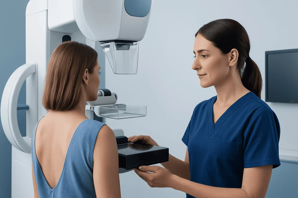
A standard 2D mammogram captures images of the compressed breast from set views. Tomosynthesis also uses breast compression, but the X-ray tube moves in a short arc over the breast to collect multiple images. These are then reconstructed into a three-dimensional view made up of thin sections.
For many patients, the experience feels very similar to a routine mammogram. The breast still needs to be positioned and compressed for a few seconds, and the entire appointment is usually brief. What changes is the type of image the radiologist receives and the amount of detail available for review.
Tomosynthesis can be especially helpful in dense breasts, where overlapping tissue may limit the visibility of abnormalities on 2D images. Dense tissue itself is common and not a disease, but it can make interpretation more challenging. Even so, tomosynthesis is not perfect, and some cancers are still best evaluated with additional imaging.
Another important difference is that tomosynthesis is an imaging tool, not a treatment. If an abnormality is found, the next step depends on the imaging appearance and clinical context. Additional MRI imaging or mammography follow-up may be recommended to clarify a finding.
Who may benefit from tomosynthesis
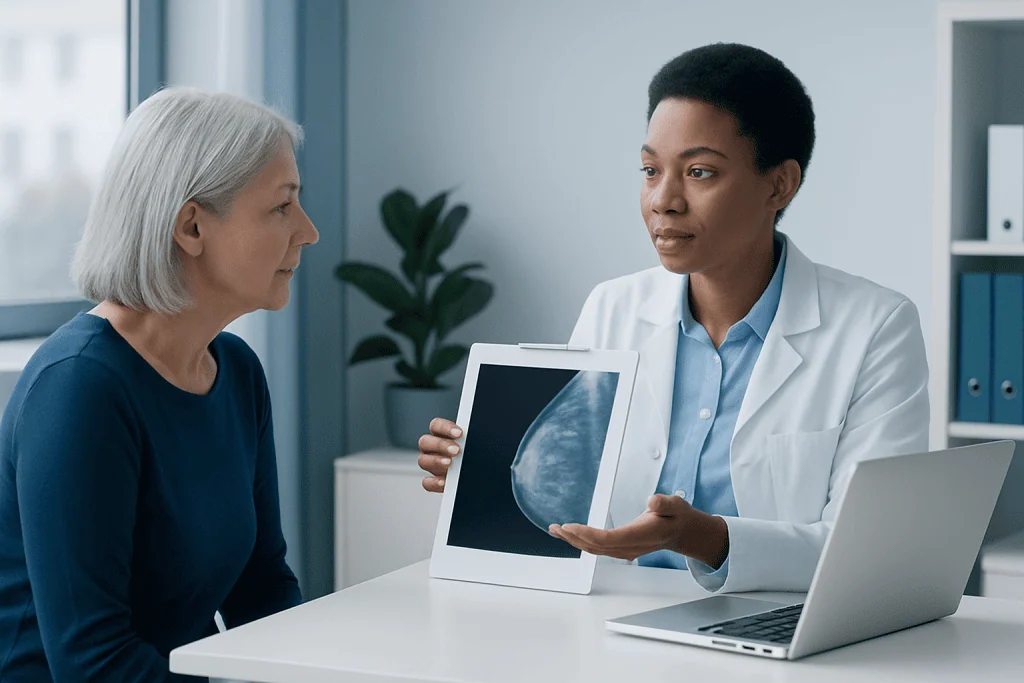
Tomosynthesis is commonly used for routine breast cancer screening in adults who are eligible for mammography according to their doctor’s advice and local screening recommendations. Many imaging centers now offer it as part of standard screening because it can improve lesion visibility and reduce some unnecessary call-backs.
It may be particularly useful for people with dense breasts, a personal history of breast abnormalities, or a prior screening result that required extra views. It can also help when a person or clinician notices a change in the breast and diagnostic imaging is needed to investigate it further.
However, not every patient has the same needs. People at higher risk for breast cancer, such as those with strong family history, certain genetic mutations, or previous chest radiation, may need a more individualized screening plan. In some cases, breast MRI is added to mammography rather than replaced by it.
Pregnancy, age, prior imaging results, current symptoms, and overall risk profile all influence which tests are most appropriate. A radiologist or breast specialist can explain whether tomosynthesis is the best choice for screening, whether supplemental imaging would be useful, or whether another pathway is more suitable.
What happens during the exam
Before the scan, the patient is usually asked to remove clothing above the waist and wear a gown. It is often recommended to avoid deodorants, powders, or lotions on the underarm and breast area on the day of the exam, because these can sometimes appear on images and interfere with interpretation.
During the exam, the technologist positions one breast at a time on the mammography unit. The breast is gently compressed to keep it still and spread the tissue for clearer images. This compression lasts only a short time for each image set. Most patients describe the sensation as pressure or brief discomfort rather than pain, though experiences vary.
The X-ray arm then moves in a small arc while taking multiple low-dose pictures. The process is repeated for the standard views of each breast. The exam itself is usually completed in a short time, though the total visit may be longer if forms, previous image comparisons, or additional views are needed.
Afterward, most people can return to normal activities immediately. Results may be reported as normal, probably benign with follow-up advice, or requiring additional imaging. If a finding needs more evaluation, that does not automatically mean cancer; many call-backs turn out to be benign changes such as cysts, normal tissue overlap, or other noncancerous findings.
Benefits, limits, and common myths
Tomosynthesis is often chosen because it can improve the detection of some breast cancers and reduce false-positive findings related to overlapping tissue. This may mean fewer patients are called back for extra imaging after screening. It can also help show the shape and margins of an abnormal area more clearly than 2D images alone in some situations.
At the same time, tomosynthesis has limits. It cannot detect every cancer, and it cannot tell with certainty whether every abnormality is benign or malignant. If an area looks suspicious, further testing is still needed. Ultrasound, MRI, short-interval imaging follow-up, or biopsy may be part of the next step depending on the case.
One common myth is that a 3D mammogram is painful or significantly more intense than a standard mammogram. In reality, the positioning and compression are very similar. Another myth is that tomosynthesis replaces all other breast imaging. It does not; rather, it complements other tools and is used as part of a broader diagnostic process.
Patients also sometimes worry about radiation. Tomosynthesis uses X-rays, but the dose is kept low and designed to stay within accepted safety standards for breast imaging. The decision to screen is based on balancing this small exposure against the benefit of early detection. A qualified clinician can help explain that balance for the individual patient.
Understanding results and what abnormal findings may mean
Breast imaging results are usually categorized using a standardized reporting system so that radiologists and clinicians can communicate clearly. A result may be negative or benign, may suggest short-term follow-up, or may recommend further diagnostic workup. The wording can feel technical, but the key point is what action, if any, is advised next.
An abnormal result on tomosynthesis does not mean a person definitely has cancer. Benign cysts, fibroadenomas, scar tissue, calcifications, and areas of normal overlapping tissue can all lead to additional evaluation. Follow-up often includes magnification views, targeted ultrasound, or comparison with older mammograms to see whether a finding is stable over time.
If a finding remains concerning, the doctor may recommend a biopsy so the tissue can be examined under a microscope. This is the only way to confirm or exclude cancer definitively. Some patients may also be referred for assessment of related breast conditions, including possible breast cancer, depending on the imaging pattern and clinical exam.
Clear communication is important after any breast imaging test. Patients can ask what category the result falls into, whether dense breast tissue affected interpretation, what additional testing is recommended, and when follow-up should occur. Understanding the plan often helps reduce uncertainty while ensuring that important findings are not overlooked.
When to seek medical care
Tomosynthesis is often done as planned screening, but medical care should be sought sooner if breast symptoms appear. A person should arrange prompt medical evaluation for a new breast lump, persistent focal breast pain, skin thickening, dimpling, nipple inversion that is new, bloody nipple discharge, or swelling in the breast or underarm.
It is also important to follow up if a screening result recommends additional imaging. Delaying further evaluation can postpone clarification of a finding that may need treatment. Even when many abnormal mammogram results prove benign, timely follow-up remains the safest approach.
Anyone with a strong family history of breast or ovarian cancer, a known inherited cancer-risk mutation, or prior chest radiation should discuss individualized screening with a qualified doctor. Their screening plan may differ from routine recommendations and may include earlier or more intensive surveillance.
Near the end of the care pathway, some patients choose evaluation in centers with coordinated imaging, pathology, and specialist review. Acibadem International’s multidisciplinary specialists and JCI-accredited hospitals provide diagnosis and treatment planning for international patients when breast imaging findings require further assessment.
Practical questions to discuss with a doctor
When considering tomosynthesis, many patients benefit from a short, focused discussion with their doctor. Helpful questions include whether screening is due now, whether the person has dense breasts, whether tomosynthesis is available and appropriate, and whether any symptoms require diagnostic imaging rather than routine screening.
It is also reasonable to ask how results will be communicated and how prior studies will be compared. Having older mammograms available can improve interpretation and may reduce unnecessary additional testing. Patients should mention any history of breast surgery, implants, prior biopsies, pregnancy, breastfeeding, or hormone therapy, since these factors can affect imaging decisions.
If a callback occurs, the next step should be clearly explained. Patients can ask whether more mammographic views, ultrasound, MRI, or biopsy is advised and how urgent that recommendation is. Understanding each step supports informed decision-making and may lessen anxiety.
Tomosynthesis is best viewed as one part of personalized breast care. Used appropriately, it can offer clearer imaging and more confident interpretation, while still working alongside clinical examination, risk assessment, and follow-up testing when needed.
Frequently asked questions
Is tomosynthesis the same as a mammogram?
Tomosynthesis is a type of mammogram, often called a 3D mammogram. It uses multiple low-dose X-ray images to create layered views of the breast, while standard mammography produces mainly 2D images.
Does tomosynthesis hurt more than a regular mammogram?
For most people, it feels very similar to a standard mammogram because the breast still needs brief compression. Some pressure or discomfort is common, but the test is usually quick and well tolerated.
Is tomosynthesis better for dense breasts?
Tomosynthesis can be especially helpful in dense breasts because it reduces the effect of overlapping tissue on the image. However, dense breast tissue may still require additional imaging in some patients, depending on risk and symptoms.
Can tomosynthesis find all breast cancers?
No imaging test can detect every breast cancer. Tomosynthesis improves visibility in many situations, but some cancers may still be missed or may need ultrasound, MRI, or biopsy for diagnosis.
Does an abnormal tomosynthesis result mean cancer?
No, an abnormal result does not automatically mean cancer. Many findings turn out to be benign, but additional imaging or biopsy may be needed to understand the cause accurately.
How should someone prepare for a tomosynthesis exam?
It is generally helpful to avoid deodorant, powder, or lotion on the breast and underarm area on the day of the test. Bringing prior breast imaging records, if available, can also help the radiologist compare current and earlier findings.
This article is for general information only and is not a substitute for professional medical advice. Please consult a qualified doctor about your individual situation.
Explore treatments in Turkey — costs, top hospitals & a free quote
JCI-accredited · board-certified surgeons · reply within 24h
Add us as a Preferred Source to see more of our trusted health content across Google Search, AI Overviews and Discover.

More from the Health Library

Dawn Phenomenon: An Evidence-Based Guide for Patients

Marcona Almonds: What Patients Need to Know

Nasal Polyps: A Complete Medical Overview

Burn From Cigarette — Explained by Medical Evidence, Not Myths

Horned Melon: An Evidence-Based Guide for Patients