Ventriculomegaly Ultrasound: Preparation, Procedure and Results

Ventriculomegaly means that one or both brain ventricles measure wider than expected on fetal ultrasound. An ultrasound can identify and measure ventriculomegaly, but further assessment may be needed to understand its cause and outlook.
Key Takeaways
- Ventriculomegaly means that one or both brain ventricles measure wider than expected on fetal ultrasound.
- An ultrasound can identify and measure ventriculomegaly, but further assessment may be needed to understand its cause and outlook.
- Isolated mild ventriculomegaly often has a favorable outlook and can resolve before birth, although follow-up scans remain important.
- The outlook depends mainly on the degree of enlargement and whether other structural, genetic, or infection-related findings are present.
- A detailed fetal brain scan, fetal MRI, and consultation with maternal-fetal medicine or pediatric specialists may be recommended in selected cases.
A ventriculomegaly ultrasound is a prenatal scan that assesses whether the fluid-filled spaces in a developing baby’s brain, called ventricles, are enlarged. It is painless and does not use radiation; the findings help the care team decide whether follow-up imaging, genetic testing, infection testing, or specialist review may be helpful.
Overview: What Is a Ventriculomegaly Ultrasound?
A ventriculomegaly ultrasound is a detailed prenatal examination used to look at and measure the ventricles, which are normal fluid-filled spaces inside a baby’s developing brain. The test is usually performed when a routine pregnancy ultrasound suggests that a ventricle may be wider than expected, or when a clinician wants a more detailed view of brain development.
During the scan, the sonographer obtains standard images of the fetal head and measures the atrium of the lateral ventricle. Ventriculomegaly is generally described when this measurement is 10 millimeters or more. It may be categorized as mild, moderate, or severe depending on the measurement, but the number alone does not determine a baby’s outcome.
The purpose of the assessment is not only to confirm the measurement. It also looks carefully for other differences in the brain or other organs, checks fetal growth and amniotic fluid, and helps guide appropriate next steps. Many pregnancies with mild, isolated ventriculomegaly progress well, especially when follow-up evaluations are reassuring.
How the Ultrasound Works and Who May Need It
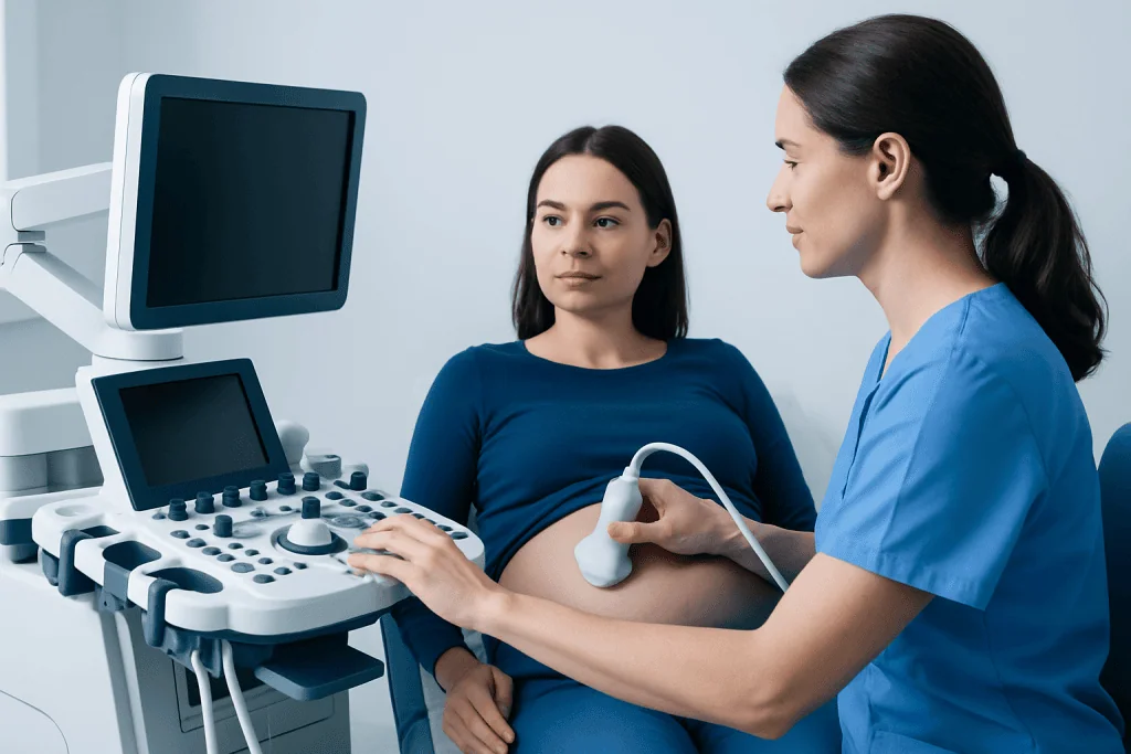
Ultrasound uses high-frequency sound waves to create real-time images of the fetus. A handheld device called a transducer sends sound waves through the abdomen and receives their echoes. A computer converts these echoes into images that allow the clinician to view the fetal brain, including the ventricles. Unlike X-rays or CT scans, ultrasound does not use ionizing radiation.
A detailed ventriculomegaly ultrasound may be recommended after a routine anatomy scan identifies a ventricle measurement of 10 millimeters or more. It may also be considered when there is a family history of certain neurological or genetic conditions, a suspected fetal infection, an unclear brain image on a standard scan, or another fetal structural finding.
The examination is usually performed by an experienced obstetric ultrasound team, often with a maternal-fetal medicine specialist. Depending on the findings, the care team may recommend a detailed anatomic scan, fetal echocardiography, genetic counseling, infection testing, or fetal magnetic resonance imaging (MRI). These steps are individualized and are not necessary for every pregnancy.
Preparation and Step-by-Step Procedure
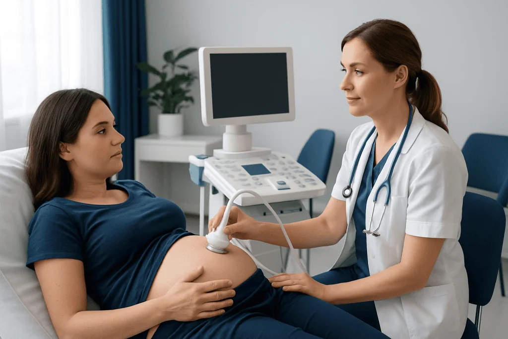
Most people do not need special preparation for a ventriculomegaly ultrasound. The clinic may provide instructions about whether to arrive with a comfortably full bladder, particularly earlier in pregnancy. Comfortable two-piece clothing can make the abdominal scan easier. Patients may wish to bring prior ultrasound reports or information about relevant medical and family history.
At the appointment, the sonographer applies a clear gel to the abdomen and moves the transducer gently across the skin. The gel helps transmit sound waves and may feel cool, but the examination should not be painful. Images are taken from several angles while the fetal head, brain structures, spine, heart, abdomen, limbs, placenta, and amniotic fluid are reviewed as appropriate.
For the ventricular measurement, the operator obtains a standardized view of the fetal head and measures the relevant part of the lateral ventricle. Fetal position, movement, gestational age, and maternal body shape can sometimes affect image quality. If images are limited, the scan may take longer or a repeat appointment may be advised.
There is no recovery period after a standard ultrasound. The patient can normally return to usual activities immediately. A clinician may discuss preliminary findings on the same day, while a formal interpretation and next-step plan may follow after review by the specialist team.
Understanding Results and Follow-Up Testing
A ventriculomegaly ultrasound report may state whether enlargement is present on one side or both sides, give the ventricular measurement, and describe whether the finding appears isolated or is accompanied by other ultrasound differences. Mild ventriculomegaly is commonly used for measurements of 10 to 12 millimeters, moderate for 13 to 15 millimeters, and severe for measurements greater than 15 millimeters.
Finding enlarged ventricles does not by itself establish a diagnosis or predict developmental outcomes with certainty. Ventricles can appear enlarged for different reasons, including normal variation, a temporary imbalance in cerebrospinal fluid flow, brain development differences, bleeding, infection, chromosomal changes, or other genetic conditions. In some cases, no cause is identified.
Follow-up often includes another ultrasound after an interval to see whether the measurement is stable, improving, or increasing. A detailed neurosonography examination may provide a closer assessment of the brain. Fetal MRI can be helpful in selected cases because it may show some brain structures differently from ultrasound, especially later in pregnancy.
Clinicians may also discuss genetic counseling and diagnostic testing, such as amniocentesis with chromosome and genetic analysis, as well as testing for selected maternal infections when clinically appropriate. These options have different benefits and limitations, and the decision should be made after informed discussion with a qualified prenatal care team.
How Serious Is Ventriculomegaly?
Ventriculomegaly can range from a mild, isolated ultrasound finding to a sign of a more complex condition. Its seriousness depends on the ventricle size, whether the measurement changes over time, whether one or both sides are affected, and whether detailed imaging identifies other differences in the brain or body.
When mild ventriculomegaly is isolated and genetic and infection evaluations are reassuring, the outlook is often favorable. However, follow-up remains important because additional findings can occasionally become clearer later in pregnancy or after birth. Moderate or severe ventriculomegaly, progressive enlargement, or associated abnormalities generally require more extensive specialist assessment and individualized counseling.
The care team should explain what is known from the scan and what remains uncertain. Maternal-fetal medicine specialists, genetic counselors, neonatologists, pediatric neurologists, and neurosurgeons may all contribute when needed. This multidisciplinary approach supports informed decisions and planning for pregnancy, birth, and newborn care.
What Mental Disorder Has Enlarged Ventricles?
Enlarged ventricles are not a mental disorder, and an ultrasound finding of fetal ventriculomegaly should not be interpreted as a psychiatric diagnosis. Ventricular size can vary for many reasons, including conditions affecting brain development or the circulation of cerebrospinal fluid. Prenatal imaging cannot diagnose a future mental health condition.
In research involving adults, enlarged ventricles have sometimes been observed in people with certain neurological or psychiatric conditions, including schizophrenia, bipolar disorder, dementia, or brain injury. These are group-level observations rather than diagnostic tests. Many people with these conditions do not have enlarged ventricles, and enlarged ventricles alone do not mean that a person has or will develop a mental disorder.
For a fetus, the clinical focus is on understanding the prenatal imaging findings, looking for associated structural or genetic causes, and arranging appropriate developmental follow-up after birth when recommended. Families should discuss individual concerns with their fetal medicine or pediatric care team rather than drawing conclusions from unrelated adult studies.
Can Mild Ventriculomegaly Resolve Itself?
Yes. Mild ventriculomegaly can decrease or return to a typical measurement on later pregnancy ultrasounds, particularly when it is isolated and no underlying cause is identified. This is why repeat imaging is commonly recommended: it provides information about the direction of change, not just a single measurement.
Resolution is reassuring, but it does not replace appropriate clinical review. The prenatal team may still consider the full anatomy scan, pregnancy history, screening results, and any recommended testing when discussing the outlook. After delivery, some babies may be offered a head ultrasound or developmental monitoring based on the prenatal findings and local care guidance.
Ventriculomegaly may also remain stable or become more pronounced, so follow-up should take place at the interval advised by the treating clinician. Regular prenatal care, attendance at scheduled scans, and open discussion of questions can help families understand the evolving picture.
What Is the Survival Rate for a Fetus With Ventriculomegaly?
There is no single survival rate that applies to every fetus with ventriculomegaly. Prognosis varies substantially according to the degree of enlargement, whether it is isolated, the presence of other brain or body abnormalities, genetic findings, possible infection, and whether ventriculomegaly progresses during pregnancy.
For isolated mild ventriculomegaly with reassuring detailed assessment, survival and childhood outcomes are generally favorable. The outlook can be more guarded when ventriculomegaly is severe, worsening, or linked to a major brain malformation, infection, chromosomal condition, or another serious health problem. A specialist can give the most meaningful counseling after reviewing all imaging and test results together.
It can be helpful for families to ask which factors in their specific case are reassuring, which findings need further evaluation, and what follow-up is planned before and after birth. Honest counseling acknowledges uncertainty while focusing on the information available and the care options that may support the baby and family.
Benefits, Limitations, and When to Seek Medical Care
The main benefit of a ventriculomegaly ultrasound is that it provides a safe, noninvasive way to assess fetal brain anatomy and guide timely follow-up. It can help identify whether the finding is isolated, monitor changes over time, and support coordinated pregnancy and delivery planning. Its limitations are that ultrasound cannot identify every cause of enlarged ventricles or predict an individual child’s development with complete certainty.
Standard prenatal ultrasound is considered safe when medically indicated and performed by trained professionals. There are no known recovery concerns from the scan itself. The most difficult aspect for many families is the waiting and uncertainty that can accompany further testing, and it is appropriate to ask the care team for clear explanations, written reports, and genetic or psychological support when needed.
Medical care should be sought promptly for urgent pregnancy symptoms such as vaginal bleeding, fluid leakage, severe or persistent abdominal pain, fever, severe headache with visual changes, or a noticeable reduction in fetal movement later in pregnancy. These symptoms are not specific to ventriculomegaly, but they need timely assessment. Otherwise, patients should attend all recommended prenatal appointments and contact their obstetric team if questions arise about scan results.
Acibadem International’s multidisciplinary specialists and JCI-accredited hospitals can evaluate fetal imaging findings and coordinate care for international patients when further assessment is required.
Frequently asked questions
Is a ventriculomegaly ultrasound painful?
No. A standard abdominal ultrasound is noninvasive and should not be painful. The transducer may apply gentle pressure, and the ultrasound gel can feel cool, but there is no injection or radiation exposure.
When is ventriculomegaly usually found during pregnancy?
It is often detected during the mid-pregnancy anatomy ultrasound, when the fetal brain can be assessed in detail. It may also be identified later in pregnancy during a scan performed for another reason or during scheduled follow-up imaging.
Does ventriculomegaly always mean hydrocephalus?
No. Ventriculomegaly means that the brain ventricles measure wider than expected, while hydrocephalus involves an abnormal buildup of cerebrospinal fluid that may increase pressure or enlarge the ventricles. Some cases of ventriculomegaly are related to fluid-flow problems, but many are not.
Will a fetal MRI always be needed after a ventriculomegaly ultrasound?
No. Whether fetal MRI is useful depends on the ultrasound findings, gestational age, local expertise, and the questions that remain after detailed neurosonography. A maternal-fetal medicine specialist can explain whether MRI may add clinically useful information in an individual pregnancy.
Can ventriculomegaly affect a child’s development?
It can, but the likelihood varies widely. Isolated mild ventriculomegaly with reassuring follow-up is often associated with favorable development, while larger measurements or associated abnormalities can increase the chance of developmental, neurological, or medical needs. Follow-up recommendations are tailored to the individual findings.
What questions should families ask after a ventriculomegaly ultrasound?
Useful questions include the exact measurement, whether one or both ventricles are affected, whether any additional findings were seen, and when repeat imaging is planned. Families may also ask whether genetic counseling, infection testing, fetal MRI, or consultations with neonatal and pediatric specialists are recommended.
References
- International Society of Ultrasound in Obstetrics and Gynecology
- Society for Maternal-Fetal Medicine
- American College of Obstetricians and Gynecologists
- Centers for Disease Control and Prevention
- National Institute of Child Health and Human Development
This article is for general information only and is not a substitute for professional medical advice. Please consult a qualified doctor about your individual situation.
Ultrasound in Turkey — costs, top hospitals & a free quote
JCI-accredited · board-certified surgeons · reply within 24h
Add us as a Preferred Source to see more of our trusted health content across Google Search, AI Overviews and Discover.

Check-up & Preventive Medicine
Comprehensive health screenings designed for early detection and prevention.
2 specialists in this unit