

What Does an EEG Show and When Is It Needed?
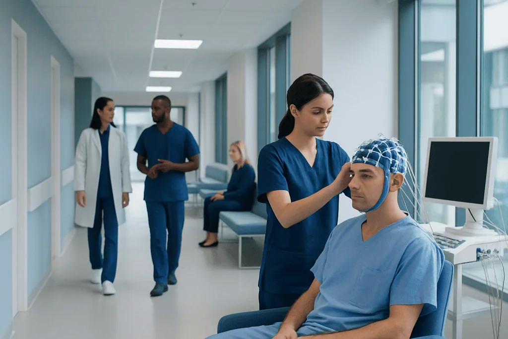
An EEG measures electrical activity in the brain using small sensors placed on the scalp. The test does not diagnose every brain condition on its own, but it provides valuable clues when interpreted with symptoms and other tests.
Key Takeaways
- An EEG measures electrical activity in the brain using small sensors placed on the scalp.
- The test does not diagnose every brain condition on its own, but it provides valuable clues when interpreted with symptoms and other tests.
- EEG is commonly used when seizures or epilepsy are suspected, but it may also be helpful for sleep disorders, confusion, fainting, or unexplained episodes.
- A normal EEG does not always rule out a seizure disorder, and an abnormal EEG does not automatically mean epilepsy.
- EEG is noninvasive, usually painless, and often completed in an outpatient setting.
An EEG, or electroencephalogram, is a painless test that records the brain’s electrical activity. It can help doctors investigate seizures, episodes of altered awareness, sleep-related concerns, and other neurological symptoms.
Overview: what an EEG is and what it shows
An EEG, short for electroencephalogram, is a test that records the brain’s natural electrical activity. Brain cells communicate through tiny electrical signals, and an EEG detects patterns in those signals using small sensors called electrodes placed on the scalp. The test does not send electricity into the body. Instead, it reads and records the activity already being produced by the brain.
Doctors use EEG to look for changes in brain wave patterns that may help explain symptoms such as seizures, staring spells, unexplained loss of awareness, sudden confusion, or sleep-related events. It can also support the assessment of certain brain disorders when symptoms suggest that electrical activity in the brain may be involved.
EEG is especially helpful because it gives real-time information about how the brain is functioning. While scans such as CT or MRI show the brain’s structure, EEG shows function. In other words, it helps doctors see whether the timing and pattern of electrical signals look typical or whether there are changes that need further evaluation.
The results are interpreted by a trained specialist, often a neurologist or neurophysiologist. An EEG is one part of the diagnostic process, and its meaning depends on the patient’s symptoms, medical history, examination, and sometimes additional tests.
What conditions can an EEG help evaluate?
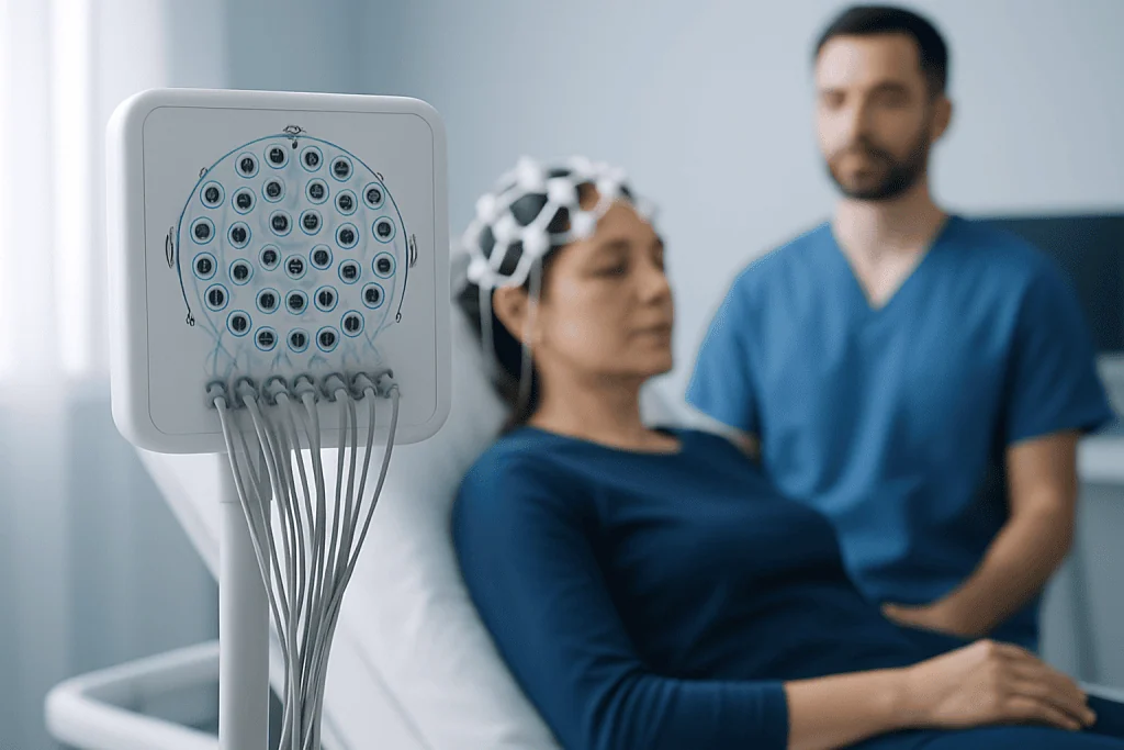
The most common reason for an EEG is to evaluate suspected seizures or epilepsy. If a person has episodes of shaking, staring, sudden confusion, memory gaps, unexplained falls, or brief periods of unresponsiveness, EEG may help identify electrical patterns linked to seizure activity. It is often used as part of the assessment for epilepsy.
EEG may also be useful in other situations. Doctors may request it for episodes that happen during sleep, unusual movements, unexplained changes in behavior, or periods of altered consciousness. In some cases, it helps distinguish seizures from other causes of fainting or spells, although it is usually not the only test needed.
Other possible uses include evaluating certain sleep disorders, confusion after illness or injury, and changes in brain function related to inflammation, infection, or metabolic disturbances. In hospital settings, continuous EEG monitoring may be used in critically ill patients to detect seizure activity that is not obvious from outward signs.
EEG can also provide supportive information after head injury, stroke, brain surgery, or unexplained encephalopathy. However, it is important to remember that EEG is not a general screening test for every neurological complaint. It is ordered when the clinical history suggests that brain electrical activity may offer useful answers.
When is an EEG needed?

A doctor may recommend an EEG when symptoms raise concern for seizures or episodic changes in brain function. Common reasons include fainting with unusual features, repeated staring spells, sudden muscle jerks, nighttime events, unexplained confusion, or episodes in which a person cannot respond normally. The test may be performed after a first suspected seizure or when someone has recurring unexplained spells.
EEG may also be needed when a doctor wants to classify a known seizure disorder, check how brain activity changes over time, or help guide treatment decisions. For example, it can support the assessment before starting or adjusting care for seizure-related conditions, often alongside imaging and a neurological evaluation.
In children, EEG is often considered when there are developmental concerns together with possible seizure-like episodes, unusual staring, or sleep-related events. In adults, it may be used after head trauma, stroke, or periods of confusion to better understand whether abnormal electrical activity is playing a role.
The timing of the test depends on the reason it is ordered. Sometimes a routine EEG is enough. In other cases, a sleep-deprived EEG, prolonged ambulatory recording, or inpatient video EEG is more useful because some abnormalities appear only during sleep, over a longer period, or during a typical event.
How the test is done and how to prepare
During a routine EEG, a technician places multiple small electrodes on the scalp using a special paste or adhesive. The person usually lies down or sits comfortably while the recording is made. The test itself is painless, and many people are surprised by how straightforward it is. A routine EEG often lasts around 20 to 40 minutes, although the appointment may be longer to allow for preparation.
During the recording, the person is asked to relax and remain as still as possible. They may be asked to open and close their eyes, breathe deeply for several minutes, or look at flashing lights. These steps can bring out brain wave patterns that help with interpretation. In some cases, the doctor may ask the patient to reduce sleep the night before, because tiredness and sleep can make certain abnormalities easier to detect.
Preparation is usually simple. Hair should be clean and free from oils, sprays, gels, or styling products so the electrodes can attach well. Patients should ask their doctor whether they should take their regular medicines as usual, since instructions can differ depending on the reason for the test.
Some people need more specialized testing. A sleep EEG records brain activity during sleep. Ambulatory EEG allows recording over many hours or days at home. Video EEG combines brain wave recording with video to compare symptoms or behaviors with EEG changes. This can be especially helpful when doctors need to tell epileptic seizures from other events.
Understanding EEG results
An EEG report describes whether the brain wave patterns look typical for the person’s age and state, such as being awake or asleep, or whether there are abnormalities. These may include slowing, asymmetry, or epileptiform discharges, which are patterns that can suggest a tendency toward seizures. The meaning of any finding depends on the whole clinical picture.
A normal EEG can be reassuring, but it does not always rule out epilepsy or another neurological disorder. Some people with seizure disorders have a normal routine EEG between events. In those cases, doctors may recommend repeat testing, sleep-deprived EEG, longer monitoring, or video EEG if symptoms continue.
An abnormal EEG also does not automatically mean that a person has epilepsy. Some abnormalities can be nonspecific and may appear with other conditions, such as sleep deprivation, medications, metabolic disturbances, or previous brain injury. That is why results are never interpreted in isolation.
Doctors often combine EEG findings with a neurological examination and structural imaging such as MRI scan or CT scan when needed. If episodes remain unclear, longer-term EEG monitoring may provide more useful information than a routine recording alone.
Benefits, limits, and possible risks
EEG has several important advantages. It is noninvasive, generally safe, and gives direct information about brain electrical function. It can detect patterns that may not appear on imaging tests, and it can help guide next steps in diagnosis, treatment planning, and follow-up.
At the same time, EEG has limitations. It records activity from the scalp, so some changes deep in the brain may be difficult to detect. Abnormal activity may come and go, which means a short routine study can miss it. This is one reason doctors sometimes order repeated or prolonged monitoring when symptoms are strongly suggestive.
The test is considered very low risk. The electrodes only record signals and do not cause electric shocks. In rare cases, flashing lights or deep breathing during the test may trigger a seizure in someone who is already prone to seizures, but the staff is trained to manage this safely.
For many patients, the greatest value of EEG is that it adds objective information to the medical history. Even when it does not provide a final diagnosis on its own, it can narrow down possibilities and help the healthcare team choose the most appropriate next tests or treatments.
After the test, next steps, and when to seek medical care
After a routine EEG, most people can return to normal activities right away unless their doctor has given other instructions. There may be some leftover paste in the hair, which can usually be washed out at home. If the test involved sleep deprivation, the person may feel tired and should plan the day accordingly.
Follow-up depends on why the EEG was ordered. If the results suggest seizure activity or another neurological concern, the doctor may recommend further evaluation, medicines, safety advice, or additional tests. In some cases, the next step may include consultation with a neurologist, sleep specialist, or another expert depending on the symptoms.
Medical attention should be sought promptly if a person has a first seizure, repeated unexplained loss of awareness, significant confusion, severe headache with neurological symptoms, or events that cause injury. Emergency care is especially important if a seizure lasts several minutes, happens repeatedly without recovery, or is associated with breathing difficulty.
For international patients who need coordinated neurological evaluation, Acibadem International’s multidisciplinary specialists and JCI-accredited hospitals diagnose and treat conditions involving EEG and related brain assessments. The goal is always to interpret the test in context and create a care plan that fits the patient’s symptoms and needs.
Frequently asked questions
What does an EEG show?
An EEG shows the brain’s electrical activity as wave patterns recorded from the scalp. It can reveal changes that may suggest seizures, abnormal slowing, or other disturbances in brain function.
Does an EEG diagnose epilepsy by itself?
No. An EEG can provide important clues, but epilepsy is diagnosed using the full clinical picture, including symptoms, medical history, examination, and sometimes imaging or longer monitoring. A normal EEG does not completely rule out epilepsy.
Is an EEG painful or dangerous?
EEG is usually painless and very safe. The electrodes only record activity and do not send electricity into the brain. Rarely, certain parts of the test, such as flashing lights, may trigger a seizure in someone already prone to seizures.
How long does an EEG take?
A routine EEG recording often takes about 20 to 40 minutes, though the full appointment may be longer because of preparation and setup. Some people need sleep EEG, ambulatory EEG, or video EEG, which can last much longer.
Can I eat and take my medicines before an EEG?
Many people can eat normally before the test, but instructions may vary depending on why the EEG is being done. Medicines should only be changed if the doctor specifically advises it, so it is important to follow the preparation guidance provided.
Why would a doctor order an EEG after fainting or a staring spell?
Some episodes that look like fainting or inattention may actually be seizures or other changes in awareness. EEG can help doctors tell whether abnormal brain electrical activity may be involved, especially when the history is unclear.
References
- World Health Organization
- National Institute of Neurological Disorders and Stroke
- American Clinical Neurophysiology Society
- International League Against Epilepsy
- MedlinePlus
This article is for general information only and is not a substitute for professional medical advice. Please consult a qualified doctor about your individual situation.
Hair transplant in Turkey — costs, graft counts & a free assessment
JCI-accredited · board-certified surgeons · reply within 24h
Add us as a Preferred Source to see more of our trusted health content across Google Search, AI Overviews and Discover.

More from the Health Library


Related Specialists
Assoc. Prof. Dr. Burak Özturan
Orthopedic Surgery & Traumatology
Dr. Sertaç Uzel
Radiology
Dr. Ulambadrakh Khenchbısh
Orthopedic Surgery & Traumatology
Dt. Okan Gültekin
Oral & Dental Health