What Does an MRI Show: Preparation, Procedure and Results
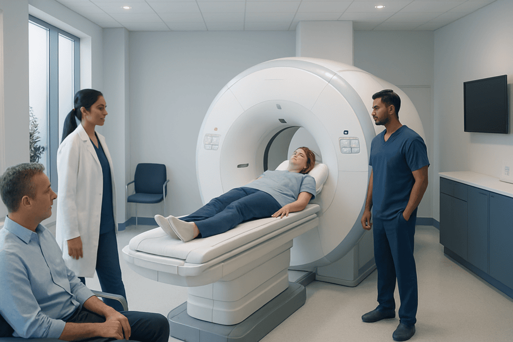
MRI uses a powerful magnetic field and radio waves rather than ionizing radiation. It can provide highly detailed views of soft tissue, the brain, spine, joints, abdominal organs and some blood vessels.
Key Takeaways
- MRI uses a powerful magnetic field and radio waves rather than ionizing radiation.
- It can provide highly detailed views of soft tissue, the brain, spine, joints, abdominal organs and some blood vessels.
- Preparation depends on the body area being scanned and whether contrast dye or sedation is planned.
- A radiologist interprets MRI images in clinical context; an abnormal finding does not always mean a serious condition.
- Most people return to usual activities immediately after a routine MRI.
An MRI shows detailed images of structures inside the body, especially soft tissues such as the brain, spinal cord, nerves, muscles, ligaments, organs and blood vessels. It helps doctors investigate symptoms, confirm or rule out certain conditions, plan treatment and monitor changes over time.
What does an MRI show?
MRI, or magnetic resonance imaging, creates detailed cross-sectional pictures of the inside of the body. What does an MRI show? It is particularly useful for showing soft tissues that may be less clearly seen on some other imaging tests, including the brain, spinal cord, nerves, muscles, tendons, ligaments, cartilage, organs and many blood vessels.
Depending on the area examined, MRI may help identify inflammation, swelling, infection, injury, bleeding, growths, changes in blood flow or structural differences. For example, it can help assess brain and spinal conditions, joint injuries, abdominal and pelvic concerns, and some heart and vascular conditions. The scan is one part of an assessment; doctors interpret it alongside symptoms, examination findings, laboratory tests and previous imaging.
An MRI does not use X-rays. Instead, it uses a strong magnet and radiofrequency energy to produce images. This makes it a valuable option when repeated imaging is needed, although it is not the best test for every symptom or body structure.
How an MRI works and what the procedure does
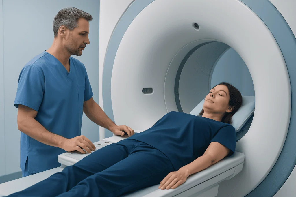
What does an MRI procedure do? It temporarily aligns tiny particles in the body called hydrogen protons using a strong magnetic field. Radio waves then alter that alignment briefly, and the MRI scanner detects the signals released as the protons return to their usual state. A computer turns these signals into detailed images.
Different tissues produce different signals, allowing radiologists to distinguish features such as fluid, fat, muscle, brain tissue and areas of inflammation. Several image sequences may be taken to examine the same region in different ways. In some cases, a contrast agent containing gadolinium is injected into a vein to make selected tissues, blood vessels or areas of active inflammation more visible.
MRI is often especially helpful when clinicians need detailed information about soft tissue. It may complement other tests rather than replace them. For instance, X-rays and CT scans may be more suitable for some bone injuries, lung concerns, emergencies or situations where speed is essential.
Do I need to do anything in preparation for an MRI?

Yes, MRI preparation steps are important because the scanner uses a powerful magnet. Before the appointment, the imaging team will ask about implanted or attached medical devices, previous surgery, metal fragments, kidney problems, pregnancy, allergies and claustrophobia. Many implants are MRI-safe or MRI-conditional, but their exact type must be checked before scanning.
What are the prep instructions for an MRI? Patients are usually asked to remove jewellery, watches, hair accessories, hearing aids, removable dental items, credit cards, clothing with metal fasteners and other metal-containing objects. They may change into a gown. It is important to tell staff about pacemakers, neurostimulators, cochlear implants, aneurysm clips, infusion pumps, joint replacements, dental work, tattoos, permanent makeup or possible metal exposure from work or an injury.
For many MRI scans, people can eat, drink and take regular prescribed medicines as usual. However, instructions differ for contrast-enhanced studies, sedation and certain examinations. The imaging centre should provide personalized directions in advance, and patients should follow those directions rather than relying on general advice.
What is the prep for an MRI of the abdomen?
The prep for an MRI of the abdomen may be more specific than preparation for a head, knee or spine MRI. The care team may ask the patient not to eat for several hours beforehand, as this can reduce movement from digestion and help produce clearer images of the liver, pancreas, bile ducts, kidneys, bowel or other abdominal structures.
Some abdominal MRI examinations involve contrast dye, medication to reduce bowel movement or special instructions about fluids. People with diabetes, kidney disease or a history of contrast reactions should inform the team before the scan so preparation can be tailored safely. Regular medication should not be stopped unless the prescribing clinician or imaging department specifically advises this.
Patients should bring relevant prior scans or reports if requested. Comparing new images with earlier studies can help radiologists identify whether a finding is new, stable or changing. Questions about fasting, medicines and arrival time are best clarified before the appointment.
Step-by-step MRI procedure, comfort and candidacy
After a safety screening, the patient lies on a padded table that moves slowly into the MRI scanner. A coil, which is a device that receives imaging signals, may be placed around or near the part of the body being examined. The technologist remains in contact through an intercom and can see the patient throughout the scan.
The MRI machine makes loud tapping, knocking or humming sounds while images are collected. Earplugs or headphones are provided. Remaining still is important because movement can blur the pictures. A typical examination may last from about 15 minutes to an hour or longer, depending on the body area and number of image sequences. If contrast is needed, it is usually given through a small intravenous line during or before part of the scan.
Most people can have MRI safely after careful screening. Open or wider-bore scanners, communication support, relaxation techniques or clinician-prescribed medication may help people who feel anxious in enclosed spaces. Sedation is sometimes considered when a person cannot remain still, but it requires additional preparation and an escort home. MRI imaging and assessment can help clinicians select and perform an appropriate examination for the clinical question.
Benefits, limitations and what will not show up on an MRI
One important benefit of MRI is its detailed soft-tissue contrast without ionizing radiation. It can show subtle changes in the brain, spinal discs, joints, muscles, ligaments, pelvic organs and abdominal organs. It may also provide information about blood vessels, heart structure or tissue activity when specialized MRI techniques are used.
What will not show up on an MRI? MRI may not clearly answer every medical question. It is generally less useful than CT for viewing air-filled lungs, some bowel conditions, very fine bone detail and certain fresh fractures. Small calcifications can also be harder to evaluate. Some abnormalities may be too small to detect, may not change the MRI signal enough to be visible, or may not be distinguishable without other tests.
Importantly, a normal MRI does not always explain or exclude every cause of symptoms. Pain, dizziness, weakness and other concerns can sometimes occur without a visible structural change on imaging. Conversely, an MRI may show an incidental finding that is unrelated to symptoms and does not require treatment. The referring doctor explains what the findings mean for the individual person.
MRI results, recovery and when to worry about MRI results
After a routine MRI without sedation, most people can return to normal activities immediately. There is no recovery period from the magnetic field itself. If contrast was used, patients are generally advised to follow the imaging centre’s instructions and remain well hydrated unless they have been told to limit fluids. People who received sedation should not drive, operate machinery, drink alcohol or make important decisions until the effects have fully worn off.
A radiologist reviews the images and sends a report to the referring clinician. The report may describe normal anatomy, findings that may explain symptoms, changes requiring follow-up or incidental findings. Results are usually discussed at a follow-up appointment or through the referring healthcare professional, rather than interpreted from images alone.
When to worry about MRI results? It is understandable to feel concerned while waiting, but wording such as “abnormal,” “lesion,” “degenerative change” or “recommend follow-up” does not by itself establish a diagnosis or emergency. Patients should contact the clinician who ordered the scan for timely explanation. Urgent medical advice is appropriate if symptoms suddenly worsen, such as new weakness, trouble speaking, loss of consciousness, severe headache, chest pain, severe breathing difficulty or loss of bladder or bowel control.
What should you not do before an MRI and when to seek medical care
What should you not do before an MRI? Do not ignore the safety questionnaire or assume an implant is safe without verification. Patients should not enter the MRI room with metal objects, electronic devices, bank cards, medication patches with metallic backing or personal items unless staff have confirmed they are safe. They should also avoid skipping fasting instructions when these have been given for an abdominal examination, contrast study or sedation.
Before the scan, patients should inform staff immediately if they are pregnant or might be pregnant, have reduced kidney function, have had a previous reaction to contrast, have metal in the eye or body, or feel unable to lie still because of pain, anxiety or claustrophobia. The team can review options and determine whether MRI is appropriate or whether another examination may be better.
Medical care should be sought promptly for severe or rapidly progressing symptoms, regardless of whether an MRI has been scheduled. These include sudden one-sided weakness, new speech or vision changes, seizures, severe head injury, fever with a stiff neck, severe abdominal pain, or new loss of bladder or bowel function. Acibadem International’s multidisciplinary specialists and JCI-accredited hospitals support international patients who require diagnostic evaluation and treatment planning.
Frequently asked questions
Can MRI detect cancer?
MRI can help detect, characterize and monitor some cancers because it provides detailed images of soft tissues and blood flow patterns. However, MRI findings alone may not confirm cancer, and further tests such as laboratory studies, other imaging or biopsy may be needed.
How long does an MRI take?
Many MRI examinations take between 15 minutes and one hour, although complex scans can take longer. The duration depends on the body part, the number of images needed, whether contrast is used and whether additional specialized sequences are required.
Is an MRI painful?
An MRI is usually not painful, but staying still on the scanning table can be uncomfortable for some people. The machine is loud, and a contrast injection may cause brief discomfort at the IV site. Patients should tell the technologist if they have pain or feel anxious.
Can I eat before an MRI scan?
For many MRI scans, eating and drinking are allowed as usual. Some examinations, especially abdominal MRI or scans involving sedation, may require fasting. The imaging centre's specific instructions should always be followed.
Why might contrast be used during an MRI?
Gadolinium-based contrast can make certain tissues, inflammation, blood vessels or abnormalities easier to see. It is not required for every MRI. The clinician and radiology team consider the potential benefit, kidney function and medical history before using it.
Can someone with a pacemaker have an MRI?
Some modern pacemakers and other implanted devices are designed to be used safely under specific MRI conditions. The device model, leads and medical circumstances must be checked carefully by trained professionals before the scan. A person should never assume that an implant is MRI-safe without confirmation.
References
- RadiologyInfo.org
- American College of Radiology
- National Institute of Biomedical Imaging and Bioengineering
- NHS
- U.S. Food and Drug Administration
This article is for general information only and is not a substitute for professional medical advice. Please consult a qualified doctor about your individual situation.
Joint and spine care in Turkey — expert assessment & treatment
JCI-accredited · board-certified surgeons · reply within 24h
Add us as a Preferred Source to see more of our trusted health content across Google Search, AI Overviews and Discover.