When Are Evoked Potentials Needed? Vision, Hearing, and Sensory Pathway Testing Explained

Evoked potentials measure the brain’s electrical response to visual, sound, or touch stimuli. These tests can help detect slowed or disrupted nerve signal conduction along specific pathways.
Key Takeaways
- Evoked potentials measure the brain’s electrical response to visual, sound, or touch stimuli.
- These tests can help detect slowed or disrupted nerve signal conduction along specific pathways.
- Common types include visual evoked potentials, brainstem auditory evoked response, and somatosensory evoked potentials.
- The tests are usually safe, painless, and performed with small electrodes placed on the skin.
- Results are interpreted together with symptoms, examination findings, and imaging or lab tests.
Evoked potentials are noninvasive neurophysiology tests that help doctors assess how well sensory signals travel to the brain. They are often used when symptoms suggest a problem in the visual, hearing, or sensory pathways, even when scans or routine exams are not enough on their own.
Overview: What evoked potentials are
Evoked potentials are specialized tests that record the nervous system’s electrical responses to sensory stimulation. In simple terms, they show how quickly and effectively signals travel from the eyes, ears, or body to the brain. Doctors use them to evaluate the function of specific nerve pathways rather than just the structure of the brain, spinal cord, or nerves.
These tests are part of clinical neurophysiology. Small electrodes are placed on the scalp and sometimes on other areas of the skin to detect very small electrical signals. A person may be asked to look at a visual pattern, listen to clicking sounds through earphones, or feel brief mild stimulation on the skin. A computer then analyzes the timing and size of the brain’s responses.
Evoked potentials are especially helpful when symptoms suggest a nervous system problem but the cause is not yet clear. They may reveal slowed conduction along a pathway, which can happen with inflammation, demyelination, compression, injury, or other disorders. Although they do not diagnose every condition by themselves, they can add important functional information that complements neurological examination and imaging studies.
When evoked potentials may be needed
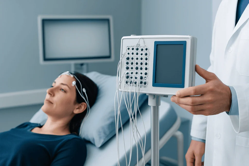
A doctor may recommend evoked potentials when someone has symptoms involving vision, hearing, balance, numbness, tingling, weakness, or changes in sensation and there is a need to assess how the relevant pathways are working. These tests can be useful when standard examination findings are subtle, when symptoms affect more than one area, or when a clinician wants objective evidence of pathway dysfunction.
For example, visual evoked potentials may be requested if there is blurred vision, unexplained visual loss, pain with eye movement, or concern about damage to the optic nerve. Auditory evoked testing may be used in cases of hearing loss, tinnitus, dizziness, suspected brainstem problems, or hearing assessment in babies and young children who cannot respond reliably to routine hearing tests.
Somatosensory evoked potentials may be considered when a person has numbness, altered sensation, weakness, or suspected disorders involving the spinal cord or sensory tracts. In some situations, these tests also help monitor nervous system pathways during surgery or support the evaluation of conditions such as multiple sclerosis when combined with other findings.
Evoked potentials are usually not the only test ordered. They are most valuable when the question is specific: whether the signal can travel normally through a visual, hearing, or sensory pathway. A neurologist, neurophysiologist, ENT specialist, ophthalmologist, or pediatric specialist may request them depending on the symptoms.
Main types: vision, hearing, and sensory pathway testing
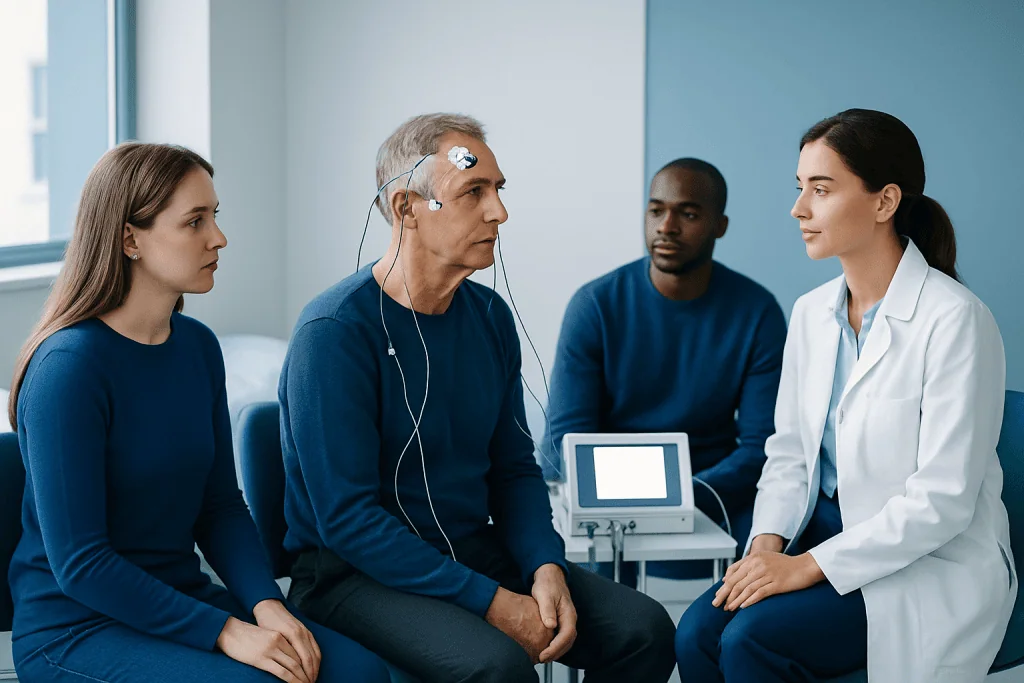
The three most commonly used types are visual evoked potentials, brainstem auditory evoked response, and somatosensory evoked potentials. Each one examines a different sensory system. The principle is similar in all of them: a stimulus is delivered, and the brain’s electrical response is recorded to see whether timing and signal pattern are normal.
Visual evoked potentials (VEP) assess the visual pathway from the eye through the optic nerve to the visual areas of the brain. During the test, the person usually looks at a checkerboard pattern on a screen while electrodes record the response. This can help detect slowed transmission due to optic nerve disease or other pathway problems, even if the eye examination is not fully conclusive.
Brainstem auditory evoked response (BAER or ABR) evaluates how sound signals travel from the ear through the auditory nerve and brainstem. Clicking sounds or tones are delivered through earphones, and scalp electrodes capture the brainstem’s responses. This test is widely used in hearing assessment, newborn screening follow-up, and evaluation of auditory nerve or brainstem disorders.
Somatosensory evoked potentials (SSEP) measure responses after mild electrical stimulation of a peripheral nerve, often at the wrist or ankle. The recordings help assess the sensory pathways that travel through peripheral nerves, the spinal cord, and up to the brain. In some settings, related neurophysiology studies may be combined with electromyography (EMG) or electroencephalography (EEG) when a broader picture of nerve or brain function is needed.
What the test can help diagnose or evaluate
Evoked potentials can help identify conduction delay or disruption in a pathway, which may happen for several reasons. Demyelinating conditions, optic nerve inflammation, spinal cord disease, auditory nerve disorders, and some congenital or acquired hearing problems are common examples. The test findings may suggest where along the pathway the signal is being slowed, although they do not always reveal the exact underlying cause.
In visual symptoms, VEP may support the assessment of optic neuritis, optic nerve injury, or other disorders affecting visual signal transmission. In auditory symptoms, BAER may contribute to the evaluation of hearing loss, auditory neuropathy, or lesions involving the auditory nerve and brainstem. In sensory symptoms, SSEP can be useful when there is concern about spinal cord compression, myelopathy, peripheral nerve involvement, or central sensory pathway dysfunction.
These tests can also play a role in follow-up and monitoring. A doctor may compare results over time to see whether pathway function is stable, improving, or changing. In selected patients, evoked potentials are used during some operations to help monitor the integrity of neural pathways in real time.
Still, normal results do not completely rule out every neurological condition, and abnormal results do not automatically point to one diagnosis. Interpretation always depends on the full clinical picture, which may include imaging such as MRI, hearing tests, ophthalmic assessment, blood tests, and neurological examination.
How evoked potentials are performed and what to expect
Evoked potential testing is usually done in a hospital neurophysiology unit or specialized outpatient clinic. It is generally noninvasive and does not require sedation in most adults. The skin is cleaned, and small recording electrodes are attached to the scalp and sometimes the neck, shoulders, arms, or legs depending on the type of study.
For visual testing, the person typically sits in front of a screen and focuses on a pattern while one eye is tested at a time. For auditory testing, earphones deliver repeated clicks or tones while the person relaxes quietly. For somatosensory testing, mild brief electrical pulses are applied to a nerve in the arm or leg; these may feel unusual or slightly tingling, but they are usually well tolerated.
The full appointment length varies depending on which tests are done, but many studies are completed within a relatively short outpatient visit. It is often helpful to arrive with clean, dry hair and avoid heavy hair products, as these can affect electrode contact. If the patient uses hearing aids, glasses, or contact lenses, the care team will explain whether they should be worn for the test.
Evoked potentials are considered very safe. The electrodes only record activity and do not send electricity into the brain. Mild discomfort can occur with skin preparation or with the brief stimulation used in SSEP, but serious complications are uncommon. In children or people who may have difficulty staying still, additional preparation or scheduling adjustments may be needed for reliable recordings.
Understanding the results
The key measurements in evoked potential testing are usually the timing of the response, called latency, and sometimes the size of the response, called amplitude. If a response arrives later than expected, it may indicate that signal conduction is slowed somewhere along the pathway. If the response is very small or absent, it may suggest reduced signal transmission or more significant dysfunction.
Normal ranges depend on the type of test, the person’s age, the equipment used, and the laboratory’s standards. For that reason, reports are interpreted by trained specialists and should not be read in isolation. A small variation from normal does not always have clinical importance, while a clearly abnormal response can be meaningful even if symptoms are mild.
Results are most useful when combined with the person’s medical history and examination. A doctor may explain that the test points to a problem in the optic nerve, the auditory brainstem pathway, or the spinal sensory tract, but further testing may still be needed to identify the cause. In some cases, the results may help guide referral to neurology, ENT, ophthalmology, or spine specialists.
Limits of the test and related evaluations
Although evoked potentials are valuable, they have limits. They test function in selected pathways, not the entire nervous system, and they cannot show all causes of symptoms. A normal test can occur even when a person has a neurological condition, especially if the affected area is outside the pathway being measured or the change is too subtle to detect.
Likewise, an abnormal test does not always mean there is a serious disease. Technical factors, reduced attention during a visual task, existing eye or ear disease, and age-related changes can all influence recordings. This is why careful preparation and expert interpretation are important.
Doctors often combine evoked potentials with other tests to answer a broader diagnostic question. Depending on the symptoms, these may include hearing assessment, eye examination, MRI, nerve conduction studies, or sleep and seizure testing. If sensory symptoms involve the spine or limbs, clinicians may also evaluate for structural, metabolic, or inflammatory causes in parallel rather than relying on one test alone.
When to see a doctor and where specialist care fits in
Medical advice is important when symptoms such as unexplained vision changes, hearing loss, persistent tinnitus, dizziness, numbness, tingling, imbalance, or weakness appear or worsen. Prompt evaluation is especially important if symptoms develop suddenly, affect one side of the body, are associated with severe pain, or interfere with daily activities. A clinician can decide whether evoked potentials are likely to be helpful or whether a different test is the better first step.
People who already have a diagnosed neurological, eye, ear, or spinal condition may also be referred for evoked potentials to clarify current function or to support treatment planning. Before the appointment, it can help to note when symptoms started, whether they come and go, and any other changes such as headaches, falls, or hearing difficulty in noisy settings.
Good self-care before evaluation includes protecting hearing from excessive noise, managing chronic conditions such as diabetes, taking medicines as prescribed, and attending regular follow-up visits. However, self-care cannot replace professional assessment when new neurological symptoms occur.
For patients who need coordinated evaluation, Acibadem International’s multidisciplinary specialists and JCI-accredited hospitals assess and manage neurological, hearing, and visual pathway disorders for international patients, using tests such as evoked potential testing as part of a broader diagnostic workup when appropriate.
Frequently asked questions
Are evoked potentials painful?
Most evoked potential tests are painless or only mildly uncomfortable. The electrodes record signals from the skin, and in somatosensory testing the brief electrical stimulation may cause a tingling feeling but is usually well tolerated.
How should a patient prepare for an evoked potential test?
Preparation is usually simple. Clean, dry hair without heavy sprays, oils, or gels can help the electrodes make good contact, and the clinic may give specific instructions about glasses, hearing aids, or medicines depending on the test.
Can evoked potentials diagnose multiple sclerosis on their own?
No. Evoked potentials can show slowed conduction in pathways that may be affected in multiple sclerosis, but they are only one part of the evaluation. Doctors interpret the results together with symptoms, neurological examination, MRI findings, and sometimes laboratory tests.
What is the difference between evoked potentials and EEG?
EEG records the brain’s ongoing electrical activity, often to assess conditions such as seizures or altered consciousness. Evoked potentials specifically measure the brain’s response to a visual, auditory, or sensory stimulus to evaluate how a pathway is conducting signals.
How long does the test take?
The timing depends on which evoked potentials are being performed and whether more than one study is scheduled. Many tests are completed during an outpatient visit, but the exact duration varies by clinic and by the complexity of the assessment.
Can children have evoked potential testing?
Yes. These tests are commonly used in children, especially for hearing assessment and selected neurological evaluations. The team may adapt the setting or timing to help the child stay calm and still enough for accurate results.
References
- World Health Organization
- National Institute of Neurological Disorders and Stroke
- American Clinical Neurophysiology Society
- American Academy of Neurology
- National Institute on Deafness and Other Communication Disorders
This article is for general information only and is not a substitute for professional medical advice. Please consult a qualified doctor about your individual situation.
Joint and spine care in Turkey — expert assessment & treatment
JCI-accredited · board-certified surgeons · reply within 24h
Add us as a Preferred Source to see more of our trusted health content across Google Search, AI Overviews and Discover.