Will a CT Scan Show Bladder Cancer: Preparation, Procedure and Results

CT urography provides detailed images of the kidneys, ureters and bladder and is often used when blood is found in urine. CT can miss small, flat or early bladder tumors, so cystoscopy may still be needed after a reassuring scan.
Key Takeaways
- CT urography provides detailed images of the kidneys, ureters and bladder and is often used when blood is found in urine.
- CT can miss small, flat or early bladder tumors, so cystoscopy may still be needed after a reassuring scan.
- Cystoscopy allows a urologist to look directly inside the bladder and take tissue samples for diagnosis.
- Visible blood in urine is an important warning sign that should be assessed promptly, even if it occurs once.
- CT uses ionizing radiation and contrast dye may not suit everyone, particularly people with certain kidney problems or severe contrast allergies.
A CT scan can show some bladder cancers, particularly larger tumors, bladder-wall thickening, enlarged lymph nodes, or cancer that may have spread. However, a normal CT scan cannot fully rule out bladder cancer, and cystoscopy with biopsy remains the main test for confirming the diagnosis.
Will a CT Scan Show Bladder Cancer?
Will a CT scan show bladder cancer? It can show many bladder tumors and may identify signs such as a mass, irregular bladder-wall thickening, blocked urine flow, enlarged lymph nodes, or spread to nearby and distant organs. A specialized examination called CT urography is often used to assess blood in the urine because it also images the kidneys and ureters.
However, CT is not the final test for bladder cancer. Small tumors, flat lesions and very early changes may not be visible on a scan. If symptoms, urine test results or imaging raise concern, a urologist usually performs cystoscopy, which uses a thin camera to inspect the bladder lining directly; suspicious tissue can then be biopsied to establish a diagnosis.
Blood in the urine can have several causes, including infection, stones and prostate conditions. It should not be assumed to mean cancer, but it deserves medical assessment, especially when it is visible, recurrent or unexplained.
How CT Imaging Works and Who May Need It
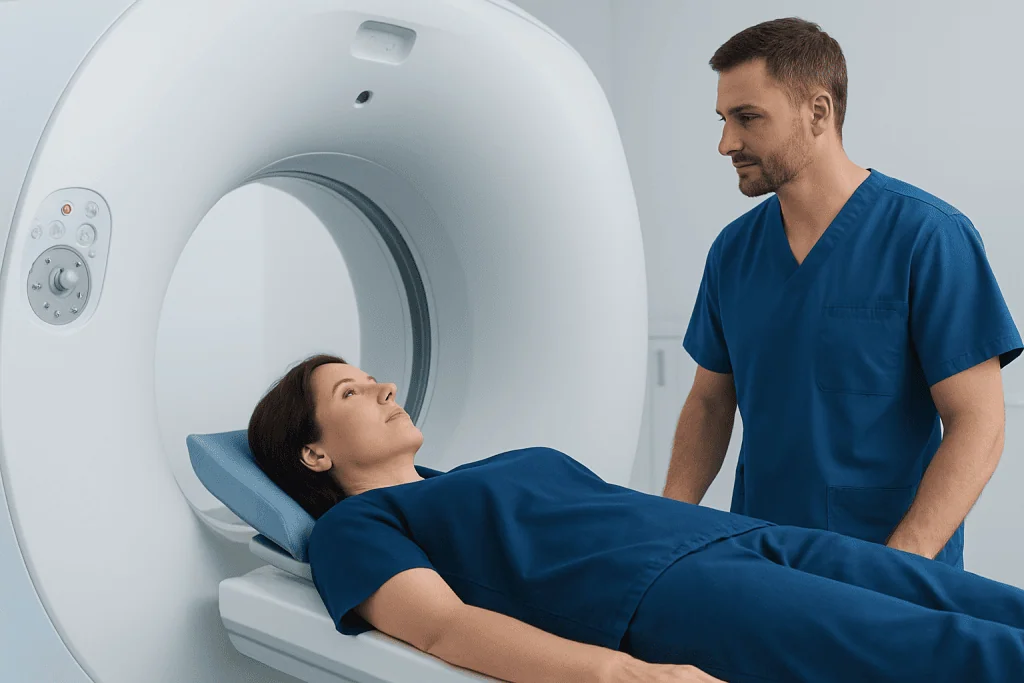
Computed tomography, or CT, combines multiple X-ray images to produce detailed cross-sectional views of the body. For a CT urogram, an iodine-based contrast material is commonly injected into a vein. The kidneys filter the contrast into the urine, helping the radiology team assess the urinary tract, including the kidneys, ureters and bladder.
A clinician may recommend CT imaging for visible or unexplained microscopic blood in the urine, persistent urinary symptoms, unexplained flank pain, or findings that need further evaluation. In people already diagnosed with bladder cancer, CT may help assess whether cancer has involved lymph nodes or other organs and can contribute to treatment planning.
CT is not always the preferred first test for every person. Pregnancy, reduced kidney function, previous serious contrast reactions and the need to limit radiation exposure may affect the imaging plan. The referring clinician and radiology team can select the safest suitable approach, which may include ultrasound, magnetic resonance imaging (MRI), cystoscopy or other tests.
Preparation, Procedure and Recovery Timeline

Preparation instructions vary by imaging center and the reason for the scan. A person should tell the care team about kidney disease, diabetes, possible pregnancy, medicines, prior contrast reactions and allergies. They may be asked to avoid food for a short time before contrast-enhanced imaging, while drinking water may be encouraged in some cases. The imaging department will provide individualized instructions.
During the procedure, the patient lies on a moving table that passes through the CT scanner. If contrast is used, it is injected through a small intravenous line. A brief warm sensation, metallic taste or feeling of needing to urinate can occur and usually passes quickly. The person will be asked to remain still and may need to briefly hold their breath while images are obtained.
The scan itself commonly takes only a short time, although the appointment may be longer because of registration, preparation and contrast timing. Most people can return to normal activities afterward. Drinking fluids, if medically appropriate, can help clear contrast from the body. A radiologist reviews the images and sends a report to the referring clinician, who explains what the results mean in the context of symptoms and other tests.
How Accurate Is a CT Scan for Bladder Cancer?
CT can be useful and informative, but its accuracy depends on the size, location and appearance of a tumor, the quality of bladder filling, the CT technique used and the experience of the interpreting team. It is generally better at finding larger growths, assessing the upper urinary tract and identifying possible spread beyond the bladder than at detecting very small or flat abnormalities of the bladder lining.
A CT report may describe a filling defect, focal wall thickening or a bladder mass that needs cystoscopic evaluation. It may also be normal despite an early cancer being present. For this reason, CT results are interpreted alongside medical history, urine testing, physical examination and direct bladder inspection.
CT cannot reliably determine the precise tumor grade or fully establish how deeply a cancer has invaded the bladder wall. Those important details typically come from tissue obtained during cystoscopy and, when needed, transurethral resection of a bladder tumor. Imaging may then be used with pathology findings to guide the next steps.
How Often Is Bladder Cancer Missed on a CT Scan?
There is no single missed-cancer rate that applies to every CT scan. Detection varies according to whether a dedicated CT urogram was performed, the bladder was adequately distended, contrast phases were appropriate, and whether the cancer is raised, flat, small or located in an area that is difficult to evaluate.
Small papillary tumors and carcinoma in situ, a flat high-grade change in the bladder lining, may be difficult or impossible to see on CT. A negative scan therefore does not completely exclude bladder cancer, particularly in a person with persistent visible blood in urine, recurrent unexplained microscopic blood, or concerning urinary symptoms.
When clinical concern continues, a urologist may recommend cystoscopy and urine cytology or other urine-based testing. This combined approach reduces the chance that an important condition is overlooked and helps ensure that symptoms are investigated appropriately.
What Is the Most Accurate Test for Bladder Cancer?
Cystoscopy with biopsy is the most definitive approach for diagnosing bladder cancer. During cystoscopy, a urologist passes a narrow flexible or rigid instrument with a camera through the urethra to view the inside of the bladder. If an abnormal area is seen, tissue can be removed or sampled and examined by a pathologist.
Pathology is essential because it confirms whether cancer is present and describes characteristics such as tumor type, grade and depth of invasion. If a tumor is identified, a transurethral resection may both remove visible tissue and provide the samples needed for accurate staging. This is commonly part of bladder cancer treatment planning.
Urine cytology can support the assessment by looking for abnormal cells shed into urine. It can be especially helpful for detecting some high-grade cancers, but it does not replace cystoscopy or biopsy. CT, cystoscopy, pathology and urine tests each provide different information and are often used together.
What Is the Biggest Indicator of Bladder Cancer?
The most common warning sign of bladder cancer is blood in the urine, known as hematuria. It may be clearly visible, making urine look pink, red or brown, or it may be found only on laboratory testing. Blood in urine may come and go and is often painless, so an episode should still be discussed with a doctor even if it resolves.
Other possible symptoms include frequent urination, urgency, burning during urination, difficulty passing urine, pelvic discomfort or recurrent urinary symptoms without a clear explanation. These symptoms are much more commonly caused by non-cancerous conditions such as urinary infection, stones or an enlarged prostate, but persistent symptoms warrant assessment.
Risk increases with age and is associated with tobacco exposure, some workplace chemical exposures, chronic bladder irritation and certain previous cancer treatments. Having a risk factor does not mean a person will develop cancer, and not having known risks does not rule it out. The appropriate evaluation depends on the individual situation.
Benefits, Risks and When to Seek Medical Care
The main benefit of CT is that it provides a broad view of the urinary tract and surrounding structures without surgery. It may identify alternative explanations for symptoms, such as kidney stones or masses in the kidneys, and helps clinicians assess possible spread when cancer is suspected or confirmed. Its main limitations are radiation exposure, imperfect detection of early bladder lesions and, in some scans, the use of contrast material.
Contrast reactions are uncommon and are often mild, but serious reactions can occur. Contrast can also require special consideration in people with significantly impaired kidney function. Patients should report any new rash, breathing difficulty, swelling or severe symptoms after contrast immediately. The imaging team reviews relevant health information before the scan to reduce risk.
When to seek medical care: Visible blood in urine should be assessed promptly, even if it happens only once. Medical advice is also important for persistent urinary burning or urgency, repeated urinary infections, unexplained microscopic blood in urine, new pelvic pain, or difficulty passing urine. Urgent care is appropriate for heavy bleeding with clots, inability to urinate, severe pain, fever with urinary symptoms, or feeling faint or unwell.
Acibadem International’s multidisciplinary specialists and JCI-accredited hospitals support international patients with investigation and management of bladder conditions, including coordinated imaging, urology assessment and cancer care when needed.
Frequently asked questions
Can a CT scan rule out bladder cancer?
No. A CT scan may make bladder cancer less likely when no abnormality is seen, but it cannot fully rule it out. Small, flat and early tumors can be missed, so cystoscopy may still be recommended when symptoms or urine findings are concerning.
Is CT urography the same as a regular CT scan?
CT urography is a CT examination designed to evaluate the urinary tract. It commonly uses contrast material and timed images to assess the kidneys, ureters and bladder. The exact protocol may differ between hospitals and according to a person's medical needs.
Does a CT scan show the stage of bladder cancer?
CT can help identify enlarged lymph nodes or possible spread outside the bladder, which supports staging. However, it cannot always show how deeply a tumor has invaded the bladder wall. Tissue examination after biopsy or tumor resection is needed for key staging and grading information.
Why is cystoscopy needed after a CT scan?
Cystoscopy lets a urologist inspect the bladder lining directly and find abnormalities that may not be visible on CT. It also allows biopsy or removal of suspicious tissue. This makes it the central test for confirming or excluding bladder cancer.
What should a person do if blood in urine goes away?
They should still contact a healthcare professional, particularly if the blood was visible or has happened more than once. Blood in urine can have many causes, and an evaluation can determine whether infection, stones, prostate conditions, kidney disease or another issue is responsible. Prompt assessment is especially important for people with smoking history or other risk factors.
Is contrast dye safe for a CT scan?
Most people receive CT contrast without serious problems. The care team will check for prior contrast reactions, kidney disease and other relevant conditions before proceeding. Anyone who has had a previous contrast reaction or has kidney concerns should mention this before the appointment.
References
- European Association of Urology
- American Urological Association
- National Cancer Institute
- American Cancer Society
- Radiological Society of North America
This article is for general information only and is not a substitute for professional medical advice. Please consult a qualified doctor about your individual situation.
Bladder Cancer in Turkey — costs, top hospitals & a free quote
JCI-accredited · board-certified surgeons · reply within 24h
Add us as a Preferred Source to see more of our trusted health content across Google Search, AI Overviews and Discover.

Hematology Department
17 specialists in this unitMore from the Health Library


Related Specialists

Dr. Meltem Batmaci
Internal Medicine
Assoc. Prof. Dr. Elif Şenocak Taşçı
Medical Oncology
Asst. Prof. Dr. Tarkan İkizoğlu
Pediatrics
Prof. Dr. Osman Güven
Orthopedic Surgery & Traumatology