Neuroendoscopic Procedures
Neuroendoscopic procedures are minimally invasive operations in which a neurosurgeon uses an endoscope, a thin tube with a light and camera, to reach deep parts of the brain through a small skull…

Quick answer
Neuroendoscopic procedures are minimally invasive brain and skull base operations performed through a small opening or the nose using an endoscope, a thin lighted camera tube. They are used for hydrocephalus, cysts, pituitary tumors, and biopsies. Hospital stays are often one to three nights, with many patients resuming light activities within weeks.
What are neuroendoscopic procedures?
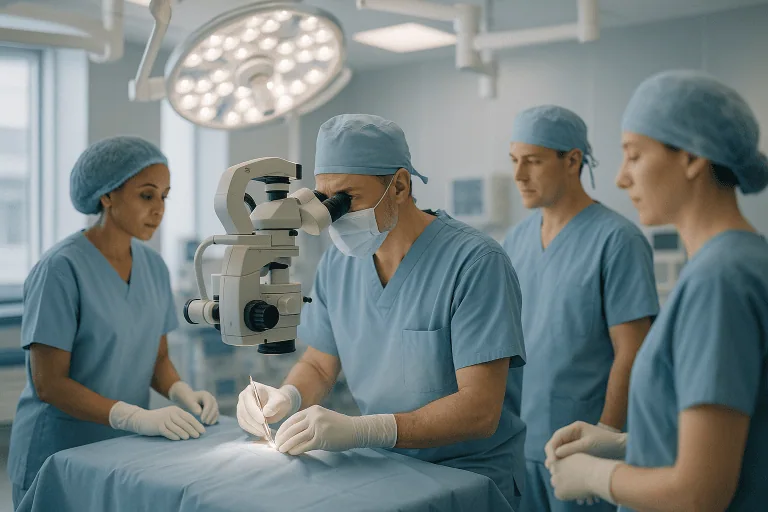
Neuroendoscopic procedures are a group of minimally invasive brain and spine operations performed with an endoscope. An endoscope is a thin tube fitted with a light and a small camera that sends magnified images to a screen. Instead of opening a large section of the skull, the surgeon works through a small opening in the bone, or through a natural passage such as the nose, and guides fine instruments alongside the camera.
Neuroendoscopy (the general term for this technique) allows the surgeon to see deep structures of the brain that would otherwise be hard to reach without moving or cutting healthy tissue. It is used for several conditions, including:
- Hydrocephalus – a buildup of cerebrospinal fluid (the clear fluid that surrounds the brain and spinal cord) inside the brain’s fluid-filled chambers, called ventricles.
- Cysts in or near the ventricles, such as colloid cysts or arachnoid cysts (fluid-filled sacs that can block fluid flow).
- Pituitary tumors – growths on the pituitary gland, a small hormone-producing gland at the base of the brain that is often reached through the nose.
- Some tumors inside the ventricles that need a tissue sample (biopsy) or removal.
- Skull base lesions, meaning abnormal tissue where the bottom of the skull meets the brain.
- Certain bleeds inside the brain, where the endoscope helps remove a blood clot through a small channel.
- Craniosynostosis in infants, a condition where skull bones fuse too early; endoscopic release is one treatment option for selected babies.
These operations are planned and carried out by a neurosurgical team. At Acibadem, they fall under the Neurosurgery department, which works together with radiology, anesthesiology, and, when hormones are involved, endocrinology.
Who is a candidate: who needs neuroendoscopic procedures
Whether a person needs neuroendoscopic procedures depends on the diagnosis, the exact location of the problem, and the person’s general health. Common reasons a surgeon may suggest this approach include:
- Obstructive hydrocephalus, where fluid is blocked at a narrow point and a small internal opening can restore flow (a procedure called endoscopic third ventriculostomy).
- A cyst or small tumor lying inside or next to a ventricle, where the fluid-filled space gives the camera room to work.
- A pituitary tumor or other lesion at the skull base that can be reached through the nasal passages.
- The need for a tissue sample from a deep lesion when imaging alone cannot give a clear diagnosis.
- A previously placed shunt (a drainage tube for hydrocephalus) that keeps failing, where an endoscopic alternative may be considered.
Neuroendoscopy is not suitable for every situation. It may not be recommended when:
- The tumor is large, solid, or spreads widely into brain tissue, so that a conventional open operation gives better control.
- The lesion is wrapped around major blood vessels or nerves that cannot be safely handled through a narrow channel.
- The ventricles are very small, leaving little space for the camera and instruments.
- There is active infection, uncontrolled bleeding tendency, or another medical condition that makes anesthesia unsafe at that time.
- Imaging suggests the fluid problem is caused by poor absorption rather than a blockage, in which case a ventriculostomy is less likely to help and a shunt may be preferred.
The final decision rests on detailed imaging, usually magnetic resonance imaging (MRI), and a discussion between the patient, family, and the surgical team about the expected benefits and alternatives.
How the neuroendoscopic procedures procedure works
The exact steps vary by condition, but most neuroendoscopic procedures follow a similar pattern.
Before the operation
You will have imaging studies so the surgeon can plan the safest path to the target. Blood tests, a heart check, and an anesthesia review are usually arranged. In pituitary surgery, hormone levels and vision are commonly tested beforehand, and an ear, nose, and throat specialist may examine the nasal passages.
During the operation
- You are given general anesthesia, so you are asleep and feel nothing.
- Your head is gently held still. Many teams use navigation software, which matches your scans to your head position like a GPS for surgery.
- For brain procedures, a small patch of hair may be shaved and a coin-sized opening (a burr hole) is made in the skull. For pituitary and skull base surgery, the surgeon works through a nostril and no external cut is needed.
- The endoscope is passed to the target. The surgeon watches the magnified image on a monitor while using fine instruments through the same or a nearby channel.
- The planned task is completed: opening a membrane to let fluid flow, draining or removing a cyst, taking a biopsy, or removing tumor tissue piece by piece.
- The area is checked for bleeding, the endoscope is withdrawn, and the small opening is closed. In nasal approaches, the surgeon may repair the base of the skull with tissue or packing to prevent fluid leakage.
Depending on the condition, the operation often takes between one and four hours, including anesthesia time.
After the operation
You wake up in a recovery area and are then observed on a neurosurgical ward or, for some cases, an intensive care unit for the first night. Nurses check your alertness, pupils, movement, and pain regularly. A scan is often done within the first day or two to confirm the result and rule out bleeding.
Preparation for neuroendoscopic procedures
Good preparation helps reduce complications and makes recovery smoother. Your team will give you personal instructions, but the following points are common:
- Medication review: tell your doctors about every medicine, supplement, and herbal product you take. Blood thinners, some pain relievers, and certain supplements may need to be paused in advance, but only under medical guidance.
- Fasting: you will usually be asked not to eat or drink for several hours before general anesthesia.
- Smoking and alcohol: stopping smoking, even for a few weeks beforehand, may support wound healing and breathing during anesthesia.
- Infections: report any cold, sinus infection, fever, or skin infection in the days before surgery, as the operation may need to be rescheduled, especially for nasal approaches.
- Practical planning: arrange for someone to accompany you home and help during the first days. Bring a list of your allergies and a copy of previous scans if they were done elsewhere.
- Questions: write down what you want to ask about the goals, alternatives, and what would happen if the endoscopic approach had to be changed to an open one during surgery.
For children, the team will also explain how parents can stay involved before and after the operation.
Recovery: neuroendoscopic procedures recovery time
Because the openings are small, recovery from neuroendoscopic procedures is often shorter than after open brain surgery, but the brain still needs time to settle. Timelines differ by diagnosis, age, and overall health.
- Hospital stay: many patients stay one to three nights. Pituitary surgery through the nose and more complex tumor cases may need a somewhat longer stay.
- First days: headache, tiredness, and mild nausea are common and usually managed with medication. After nasal surgery, a stuffy nose, reduced sense of smell, and mild nosebleeds are expected for a while.
- First two weeks: most people can walk, wash, and manage light tasks at home. Heavy lifting, straining, bending, and vigorous exercise are typically discouraged. After nasal approaches, patients are usually told not to blow the nose or use straws for a set period.
- Two to six weeks: many patients return to desk work, school, or light routines during this window, depending on how they feel and what the surgeon advises.
- Driving: you should not drive until your doctor confirms it is safe, and never while taking strong pain medication.
Follow-up usually includes a wound or nose check, review of symptoms, and repeat imaging at planned intervals. People treated for hydrocephalus are typically monitored over the long term because the opening created can occasionally narrow again. Those who had pituitary surgery often need repeat hormone tests, since gland function can change after the operation.
Neuroendoscopic procedures risks and benefits
Every operation carries risk, and it is important to weigh neuroendoscopic procedures risks and benefits with your surgeon in the context of your own condition.
Potential benefits
- Smaller openings in the skull or none at all with nasal approaches.
- Less disturbance of healthy brain tissue compared with wider open surgery.
- Direct, magnified view of deep structures that are otherwise hard to see.
- Often a shorter hospital stay and quicker return to routine activities.
- In hydrocephalus, the chance of avoiding a permanent shunt, which has its own long-term failure and infection risks.
Possible risks and side effects
- Bleeding inside the brain or ventricles, which in rare cases can be serious.
- Infection of the wound, the fluid spaces (meningitis), or the sinuses after nasal surgery.
- Cerebrospinal fluid leak, especially after skull base surgery, which may cause clear fluid to drip from the nose and sometimes needs further repair.
- Injury to nearby structures, such as the hypothalamus (which regulates temperature, thirst, and appetite), the optic nerves, or small blood vessels.
- Hormone disturbance after pituitary surgery, including temporary or lasting changes in fluid balance or the need for hormone replacement.
- Failure or recurrence, where the new fluid opening closes, a cyst refills, or tumor tissue remains and needs more treatment.
- Seizures, memory change, or short-term confusion, more often in the early period.
- Anesthesia-related risks, such as reactions to medication, breathing difficulties, or blood clots in the legs.
In some cases the surgeon may need to change to an open approach during the operation if the view is unclear or bleeding is difficult to control. This possibility is normally discussed and consented to in advance.
Results and outlook
The outlook after neuroendoscopic procedures depends mainly on the underlying diagnosis rather than on the technique itself. In general terms, the clinical evidence shows that:
- Endoscopic third ventriculostomy can relieve symptoms of obstructive hydrocephalus in many suitable patients and may allow them to avoid a shunt, though it does not succeed in everyone and a shunt may still be needed later. Success tends to be lower in very young infants and in hydrocephalus caused by infection or bleeding.
- Endoscopic removal of colloid cysts and drainage of arachnoid cysts often relieves pressure symptoms, with a small chance of the cyst returning.
- Endoscopic pituitary surgery can remove or reduce many tumors, with results influenced by the tumor’s size, its hormone activity, and how far it extends into surrounding tissue. Some patients need additional medication or radiation therapy afterward.
- For biopsies, the endoscope usually provides enough tissue for a diagnosis that then guides further treatment.
Symptoms caused by pressure, such as headache or vision changes, often improve over weeks to months, but problems present for a long time before surgery may recover only partly. Regular follow-up imaging and clinical review are the best way to detect recurrence early.
Cost considerations
The cost of neuroendoscopic procedures varies widely and no single figure applies to all patients. The main factors that influence the total are:
- Type and complexity of the operation, for example a single fluid opening compared with a multi-hour tumor removal.
- Hospital stay, including whether intensive care monitoring is needed and how many nights are spent on the ward.
- Devices and materials, such as navigation systems, disposable endoscope parts, skull base repair materials, or a shunt if one is placed.
- Imaging and laboratory tests before and after surgery, including MRI scans and hormone panels.
- Specialist involvement, such as an ear, nose, and throat surgeon in nasal approaches or an endocrinologist afterward.
- Follow-up care, including repeat scans, clinic visits, and any additional treatment.
- Insurance coverage and the country or region where treatment takes place.
Asking for a written treatment plan that lists what is and is not included can help you understand the expected expenses.
Frequently asked questions
Who needs neuroendoscopic procedures instead of open surgery?
People whose problem lies inside or next to the fluid-filled ventricles, or at the skull base near the nose, are the most common candidates. The surgeon decides based on imaging, the size and nature of the lesion, and your overall health. Some conditions still require a conventional open operation, and in certain cases both approaches are combined.
How long does the neuroendoscopic procedures procedure take?
Most operations take roughly one to four hours, including the time needed for anesthesia and positioning. Simple fluid openings are usually at the shorter end, while tumor removals through the nose or complex cyst work can take longer. Your surgeon can give an estimate for your specific case.
What is the typical neuroendoscopic procedures recovery time?
Many patients leave the hospital within one to three nights and resume light daily activities within about two weeks. A return to work, school, or exercise often happens between two and six weeks, depending on the procedure and how you feel. Full recovery of symptoms such as headache or vision change can take longer.
Are neuroendoscopic procedures painful?
You are asleep under general anesthesia during the operation. Afterward, headache, scalp soreness, or nasal discomfort are common but usually controlled with standard pain relief. Severe or worsening pain is not expected and should be reported to your care team.
What are the main neuroendoscopic procedures risks and benefits I should weigh?
The main benefits are smaller openings, less disturbance of healthy tissue, and often a quicker recovery. The main risks include bleeding, infection, cerebrospinal fluid leak, injury to nearby structures, hormone changes after pituitary surgery, and the chance that the problem returns. Your surgeon will explain how these apply to your diagnosis.
Will I need a shunt after endoscopic hydrocephalus surgery?
The aim of an endoscopic third ventriculostomy is to avoid a shunt, and in many suitable patients that goal is achieved. However, the opening can close in some people, and a shunt may still be needed later. Long-term follow-up is important so that any return of symptoms is caught early.
Can children have neuroendoscopic procedures?
Yes. Neuroendoscopy is widely used in children for hydrocephalus, cysts, certain tumors, and selected cases of craniosynostosis. Age matters for some procedures, and pediatric neurosurgeons consider the child’s development and skull anatomy when planning.
When to see a doctor
You should be assessed by a specialist, such as a neurologist or neurosurgeon, if you or your child develop symptoms that could point to raised pressure in the brain or a growth near the pituitary gland. These include:
- Persistent or worsening headaches, especially in the morning or with vomiting.
- Blurred, double, or narrowing vision, or loss of side vision.
- Unsteady walking, memory decline, or problems with bladder control in adults.
- In infants, a rapidly enlarging head, a bulging soft spot, irritability, poor feeding, or downward-turned eyes.
- Unexplained hormone-related changes, such as menstrual irregularity, milk production without pregnancy, unusual growth of hands and feet, or persistent fatigue.
After neuroendoscopic procedures, seek urgent medical attention if you notice any of the following:
- Severe or rapidly worsening headache, repeated vomiting, or increasing drowsiness and confusion.
- New weakness, numbness, speech difficulty, or a seizure.
- Fever, stiff neck, or sensitivity to light, which may signal infection.
- Clear, watery fluid dripping from the nose or wound, or a salty taste at the back of the throat, which may indicate a cerebrospinal fluid leak.
- Heavy nosebleed that does not stop, or redness, swelling, and discharge at the incision.
- Extreme thirst with very frequent urination after pituitary surgery, which may reflect a fluid-balance hormone problem.
Your discharge instructions will list the contact route for your surgical team; these warning signs should not wait for the next scheduled visit.
Preparation
- Have the planned MRI, blood tests, and anesthesia review completed and share a full list of your medicines and supplements, since blood thinners may need to be paused under medical guidance. Fast as instructed before general anesthesia and report any cold, sinus, or skin infection beforehand. Arrange for someone to accompany you home and help during the first days.
Aftercare
- Expect headache, tiredness, and, after nasal approaches, a blocked nose for a period; take pain relief as prescribed and avoid heavy lifting, straining, or nose blowing until cleared. Keep wound or nasal care instructions, attend follow-up imaging and hormone checks if advised, and do not drive until your doctor confirms it is safe. Seek urgent care for worsening headache, fever, stiff neck, clear nasal fluid, new weakness, or seizures.
Medically reviewed by the Acıbadem International Medical Board September 13, 2026
See our medical review board →
Update history
- PublishedSeptember 13, 2026
- Medical review approvedSeptember 13, 2026
- Last content updateSeptember 13, 2026
References3
From the Acibadem Blog
Neuroendoscopic vs Open Brain Surgery: When the Smaller Opening Is the Right Choice
Endoscopic brain surgery reaches a target through a small opening, often the nostril or a coin-sized burr hole, using a thin camera, while open…

What to Expect After a Neuroendoscopic Procedure: Blocked Nose, Headache and No-Lifting Weeks
Recovery after neuroendoscopic surgery, most often performed through the nose to reach the pituitary gland or skull base, typically involves a hospital stay of…