

Neuroendoscopic vs Open Brain Surgery: When the Smaller Opening Is the Right Choice
Key Takeaways
- Endoscopic brain surgery travels through existing corridors, chiefly the nostril to the pituitary region or a burr hole into the fluid-filled ventricles, rather than through functioning brain tissue.
- Most pituitary tumors are noncancerous adenomas, and the nasal route is the standard approach for those that need removal, according to Mayo Clinic.
- Endoscopic third ventriculostomy creates a new drainage path for cerebrospinal fluid and is described by the NHS as an alternative to a shunt for suitable people with hydrocephalus.
- Systematic reviews comparing endoscopic and microscopic pituitary surgery mostly find broadly similar outcomes with a different mix of complications, and the evidence is largely observational.
- Awake craniotomy works because the brain has no pain receptors, allowing real-time mapping of speech and movement that is only possible through an open approach.
- The shared risks of bleeding, infection, seizures and stroke apply to both routes; the endonasal approach adds a specific risk of cerebrospinal fluid leaking through the nose.
Endoscopic brain surgery reaches a target through a small opening, often the nostril or a coin-sized burr hole, using a thin camera, while open surgery (craniotomy) removes a section of skull for direct access. Neither is universally better. Surgeons choose based on where the problem sits, its size, and whether fluid-filled spaces or natural corridors lead to it. The decision always rests with the treating team.
The sketch is on the back of a scan report: a rough circle for a skull, a small dot for the growth, and two arrows. One curves in through the nose. The other comes straight down through the top of the head. The person holding the paper has been told both routes could work, and now has a week to think about which one.
That sketch is the whole endoscopic vs open brain surgery question in miniature. It is not a contest between old and new medicine, and it is rarely a question of which surgeon is braver. It is a question of geometry: where the target sits, what surrounds it, and whether there is a natural corridor to it that does not have to be created.
This explainer walks through what each approach actually involves, which conditions usually suit a smaller opening, why some people are steered firmly toward a craniotomy, and what the days and weeks afterward tend to look like, using guideline-level evidence rather than reassurance.
What does endoscopic vs open brain surgery actually mean?
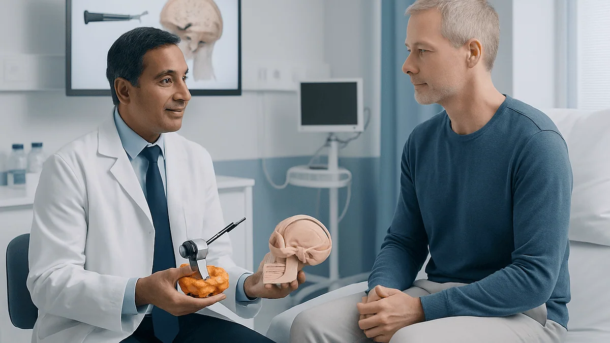
Two words carry most of the weight here. A craniotomy is an operation in which a piece of skull bone is temporarily removed so the surgeon can work directly on the brain, then replaced at the end. Neuroendoscopy is surgery performed through a narrow tube that carries a light and a camera, so the surgeon watches a screen rather than looking down into the head.
The difference is not the size of the incision alone. Open surgery gives a wide, direct, three-dimensional view and room for two hands and several instruments. The endoscope gives a bright, magnified view from inside a cavity, and can look around corners, but it works through a corridor no wider than a finger, and the picture is two-dimensional on most systems.
The two approaches also overlap more than the framing suggests. Surgeons routinely pass an endoscope through a craniotomy to peer behind a structure they cannot see with the microscope. Many operations are described as endoscope-assisted rather than purely endoscopic. And some purely endoscopic operations, especially through the nose, are large procedures in every sense except the visible scar.
So the real question a treating team asks is not smaller or bigger. It is: which route lets us see the whole problem, protect the tissue around it, and control bleeding if it happens? For a pituitary growth sitting just above the back of the nasal cavity, the answer is usually the endoscope. For a tumor lodged in the outer layer of the brain near the movement area, the answer is usually open surgery with the brain mapped in real time. The following sections explain why.
What is neuroendoscopy, and how does the camera get inside?
An endoscope is a rigid or flexible tube, typically a few millimeters across, with a lens at one end, a light source, and a channel for tiny instruments. In the brain it travels along spaces that already exist. Two corridors account for most cases.
The first is the nose. Behind the nasal cavity sits a thin plate of bone called the sphenoid sinus, and directly above it lies the pituitary gland in its bony pocket. By passing the scope through a nostril and opening that plate, the surgeon reaches the gland without touching the brain’s surface at all. This is called the endoscopic endonasal approach, and it is the route Mayo Clinic describes for most pituitary tumors, which are mostly noncancerous growths called adenomas.
The second corridor is the ventricles, the fluid-filled chambers deep inside the brain. A burr hole roughly the width of a large coin is drilled in the skull, and the scope is guided into a ventricle, often with a navigation system that matches the scan to the patient’s head like a GPS. From there the surgeon can open a blocked passage for fluid, take a biopsy of a growth on the ventricle wall, or remove a cyst.
Newer variants extend the idea. Tubular retractors, which are slim clear cylinders inserted through a small opening, let a surgeon reach deeper tissue while pushing brain aside gently rather than cutting through it. In some centers, blood clots deep in the brain are drained through similar small openings.
What all of these share is the trade: less disturbance on the way in, in exchange for a narrower field of view and less room to maneuver once there.
What actually happens in open brain surgery (craniotomy)?
Open surgery follows a sequence that has been refined for decades and that MedlinePlus describes plainly. Hair over the planned incision is clipped, though rarely the whole head. The scalp is opened and folded back. A drill and a fine saw remove a window of bone, often the size of a playing card, sometimes much smaller. The tough membrane under the bone, the dura, is opened, and the brain itself is visible.
The surgeon then works under an operating microscope, which magnifies the field and lights it intensely. Modern craniotomies usually also use image guidance, ultrasound, or, in some tumors, a dye that makes abnormal tissue glow under a special light. Monitoring electrodes may track nerve pathways so that a wrong move triggers an alarm before it causes lasting harm.
At the end the dura is stitched closed, the bone flap is returned and fixed with small plates, and the scalp is closed. In some emergencies, such as severe swelling after trauma or stroke, the bone is deliberately left out for weeks to give the brain room, and replaced later.
It sounds dramatic, and it is major surgery. Yet the brain itself has no pain sensors, which is why some of these operations can be done with the patient awake, a subject covered later in this article. The parts that hurt afterward are the scalp, the muscles of the temple and jaw, and the headache that comes from air and blood irritating the coverings of the brain.
The great advantage of the open route is control. If a vessel bleeds, the surgeon can see it and stop it. If the tumor turns out to be stuck to something vital, there is room to change plan.
Which conditions are usually treated with the smaller opening?
Neuroendoscopy earns its place where a natural corridor leads straight to the problem. The clearest examples fall into four groups.
Pituitary and nearby skull-base growths come first. Because the gland sits directly above the sphenoid sinus, the nose is a straight line to it. Mayo Clinic notes that most pituitary tumors that need removal are approached this way, and the same corridor can reach some growths on the bone at the base of the skull, such as certain meningiomas and craniopharyngiomas, when they are positioned favorably.
Hydrocephalus is the second. In this condition, cerebrospinal fluid, the clear liquid that cushions the brain, builds up because its flow is blocked. The NHS describes an operation called endoscopic third ventriculostomy, in which the scope is used to make a small opening in the floor of one chamber so fluid can drain by a new route, offering an alternative to a shunt tube for suitable people.
Cysts and intraventricular tumors are the third. Colloid cysts, arachnoid cysts, and some growths that hang into the ventricles can be sampled or removed under direct endoscopic view.
Deep blood clots are the fourth and most debated. Trials of minimally invasive drainage for bleeding inside the brain have been indexed in PubMed, and the evidence is genuinely mixed: some suggest lower death rates, but a clear improvement in day-to-day function has been harder to show. Guideline language remains cautious.
Common to all four is that the target is either at the end of a hollow space or reachable through one. The endoscope does not do well when it has to be pushed through solid, functioning brain to get somewhere.
Who is usually asked to wait, or steered toward open surgery?
Being told the smaller operation is not the right fit can feel like a loss. Understanding the reasons usually helps.
Size and shape come first. A growth that spreads sideways around the arteries at the base of the brain, or one that wraps around the nerves to the eyes, may need the wide view and two-handed dissection an open approach allows. A surgeon working through a nostril has limited ability to peel tumor off a vessel that is out of the direct line of sight.
Location on the surface comes second. Tumors in the outer layers of the brain, particularly near areas controlling speech or movement, are approached openly so the surgeon can map function with electrical stimulation and stop exactly where safety demands.
Bleeding risk comes third. Some growths are richly supplied with blood. If heavy bleeding starts at the end of a narrow tube, controlling it is far harder than in an open field, and many teams will not accept that risk.
Anatomy of the corridor comes fourth. Narrow nasal passages, prior sinus surgery, or unusual bone shape can make the endonasal route unsafe.
And then there are those asked simply to wait. Many small pituitary adenomas that make no hormones and press on nothing are monitored with scans, as Mayo Clinic describes, because the risk of operating can exceed the risk of watching. Small meningiomas found by chance in older adults are often observed for the same reason. Being placed on surveillance is not neglect; it is a decision that the disease is currently less dangerous than the knife. That judgment belongs to the treating team, who weigh scan findings, hormone tests, vision and overall health together.
Endoscopic vs open brain surgery at a glance
Numbers and labels flatten a nuanced choice, but a side-by-side view clarifies what each route trades away. Read the table as typical tendencies described in patient resources from Mayo Clinic, MedlinePlus and the NHS, not as guarantees for any individual.
| Feature | Endoscopic approach | Open craniotomy |
|---|---|---|
| Route in | Nostril, or a burr hole into a fluid space | Window of skull bone, temporarily removed |
| What the surgeon sees | Magnified, angled camera view on a screen | Direct, three-dimensional view under a microscope |
| Room to work | Narrow corridor, fine instruments | Wide field, two hands, multiple instruments |
| Best-suited targets | Pituitary region, ventricles, cysts, blocked fluid pathways | Surface tumors, growths wrapped around vessels, most aneurysms |
| Typical anesthesia | General | General, or awake for parts of selected operations |
| Visible scar | None (nasal) or small scalp incision | Curved scalp incision, usually within the hairline |
| Distinctive risks | Fluid leak through the nose, sinus symptoms, loss of smell | Larger wound, more muscle pain, bone flap issues |
| Shared risks | Bleeding, infection, seizures, stroke, damage to nearby nerves or hormone function |
One row deserves emphasis. The shared risks are shared. A smaller opening does not remove the possibility of stroke, bleeding or infection; it changes the mix. Endonasal surgery, for instance, carries a specific risk that cerebrospinal fluid leaks through the repair into the nose, which sometimes requires a second procedure. Craniotomy carries specific risks of wound and bone problems that the nasal route avoids.
The other row worth pausing on is room to work. That single constraint explains most of the decisions in the previous two sections.
Is endoscopic brain surgery safer? What the evidence really shows
The honest answer is that safety depends on matching the approach to the problem, and the studies that exist mostly compare the two routes for the same problem, which is a narrower question.
The best-studied comparison is pituitary surgery, where the endoscope replaced the microscope through the same nasal corridor over the past two decades. Systematic reviews indexed in PubMed have generally found the two techniques similar in how completely they remove tumors and in major complication rates, with some analyses suggesting a small advantage for the endoscope in visualization and in certain leak or sinus outcomes, and others finding no meaningful difference. The quality of that evidence is mostly observational rather than randomized, which means it can be swayed by which patients were selected for each technique.
For hydrocephalus, the NHS presents endoscopic third ventriculostomy and shunting as alternatives, each with its own failure and complication profile, rather than one being safer across the board. Shunts can block or become infected; the endoscopic opening can close over. Which is preferable depends on the cause of the blockage and the patient’s age.
For deep brain hemorrhage, as noted earlier, minimally invasive drainage trials have not produced the clear functional benefit that would settle the question, so guideline language remains conditional.
There is no credible evidence that endoscopic surgery is safer for tumors that were never suitable for it in the first place. A surface tumor near the speech area is not made safer by a smaller hole; it is made safer by awake mapping through an open one.
Where a surgeon says the approaches are roughly equivalent for your particular lesion, that statement usually reflects the evidence accurately, and the choice may reasonably come down to the team’s experience with each route.
Endoscopic pituitary surgery: the most studied example
If one operation defines the modern endoscopic era, it is endonasal pituitary surgery, so it is worth walking through in detail.
The pituitary is a pea-sized gland at the base of the brain that directs the thyroid, adrenal glands, growth, fertility and water balance. Mayo Clinic explains that most pituitary tumors are adenomas, which are noncancerous, and that trouble arises either because the tumor overproduces a hormone or because it grows large enough to press on the nerves carrying vision. Surgery is considered when a tumor is causing symptoms or threatening sight, and some hormone-producing tumors are managed with medication first; that sequencing is decided by an endocrinologist and surgeon together.
Under general anesthesia, the surgeon, often working alongside an ear, nose and throat colleague, passes the endoscope through one or both nostrils, opens the sphenoid sinus, and removes the thin bone in front of the gland. The tumor is then removed piece by piece with curved instruments while the camera looks in from beside them. Angled lenses let the surgeon check the corners of the cavity where residual tumor hides.
Closing is where this approach demands the most care. The barrier between nose and brain has been opened, so it must be rebuilt, sometimes with a flap of tissue from inside the nose, to prevent cerebrospinal fluid leaking downward.
Afterward the nose feels blocked and crusty for weeks, and the sense of smell can be dulled. Some people temporarily lose the ability to concentrate urine and need close monitoring of fluid balance. Hormone levels are checked before and after, because the normal gland can be affected either way.
Not every pituitary tumor fits this route. Very large ones that extend far above or to the sides may still need an open approach, or a staged combination of both.
How long does it take to recover from endoscopic brain surgery?
People searching for endoscopic brain surgery recovery time usually want a number, and the honest reply is a range shaped by which operation was done.
For endonasal pituitary surgery, hospital stays of a few days are typical in patient resources from Mayo Clinic, with the first night often spent under closer observation to watch fluid balance and vision. Many people are walking the next day. What lingers is the nose: congestion, crusting and a dulled sense of smell that can persist for several weeks while the lining heals. Most teams ask people not to blow their nose forcefully, strain, or bend head-down for a period afterward to protect the repair, and the exact restrictions come from the surgical team, not a magazine.
For endoscopic third ventriculostomy or cyst surgery through a burr hole, the wound is small and the stay is often similarly short, though recovery is governed less by the incision and more by how the brain responds to the change in fluid pressure. Headache and tiredness are common in the early days.
For minimally invasive drainage of a hemorrhage, recovery is dominated by the stroke itself rather than the surgery, and rehabilitation can extend over months.
The pattern across all of these is the same: the shorter, smaller-scar recovery applies to the wound, not necessarily to the underlying condition. Someone whose hormones were disturbed by a pituitary tumor may need weeks of adjustment and follow-up blood tests regardless of how neatly the surgery went.
Return to work and driving are individual decisions. Many people are back to desk work within a few weeks after endonasal surgery, but seizures, vision changes or hormone issues can extend that, and licensing rules on driving after brain surgery vary by jurisdiction, so the treating team’s advice is the one that counts.
What the first weeks after open brain surgery usually look like
Craniotomy recovery has a shape that most people recognize once it is described, and knowing it in advance takes some of the fear out of it.
The first night is often spent in an intensive care or high-dependency bed, not because something is expected to go wrong but because the hours immediately after surgery are when swelling or bleeding would show, and frequent neurological checks catch it early. Expect to be woken repeatedly and asked to squeeze fingers, name the month, and follow a light with your eyes.
Over the following days, MedlinePlus describes a hospital stay that commonly runs from a few days to about a week, longer if the reason for surgery was complex. Headache is nearly universal and usually manageable; the scalp and the chewing muscles on the operated side ache, so soft foods help. Steroids given around the operation to reduce swelling can disturb sleep and mood for a short period.
Home recovery is measured in weeks rather than days. MedlinePlus discharge guidance describes a gradual return of energy, with many people needing several weeks before feeling like themselves, and advises against heavy lifting, contact sports and driving until the surgical team clears each one. Fatigue that arrives mid-afternoon like a wall is normal and improves.
Wound care is simple: keep it clean and dry as instructed, and watch for redness, swelling, or fluid. Staples or stitches come out at a follow-up visit, typically a week or two after surgery.
Seizures are a known risk after any operation on the brain’s surface, so some people are prescribed an anti-seizure medicine for a period; the decision to start it, and when to stop, belongs to the neurosurgical team.
The timeline above describes a straightforward course. Where a tumor needs radiotherapy or chemotherapy afterward, as the NHS outlines for some brain tumors, recovery from surgery overlaps with the start of the next treatment.
Are people awake during open brain surgery?
Sometimes, yes, and the idea unsettles people far more than the experience usually does.
Awake craniotomy is used when a tumor or seizure focus sits close to areas of the brain that control speech, language or movement. Mayo Clinic explains the logic: because the brain itself has no pain receptors, once the scalp and skull are numbed and opened under sedation, the patient can be gently woken. A neuropsychologist or speech specialist then asks them to name pictures, count, or move a hand while the surgeon applies a tiny electrical current to spots on the brain’s surface. If speech falters or the hand stops, that spot is marked as off-limits. The surgeon removes what can be removed and leaves the rest.
The patient is not lying there wide-eyed for the whole operation. The opening and closing are done under sedation or general anesthesia; the awake portion is the mapping and the removal near critical tissue. People describe hearing voices, feeling pressure or vibration, and being asked strange questions. Pain is not the typical report.
Awake surgery is not offered for everything, and it is never offered through the nose, since there is nothing to map there. It requires a cooperative, calm patient who can lie still for a long period, so it is generally not used for young children, for people with severe anxiety, or when the airway would be difficult to manage if things changed suddenly.
Why do it at all? Because mapping while awake gives real-time information that no scan can, and it lets the surgeon take a tumor closer to a language area than would be safe blind. That is the reason it is a technique of open, not endoscopic, surgery: mapping needs a wide view of the cortex.
What is the survival rate of open brain surgery?
This is one of the most searched questions about brain surgery, and the most honest answer is that no single figure exists, because open brain surgery is not one condition.
A craniotomy to remove a benign meningioma from a healthy adult, a craniotomy to clip a ruptured aneurysm, and an emergency craniotomy for a traumatic bleed are entirely different situations with entirely different outcomes. Quoting one average would mislead everyone in each group. That is why the NHS, MedlinePlus and Mayo Clinic describe risks in terms of what can go wrong rather than a headline percentage.
What patient resources do agree on is the list of serious risks that apply across the board: bleeding into the brain, infection, seizures, stroke, swelling, blood clots in the legs or lungs, and injury to nearby nerves that can affect vision, speech, movement or hormone function. How likely each is depends on the location and nature of the problem, the patient’s age and other illnesses, and whether the operation is planned or urgent.
If you want a meaningful number, ask a narrower question. Your surgeon can tell you, for your specific diagnosis and your specific approach, roughly how often serious complications occur in their experience and in published series. That number will mean something. A generic survival rate for brain surgery does not.
One more distinction matters. For malignant brain tumors, long-term outlook is driven mostly by the tumor’s biology and the treatments that follow, as the NHS explains in its brain tumor guidance, not by the operation itself. Surgery removes what it safely can and provides tissue for diagnosis; it is the beginning of a treatment pathway, not a verdict on it.
What is the hardest brain surgery?
Surgeons do not keep a ranked list, but they do agree on what makes an operation difficult, and the answer has almost nothing to do with the size of the opening.
Depth and neighbors drive difficulty. Operations at the base of the skull, where tumors sit tangled among the arteries that feed the brain and the nerves that control the eyes, face and swallowing, are among the most demanding because there is no safe margin in any direction. The brainstem, the thumb-sized column that runs breathing, heart rate and consciousness, is the other region surgeons approach with the greatest caution; even small growths there may be judged inoperable or approached only for biopsy.
Blood supply is the second factor. Aneurysms, which are weak bulges in artery walls, and arteriovenous malformations, which are tangles of abnormal vessels, can bleed catastrophically if handled wrongly, and the margin for error is measured in millimeters.
Function is the third. A tumor in the part of the brain that generates speech may be technically easy to reach but hard to remove without harm, which is why awake mapping exists.
Interestingly, some of the hardest operations in this sense are now done endoscopically. Extended endonasal approaches to tumors at the very center of the skull base require a surgeon to work at the end of a long, narrow corridor, next to the carotid arteries and the optic nerves, with no ability to see behind them. A small opening does not make the surgery small.
The practical takeaway is that difficulty is a property of the lesion and its address, not of the technique. When a team explains that your operation is complex, they are describing the anatomy, not warning you off.
What people often get wrong about minimally invasive brain surgery
Several myths follow this topic around, and each one can distort a real decision.
The first is that minimally invasive brain surgery means minor surgery. The incision is minimal; the operation may not be. An extended endonasal procedure can take many hours and disturb hormone and fluid balance for weeks. The label describes the entrance, not the journey.
The second is that a smaller opening always means faster recovery. For the wound, yes. For the condition, not necessarily. Someone whose hydrocephalus was relieved through a burr hole may still need weeks to adjust; someone whose pituitary tumor was removed through the nose may need months of hormone follow-up.
The third is that open surgery is outdated. The NHS still describes craniotomy as the main surgical treatment for most brain tumors, and it remains the only route that allows awake functional mapping. The endoscope added a tool; it did not retire the older ones.
The fourth is that the endoscope reaches deeper. It reaches through existing spaces. A target buried in solid brain tissue with no adjacent cavity is often better approached with a small open corridor and a tubular retractor than with an endoscope forced through functioning brain.
The fifth is that whichever approach has the nicer brochure is the better one. The comparative evidence for the same lesion, where it exists, tends to show broadly similar outcomes and a different mix of complications, as the pituitary literature indexed in PubMed illustrates.
And the sixth is that the choice belongs to the patient alone. It is shared. Patients rightly weigh scars, downtime and personal risk tolerance, but the anatomical judgment about which corridor is safe is one only the operating team can make.
Questions to ask your care team
A consultation about brain surgery is short and the vocabulary is thick, so having questions written down changes the conversation. These are the ones that tend to surface the reasoning behind an approach.
- Why is this route, open or endoscopic, the right one for where my lesion sits, and what would make you switch to the other?
- Is the goal complete removal, partial removal for safety, or a biopsy for diagnosis? How will I know afterward which was achieved?
- Which structures are closest to the target, and what could happen if they are affected: vision, speech, movement, hormones, smell?
- Will any part of the operation be done awake, and if so, what will I be asked to do?
- What is the specific complication you worry about most for this operation, and how often does it happen in your experience and in published series?
- If bleeding or a fluid leak occurs, what is the plan, and does that plan differ between the two approaches?
- How long do you expect me to be in hospital, and what will the first two weeks at home look like?
- What restrictions apply afterward: nose-blowing, lifting, driving, flying, returning to work?
- What follow-up scans or blood tests are planned, and for how long?
- What happens if we choose to monitor rather than operate right now, and what would prompt a change?
Notice that none of these asks which approach is best in general. They ask why this approach is right for this lesion. That framing gets you an answer grounded in your own scan rather than in a marketing comparison.
Bring someone with you if you can. People retain a fraction of what they hear in these appointments, and a second set of ears, or permission to record the conversation, helps with the questions that only occur to you in the car park.
When to call your doctor
After either kind of brain surgery, the team will give written instructions on what to watch for. The following signs, drawn from MedlinePlus discharge guidance and NHS resources, warrant an urgent call to the surgical team or, where noted, emergency services.
Call emergency services immediately for a seizure, sudden weakness or numbness of the face, arm or leg, sudden difficulty speaking or understanding speech, sudden severe headache unlike your usual post-operative headache, a drop in alertness or confusion that is new, or difficulty breathing or chest pain, which can signal a clot that has traveled to the lung.
Contact the surgical team the same day for a fever, increasing redness, swelling or discharge from the scalp wound, a wound edge that opens, persistent vomiting, a headache that steadily worsens rather than improves, new vision changes, or, after endonasal surgery, a clear watery drip from the nose that increases when you lean forward, a salty taste at the back of the throat, or a stiff neck with light sensitivity, which can indicate a fluid leak or infection of the brain’s coverings.
Also report calf pain or swelling in one leg, unusual thirst with very frequent urination after pituitary surgery, and any mood or behavior change that worries those around you.
None of these signs confirms a complication on its own, and many turn out to be manageable. The point is speed: the treatments for a leak, a bleed or an infection work best when started early, and the surgical team would always rather take an unnecessary call than a late one.
Keep the team’s direct number where you and your household can find it, and use it.
Frequently asked questions
How long does it take to recover from endoscopic brain surgery?
Recovery depends on the operation, but for endonasal pituitary surgery a hospital stay of a few days is typical, with nasal congestion and a dulled sense of smell lasting several weeks while the lining heals. Return to desk work often happens within a few weeks. Hormone follow-up can continue for months. Your surgical team sets the specific restrictions and timeline.
What is the survival rate of open brain surgery?
There is no single survival rate, because open brain surgery covers everything from removing a benign meningioma to emergency treatment of a traumatic bleed. Outcomes vary enormously by diagnosis, urgency, age and overall health. Ask your surgeon for figures specific to your condition and approach; those numbers are meaningful, whereas a generic average for all brain surgery is not.
What is the hardest brain surgery?
Surgeons judge difficulty by location and neighbors rather than technique. Operations at the skull base among the major arteries and cranial nerves, surgery near the brainstem, and repair of aneurysms or vascular malformations are widely regarded as the most demanding. Some of these are now performed endoscopically, which shows that a small opening does not make an operation small.
Are people awake during open brain surgery?
Sometimes. Awake craniotomy is used when a tumor lies near speech or movement areas. The scalp and skull are opened under sedation, the patient is woken for a period of mapping while a specialist asks them to speak or move, and sedation resumes for closing. The brain itself has no pain receptors, so pain is not the typical experience.
What is neuroendoscopy in simple terms?
Neuroendoscopy is brain surgery performed through a thin tube carrying a light and camera, so the surgeon watches a magnified image on a screen. The tube travels along natural spaces such as the nasal passages or the fluid-filled ventricles, avoiding a large opening in the skull. It is best suited to targets at the end of those corridors.
Is minimally invasive brain surgery always better than a craniotomy?
No. It is better for lesions it can reach safely, such as many pituitary tumors and blocked fluid pathways. For surface tumors near speech or movement areas, growths wrapped around blood vessels, and most aneurysms, an open approach offers the view and control needed. The comparative evidence for the same lesion generally shows similar outcomes with different complication profiles.
What are the risks of endoscopic pituitary surgery?
Shared risks include bleeding, infection, vision or nerve injury, and disturbance of normal hormone production. Risks specific to the nasal route include leakage of cerebrospinal fluid through the repair, sinus problems, and reduced sense of smell. Temporary difficulty concentrating urine can occur and is monitored closely after surgery. Your team will explain how these apply to your case.
Why was I told to wait and monitor instead of having surgery?
Many small pituitary adenomas and meningiomas that produce no symptoms and press on nothing grow slowly or not at all, so the risk of operating can exceed the risk of watching. Surveillance with repeat scans, and hormone or vision checks where relevant, is a recognized strategy. Your team will explain what change on a scan would prompt reconsidering surgery.
Can an endoscope be used during open brain surgery too?
Yes, often. Surgeons frequently pass an endoscope through a craniotomy to inspect corners the microscope cannot see, such as behind a tumor or around a nerve. These operations are called endoscope-assisted. The two techniques are complementary tools rather than rival philosophies, and many teams plan a route that combines them when the anatomy calls for it.
When should I worry about a runny nose after endonasal surgery?
A clear, watery drip that increases when you lean forward, a salty taste at the back of the throat, or a headache with stiff neck and light sensitivity can indicate a cerebrospinal fluid leak or infection and should be reported to the surgical team the same day. Ordinary congestion and crusting are expected for weeks and are different in character.
References
This article is for general information only and is not a substitute for professional medical advice. Please consult a qualified doctor about your individual situation.

More from the Blog

What Epilepsy Surgery Results Usually Look Like and Why Medicines Continue Afterward
Epilepsy surgery outcomes are usually described in stages rather than as one verdict: an early period when the brain settles, a first-year review, and…

Why Do Ischemic and Hemorrhagic Strokes Need Different Stroke Medicine?
Ischemic and hemorrhagic strokes need different medicines because they are opposite problems in the same organ. An ischemic stroke is caused by a clot…

Swallowing Therapy, Endoscopy or Surgery for Dysphagia: How Each Option Is Chosen
Treatment for dysphagia is chosen by cause, not by severity alone. Swallowing therapy with a speech-language pathologist is usually first when the problem sits…

Peripheral Nerve Surgery Recovery: Splints, Physical Therapy and How Nerve Healing Is Tracked
Recovery after peripheral nerve surgery is slow because nerve fibers regrow from the repair site at roughly an inch a month. A splint protects…

Acute Flaccid Myelitis Treatment: How Supportive Care and Rehabilitation Are Planned
Acute flaccid myelitis treatment centers on supportive care, because no medicine has been proven to reverse the spinal cord injury. Hospital teams protect breathing…

When Do Tics Need Treatment? How a Tourette Syndrome Care Plan Decides
Tics usually need treatment when they cause pain or injury, real distress, or interfere with school, work, sleep or friendships, not simply because other…