Diastasis Recti — Explained by Medical Evidence, Not Myths

Diastasis recti means the rectus abdominis muscles have separated along the midline connective tissue. It commonly develops in pregnancy and after childbirth, but it can also affect men and newborns.
Key Takeaways
- Diastasis recti means the rectus abdominis muscles have separated along the midline connective tissue.
- It commonly develops in pregnancy and after childbirth, but it can also affect men and newborns.
- Core weakness, abdominal bulging, back discomfort, and poor trunk support are more typical than severe pain.
- Diagnosis is usually clinical, though imaging may be used when a hernia or other abdominal wall problem is suspected.
- Treatment often begins with physiotherapy and activity modification; surgery may be considered for selected persistent cases.
Diastasis recti is a widening of the connective tissue between the left and right abdominal muscles, most often seen during or after pregnancy but also possible in men and infants. It is not the same as a hernia, and while many cases improve with time and guided exercise, persistent symptoms may need medical assessment.
What diastasis recti is — and what it is not
Diastasis recti is the widening and thinning of the linea alba, the band of connective tissue that runs down the middle of the abdomen. As this tissue stretches, the two sides of the “six-pack” muscles move farther apart. This can make the abdomen look rounded, domed, or less supported, especially when sitting up, coughing, or straining.
Medical evidence does not support the idea that diastasis recti is simply a cosmetic issue or a sign that a person has not exercised enough. It is a structural change in the abdominal wall that often happens because pressure inside the abdomen increases and the connective tissue adapts. Pregnancy is the most common setting, but it may also occur with significant weight changes, repeated heavy lifting, chronic straining, or abdominal surgery.
It is also important to separate diastasis recti from a hernia. In diastasis recti, the tissue is stretched but there is no true hole in the abdominal wall. A hernia involves a defect through which tissue can push. Because the two conditions can sometimes exist together, a medical exam is useful if there is a painful lump, worsening bulge, or concern about abdominal wall weakness. Related abdominal wall problems may overlap with hernia in some patients.
Symptoms and how it may feel in daily life
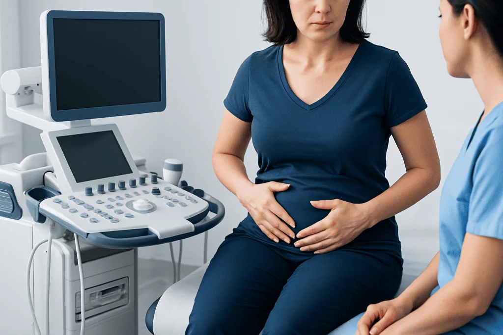
The most noticeable sign of diastasis recti is a bulge or ridge along the midline of the abdomen. This may appear when getting out of bed, doing a sit-up, lifting a child, or standing after lying down. Some people mainly notice that their stomach still looks prominent months after pregnancy despite general recovery.
Symptoms are not always dramatic. Many people report a feeling of core weakness, reduced trunk stability, poor posture, or low back discomfort rather than pain in the abdominal wall itself. Others describe difficulty generating abdominal tension during exercise or a sensation that the middle of the abdomen is soft or unsupported.
Diastasis recti can also affect daily function. Rolling in bed, carrying groceries, lifting a baby seat, or returning to exercise may feel harder because the abdominal wall is not transferring force as efficiently. In some people, pelvic floor symptoms can coexist, such as pressure or leakage with coughing, although these symptoms can have several causes and should not be automatically blamed on diastasis alone.
Severe pain is not typical. If a bulge is tender, suddenly enlarges, becomes firm, or is associated with nausea or vomiting, a different problem such as a hernia should be considered and prompt medical assessment is important.
Why diastasis recti happens: causes and risk factors

Pregnancy is the best-known cause because the growing uterus increases tension across the abdominal wall, while hormones also affect connective tissue. This is a normal adaptation to pregnancy, and some separation is very common in late pregnancy and early postpartum recovery. What differs from person to person is how much the tissue stretches and how well it regains tension afterward.
Several factors may increase the likelihood of persistent diastasis recti. These include multiple pregnancies, carrying twins or higher-order multiples, a larger baby, higher maternal age, prior abdominal wall weakness, and significant fluctuations in body weight. Repeated activities that sharply increase abdominal pressure may also contribute.
Diastasis recti is not limited to postpartum women. Men can develop it, especially with central weight gain, repeated heavy straining, or previous abdominal surgery. Infants may also show a visible midline separation because their abdominal wall is still developing; this often improves naturally as they grow.
Many myths suggest that a specific movement or one “wrong” exercise alone causes diastasis recti. In reality, it is usually the result of combined tissue, pressure, hormonal, and mechanical factors. Certain exercises may worsen visible doming in some people, but they are not the sole explanation for the condition.
How doctors diagnose it and when tests are needed
Diagnosis usually starts with a medical history and physical examination. A clinician may ask about pregnancy history, abdominal surgery, changes in body weight, discomfort, back symptoms, and functional limitations. During the exam, the patient may be asked to lift the head slightly or tense the abdomen so the doctor can assess the width and tension of the midline tissue.
Self-checks at home are common, but they are not always reliable. Measuring the gap with fingers can give a rough impression, yet the quality of the connective tissue and the person’s symptoms are often more important than the exact width alone. A narrow gap with very weak tissue may be more troublesome than a wider gap with good tension and function.
Imaging is not always necessary. Ultrasound can help measure the separation and assess the abdominal wall more precisely, especially when planning rehabilitation or evaluating persistent concerns. In selected cases, other imaging may be used if a hernia, post-surgical change, or another abdominal wall condition is suspected.
When the diagnosis is unclear, doctors may assess related issues such as pelvic floor dysfunction, low back mechanics, or abdominal wall pain from other causes. If a structural defect is suspected, advanced evaluation through diagnostic imaging may help clarify the problem and guide treatment.
Treatment: from targeted rehabilitation to surgery
Treatment depends on symptoms, function, and the goals of the patient. For many people, the first step is conservative care, especially postpartum. This often includes education about posture, breathing, movement patterns, and safe ways to increase abdominal loading while the tissues recover.
Physiotherapy is a common and evidence-informed option. A therapist may focus on deep core coordination, transverse abdominal activation, breathing control, pelvic floor integration, and progressive strengthening rather than high-pressure movements too early. The goal is not simply to “close the gap” but to improve support, strength, and function in daily activities. When appropriate, a personalized physical therapy and rehabilitation plan can help guide this process.
There is no single exercise program proven to cure every case. Some people benefit from avoiding movements that create strong midline doming at first, while others can gradually return to these activities with supervision. Bracing garments may provide comfort or support for a short time, but they do not by themselves restore connective tissue strength.
Surgery may be considered when symptoms persist despite rehabilitation, when there is marked functional impairment, or when diastasis coexists with a hernia or excess abdominal wall laxity. Surgical options are individualized and may be discussed with specialists in general surgery or, in selected cases, plastic and reconstructive surgery. The decision is usually based on function, anatomy, and patient priorities rather than appearance alone.
What helps at home: self-care, exercise choices, and prevention
Self-care begins with realistic expectations. Tissue recovery often takes time, particularly after childbirth, and improvement is usually gradual rather than immediate. Gentle walking, comfortable posture changes, supported coughing, and a gradual return to exercise are often reasonable starting points, but individual needs differ.
Helpful strategies may include learning to exhale during effort, avoiding breath-holding when lifting, and building strength progressively rather than jumping into intense abdominal training. Many people do well with exercises that emphasize alignment, controlled core engagement, and hip and glute strength. A clinician or physiotherapist can help decide when to resume planks, crunch-type movements, running, or heavy lifting.
It may also help to reduce factors that increase ongoing abdominal pressure. Managing constipation, treating a chronic cough, using proper lifting mechanics, and working toward a healthy weight can all support the abdominal wall. These steps do not guarantee prevention, but they may reduce strain on the connective tissue.
Prevention is not always fully possible, especially in pregnancy, because abdominal wall changes are often part of normal physiology. A more practical goal is to support recovery, protect function, and seek guidance early if symptoms interfere with movement, exercise, or daily life.
When to seek medical care
Medical assessment is a good idea if abdominal bulging continues well beyond the early recovery period, if there is ongoing weakness that affects daily activities, or if exercise seems to worsen symptoms despite careful technique. A professional evaluation can help determine whether the issue is diastasis recti, a hernia, pelvic floor dysfunction, or another abdominal wall condition.
Urgent medical attention is needed if there is a new painful lump, skin changes over a bulge, vomiting, fever, sudden severe abdominal pain, or inability to pass stool or gas. These are not typical features of uncomplicated diastasis recti and may point to a different problem that needs prompt care.
People who have had abdominal surgery, high-level athletic demands, or persistent postpartum symptoms may benefit from multidisciplinary care. Near the end of the treatment journey, if specialist input is needed, Acibadem International’s multidisciplinary teams in JCI-accredited hospitals evaluate and treat abdominal wall conditions for international patients.
Frequently asked questions
Can diastasis recti heal on its own?
Yes, some cases improve naturally over time, especially in the months after pregnancy. However, improvement may be incomplete, and persistent weakness or bulging can benefit from medical or physiotherapy assessment.
Is diastasis recti the same as a hernia?
No. Diastasis recti is a stretching and widening of the midline connective tissue, while a hernia is an actual defect or opening in the abdominal wall. The two can occur together, which is why a proper examination matters.
Does everyone with diastasis recti need surgery?
No, many people do not need surgery. Conservative treatment with targeted exercise, movement education, and time is often the first approach, and surgery is usually reserved for selected persistent or complicated cases.
What exercises should be avoided?
There is no universal banned exercise list for every person. In general, movements that cause strong midline doming, breath-holding, or worsening symptoms may need to be modified until better core control is developed under professional guidance.
Can men get diastasis recti?
Yes. Men can develop diastasis recti, especially with increased abdominal pressure, central weight gain, repeated heavy straining, or previous abdominal surgery. The diagnosis and treatment principles are similar, though the underlying causes may differ from postpartum cases.
How long after childbirth should someone wait before getting checked?
A brief separation is common soon after delivery, so early changes do not always need urgent evaluation. If symptoms persist for several weeks to months, interfere with movement, or are associated with pain or a suspicious lump, it is reasonable to consult a qualified clinician sooner.
This article is for general information only and is not a substitute for professional medical advice. Please consult a qualified doctor about your individual situation.
Explore treatments in Turkey — costs, top hospitals & a free quote
JCI-accredited · board-certified surgeons · reply within 24h
Add us as a Preferred Source to see more of our trusted health content across Google Search, AI Overviews and Discover.

More from the Health Library

Obturator Nerve: An Evidence-Based Guide for Patients

Flipper Tooth: A Complete Medical Overview

Tick Repellent: An Evidence-Based Guide for Patients

Lacrimal: A Complete Medical Overview

Emotional Intelligence: What Patients Need to Know