Hida Scan: An Evidence-Based Guide for Patients

A hida scan shows how well the gallbladder and biliary system function, not just what they look like. It is commonly used for suspected gallbladder inflammation, bile duct blockage, bile leaks, or poor gallbladder emptying.
Key Takeaways
- A hida scan shows how well the gallbladder and biliary system function, not just what they look like.
- It is commonly used for suspected gallbladder inflammation, bile duct blockage, bile leaks, or poor gallbladder emptying.
- Preparation may include fasting for a few hours and reviewing current medicines with the care team.
- The test usually involves a small radioactive tracer, lying still for imaging, and sometimes an additional medicine to assess gallbladder function.
- Results are interpreted together with symptoms, blood tests, ultrasound, and other imaging.
A hida scan is a nuclear medicine imaging test used to evaluate how bile moves through the liver, gallbladder, bile ducts, and small intestine. It can help doctors investigate abdominal pain, suspected gallbladder disease, bile duct blockage, or bile leaks when other tests do not fully explain symptoms.
Overview: what a HIDA scan is and why it is used
A hida scan is a nuclear medicine test that helps doctors assess how bile flows through the liver, gallbladder, bile ducts, and small intestine. HIDA stands for hepatobiliary iminodiacetic acid, which refers to the type of tracer used during the scan. Unlike an ultrasound or CT scan, which mainly show anatomy, a hida scan focuses on function. This can make it especially useful when symptoms suggest a biliary problem but the cause is still unclear.
During the test, a small amount of radioactive tracer is injected into a vein. The tracer travels through the bloodstream to the liver, where it is taken up into bile and then moves into the gallbladder and bile ducts. A special camera tracks this movement over time and creates images that show whether bile is flowing normally.
Doctors often request this scan when a person has right upper abdominal pain, nausea after meals, fever with suspected gallbladder inflammation, or abnormal liver-related test results. It may also be used after surgery if there is concern about a bile leak. In some situations, a hida scan helps assess gallbladder emptying, sometimes called the gallbladder ejection fraction.
Because it adds functional information, a hida scan is often considered alongside other digestive evaluations such as gallstones or cholecystitis. It does not replace a clinical examination or other tests, but it can help clarify the diagnosis and guide treatment decisions.
When a doctor may recommend a HIDA scan
A hida scan is not needed for every episode of abdominal discomfort. It is usually recommended when a doctor needs more information about the biliary system after taking a history, performing an examination, and often arranging an ultrasound. If symptoms strongly suggest that the gallbladder is not functioning properly, the scan can provide useful evidence.
Common reasons for ordering a hida scan include suspected acute or chronic gallbladder inflammation, concern about a blockage in the bile ducts, unexplained upper abdominal pain, or evaluation of the gallbladder’s ability to empty. In hospital settings, it may also be used when ultrasound findings are uncertain but infection or inflammation remains a concern.
After gallbladder or liver-related surgery, the test may help detect a bile leak or show whether bile is moving normally through the ducts. In newborns and infants, specialized forms of hepatobiliary imaging may sometimes be used in the evaluation of prolonged jaundice, but the interpretation and preparation are different and require pediatric expertise.
A hida scan is only one part of the diagnostic process. Doctors may combine it with blood tests, abdominal ultrasound, MRI-based imaging, or procedures such as endoscopy when symptoms point to digestive or biliary disease.
How to prepare and what happens during the test

Preparation instructions can vary, so patients should follow the guidance given by their own imaging department. In many cases, fasting for several hours before the test is recommended. This helps the gallbladder fill appropriately and can improve the accuracy of the images. Drinking water may still be allowed, but instructions differ between centers.
It is important to tell the medical team about all medicines, pregnancy, breastfeeding, allergies, and any recent imaging tests involving contrast or nuclear tracers. Some medicines, especially certain pain medicines or drugs that affect gallbladder contraction, may influence the results. The team may advise whether any medicine should be adjusted before the scan.
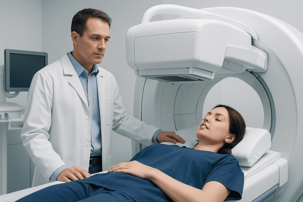
At the appointment, a tracer is injected into a vein in the arm or hand. The patient then lies on an imaging table while a gamma camera takes pictures over time. The scan is painless, although staying still for an extended period may be uncomfortable for some people. In certain cases, an additional medicine is given to help the gallbladder contract so its emptying can be measured more clearly.
The test often takes about one to two hours, but some scans last longer if bile flow is slow or delayed images are needed. Most people can go home the same day and return to normal activities unless their doctor gives different advice.
What a HIDA scan can show
The main value of a hida scan is that it shows movement. If the tracer enters the liver normally but does not reach the gallbladder, this may suggest acute gallbladder inflammation caused by blockage of the cystic duct. If bile flow is delayed throughout the system, a doctor may consider problems affecting the bile ducts or liver function.
When the scan is used to measure gallbladder emptying, the report may include an ejection fraction. This estimate reflects how much bile the gallbladder releases after stimulation. A low value can support the diagnosis of poor gallbladder function in the right clinical setting, but the number is not interpreted in isolation. Symptoms, examination findings, and other tests remain important.
A hida scan can also help detect a bile leak after surgery or injury if tracer is seen collecting outside the normal biliary pathway. In some people, the test may suggest blockage of the common bile duct or another flow problem that needs further assessment with blood tests, ultrasound, MRI, or ERCP.
Like any diagnostic test, it has limits. A hida scan does not always identify the exact cause of symptoms, and abnormal results do not automatically mean surgery is needed. Careful interpretation by the treating doctor and nuclear medicine specialist is essential.
Understanding the results and possible next steps
Results are usually described as normal, delayed, absent, or reduced in certain phases of bile flow. A normal study generally means the tracer moved through the liver, into the gallbladder, through the bile ducts, and into the small intestine in an expected way. This can make some biliary conditions less likely, although it may not fully explain ongoing symptoms.
If the scan suggests acute inflammation, blockage, or poor gallbladder emptying, the doctor will compare this with the person’s symptoms, blood tests, and previous imaging. Further testing may still be needed because gallbladder disease can overlap with other digestive conditions such as ulcers, liver disease, pancreatitis, or gastroesophageal reflux disease.
Treatment depends on the underlying diagnosis rather than the scan alone. Some people may be managed with observation, dietary adjustments, or treatment of another digestive problem. Others may need referral to a gastroenterologist, surgeon, or hepatobiliary specialist for more targeted care.
When surgery is appropriate, the scan may support decisions about gallbladder surgery. Near the end of the care pathway, patients may also value coordinated review by multidisciplinary teams; Acibadem International’s specialists in JCI-accredited hospitals diagnose and treat biliary disorders for international patients when this is needed.
Benefits, limitations, and safety considerations
One important benefit of a hida scan is that it evaluates function rather than structure alone. This makes it helpful when an ultrasound shows no stones or obvious inflammation but symptoms continue. It can also clarify whether bile is flowing normally after an operation or injury.
The test uses a small amount of radiation, which is generally considered low and medically acceptable when the scan is appropriately indicated. The tracer is different from contrast used in standard CT scans, and severe reactions are uncommon. Still, the imaging team should be told about prior reactions, pregnancy, or breastfeeding so that the benefits and risks can be weighed carefully.
There are also practical limitations. The scan can take time, requires the patient to remain relatively still, and may be less informative if preparation instructions are not followed. Certain medicines, prolonged fasting, severe liver disease, or recent eating can affect how bile flows and may complicate interpretation.
For these reasons, a hida scan works best as part of a broader diagnostic plan. It is most useful when the question is clear, such as whether the gallbladder is inflamed, whether it empties poorly, or whether bile is leaking or obstructed.
After the scan, self-care, and when to seek medical care
Most people feel well enough to resume usual activities after the test. Drinking fluids afterward may be advised as part of normal hydration, and patients should follow any specific instructions from the imaging department. If a medicine was used during the scan to stimulate the gallbladder, temporary cramping or nausea can occasionally occur, but this is usually short-lived.
A hida scan itself does not treat symptoms, so ongoing care depends on the diagnosis. While waiting for results, it may help to note when pain occurs, whether meals trigger symptoms, and whether fever, vomiting, jaundice, or pale stools develop. This information can help the doctor interpret the scan in context.
Medical review should be sought promptly for severe or persistent right upper abdominal pain, fever, repeated vomiting, yellowing of the skin or eyes, dark urine, fainting, or signs of dehydration. These symptoms can suggest urgent gallbladder, liver, pancreatic, or bile duct problems and should not be ignored.
Patients should also contact their doctor if they are unsure how to prepare for the test, have questions about medicines before the scan, or do not understand the results afterward. A clear explanation from a qualified clinician is the best way to connect the findings with the next step in care.
Frequently asked questions
Is a HIDA scan painful?
A hida scan is usually not painful. Most people only feel a brief needle stick when the tracer is injected, then lie still while images are taken. If an extra medicine is used to make the gallbladder contract, it may briefly cause cramping or nausea.
How long does a HIDA scan take?
Many hida scans take about one to two hours, but some take longer. Extra time may be needed if bile flow is slow or if delayed images are required. The imaging team can explain the expected timing before the test begins.
Do patients need to fast before a HIDA scan?
Many patients are asked to fast for several hours before the scan, but instructions vary. Fasting helps the gallbladder fill normally and can improve the accuracy of the test. It is important to follow the exact guidance provided by the imaging center.
What does a low gallbladder ejection fraction mean?
A low ejection fraction means the gallbladder did not empty as well as expected during the test. This can support poor gallbladder function in someone with typical symptoms, but it is not a diagnosis by itself. Doctors interpret the number together with symptoms, examination findings, and other test results.
Is the radiation from a HIDA scan dangerous?
A hida scan uses a small amount of radiation that is generally considered acceptable when the test is medically indicated. The goal is to gain important diagnostic information while keeping exposure as low as reasonably possible. Patients who are pregnant or breastfeeding should discuss this with their doctor beforehand.
Can a HIDA scan diagnose gallstones?
A hida scan is not the main test for directly seeing gallstones. Ultrasound is usually better for detecting stones, while a hida scan helps show whether the gallbladder and bile ducts are functioning properly. Sometimes both tests are used because they answer different questions.
This article is for general information only and is not a substitute for professional medical advice. Please consult a qualified doctor about your individual situation.
Persistent digestive symptoms? Get evaluated in Turkey
JCI-accredited · board-certified surgeons · reply within 24h
Add us as a Preferred Source to see more of our trusted health content across Google Search, AI Overviews and Discover.

More from the Health Library

Yellow Pear Tomato: An Evidence-Based Guide for Patients

Ultra Tampons: A Complete Medical Overview

Fournier’s Gangrene: What Patients Need to Know

Warfarin — Explained by Medical Evidence, Not Myths

Agua De Jamaica: What Patients Need to Know