

Bicornuate Uterus: What Is Normal, What Is Not, and When to Seek Care

A bicornuate uterus is a structural difference present from birth, not something a person causes. Many people with a bicornuate uterus have no symptoms and may only learn about it during imaging or pregnancy care.
Key Takeaways
- A bicornuate uterus is a structural difference present from birth, not something a person causes.
- Many people with a bicornuate uterus have no symptoms and may only learn about it during imaging or pregnancy care.
- It can be associated with pelvic pain, painful periods, miscarriage, preterm birth, or fetal positioning issues in some cases.
- Diagnosis usually relies on pelvic ultrasound, 3D ultrasound, MRI, or hysterosalpingography to distinguish it from similar uterine anomalies.
- Treatment depends on symptoms and reproductive history; many people need monitoring rather than surgery.
- Medical advice is important for severe pain, repeated miscarriage, difficulty conceiving, or pregnancy concerns.
A bicornuate uterus is a congenital variation in which the uterus has two upper cavities, creating a heart-like shape rather than the usual pear shape. Many people never notice it, but some may have painful periods, recurrent pregnancy loss, or pregnancy complications that deserve medical evaluation.
Overview: what a bicornuate uterus means
A bicornuate uterus is a type of congenital uterine anomaly. This means the uterus formed differently before birth, developing with two upper “horns” instead of one smooth upper cavity. The result is often described as a heart-shaped uterus.
In a typical uterus, the upper portion is rounded and the cavity is more uniform. In a bicornuate uterus, the outer contour has a deeper indentation at the top, and the cavity may be partly divided into two sections. This is different from normal anatomical variation because it reflects a change in the way the uterus developed, but it does not always cause health problems.
Many people with a bicornuate uterus feel well and have regular menstrual cycles. Some only find out after a pelvic ultrasound, a fertility evaluation, or imaging during pregnancy. Others may have symptoms such as pelvic discomfort, painful periods, or reproductive challenges.
It is also important to distinguish a bicornuate uterus from related conditions, such as a septate uterus or endometriosis, because the causes, risks, and treatment approaches are different. Careful imaging helps doctors identify what is normal for that individual and what may need follow-up.
What is normal, and what is not
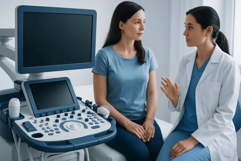
For many patients, a bicornuate uterus is simply an anatomical variant that does not interfere with day-to-day life. Normal experiences can include having no symptoms, normal menstrual bleeding, and the ability to become pregnant naturally. Some people also carry pregnancies successfully without needing any procedure on the uterus itself.
What is not considered normal is ongoing or disruptive symptoms that affect quality of life or reproductive health. Examples include unusually painful periods, repeated pregnancy loss, preterm labor, or a history of babies being in breech position late in pregnancy. These situations do not automatically mean the bicornuate uterus is the only cause, but they do deserve evaluation.
Another point that can be confusing is that not every “heart-shaped” appearance on basic imaging means a true bicornuate uterus. Similar findings can occur with other uterine differences, and the distinction matters. A septate uterus, for example, may look somewhat similar internally but often has a different treatment pathway.
Because of this, the main question is not whether the uterus looks perfectly typical. The key question is whether the shape is causing symptoms, affecting fertility, or changing how a pregnancy should be monitored. That is why precise diagnosis is more useful than assumptions.
Symptoms and possible effects on fertility or pregnancy
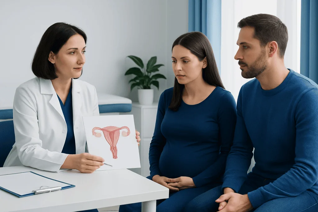
A bicornuate uterus often causes no symptoms at all. When symptoms do occur, they may include pelvic pain, pressure, painful menstruation, or abnormal bleeding patterns. Some people also report discomfort during intercourse, although these symptoms can have many other gynecologic causes as well.
Fertility is a common concern. A bicornuate uterus does not always prevent conception, and many patients become pregnant without difficulty. However, in some cases it may be linked with infertility evaluations, especially when it exists alongside other reproductive conditions such as ovarian cysts or hormonal issues.
Pregnancy is often the stage when a bicornuate uterus becomes most relevant. Because the uterine cavity may have less space or a different shape, there can be a higher chance of miscarriage, preterm birth, fetal growth concerns, or an abnormal fetal position such as breech presentation. Some pregnancies also need closer surveillance for cervical shortening or early contractions.
Even so, a bicornuate uterus does not mean a healthy pregnancy is impossible. Many people have successful outcomes with early prenatal care, regular scans, and individualized planning. The level of monitoring depends on symptoms, prior pregnancy history, and whether other medical factors are present.
- No symptoms in many patients
- Possible painful periods or pelvic discomfort
- May be associated with recurrent miscarriage in some cases
- Can increase the chance of preterm birth or breech presentation
- Often discovered during fertility work-up or prenatal imaging
Causes and risk factors
A bicornuate uterus develops before birth, during the formation of the reproductive tract. The uterus normally forms when two embryologic structures called the Müllerian ducts fuse together. If this fusion is incomplete, the uterus may develop with two upper sections, creating a bicornuate shape.
This condition is not caused by exercise, stress, sexual activity, contraception, or anything a patient did later in life. It is present from birth. In many cases, there is no clear single reason why the developmental difference occurred.
Some people with one uterine anomaly may also have other structural differences in the reproductive or urinary system, because these organs develop around the same time. For that reason, a doctor may occasionally recommend additional imaging if there is a reason to suspect an associated anomaly.
The main “risk factors” are therefore not lifestyle factors but reproductive history and associated findings. A history of repeated miscarriage, preterm birth, infertility evaluation, or unexplained pelvic symptoms may increase the likelihood that a uterine anomaly will be discovered and investigated more closely.
How a bicornuate uterus is diagnosed
Diagnosis starts with medical history and imaging rather than symptoms alone. A clinician may suspect a uterine anomaly after recurrent pregnancy loss, preterm delivery, infertility evaluation, or an unusual appearance on a routine pelvic scan. A pelvic examination by itself usually cannot confirm the diagnosis.
Ultrasound is often the first test. In many cases, a transvaginal ultrasound can suggest the diagnosis, while a 3D ultrasound provides a clearer view of the shape of both the uterine cavity and the outer contour. This helps distinguish a bicornuate uterus from a septate uterus, which is an important step because management differs.
Magnetic resonance imaging, or MRI, may be used when more detail is needed. MRI can show the uterus in multiple planes and is particularly useful when ultrasound findings are not fully conclusive. In some fertility assessments, hysterosalpingography may also be used to outline the uterine cavity and fallopian tubes.
Accurate diagnosis matters because treatment decisions should be based on the exact anomaly, symptoms, and pregnancy history. When a patient is being evaluated for fertility or recurrent loss, the doctor may also recommend other tests to rule out additional causes. If imaging is needed, this may be coordinated with MRI or ultrasound evaluation as part of a broader gynecologic assessment.
Treatment options and pregnancy care
Treatment for a bicornuate uterus is individualized. If a person has no symptoms and no history of infertility or pregnancy complications, treatment may not be needed. In these situations, reassurance and routine gynecologic follow-up are often enough.
When there are reproductive concerns, management focuses first on careful monitoring and confirming that the diagnosis is correct. During pregnancy, this may include more frequent prenatal visits, ultrasound scans to track fetal growth and position, and observation for signs of preterm labor. Delivery planning depends on the course of the pregnancy and the baby’s presentation.
Surgery is not routine for every bicornuate uterus. In select cases, a reconstructive procedure called metroplasty may be considered, usually for patients with repeated pregnancy loss or poor obstetric outcomes clearly linked to the uterine shape. Because surgery carries risks, the potential benefits must be weighed carefully by specialists in reproductive medicine and gynecology.
Some patients may also need treatment for related problems rather than the uterine shape itself. For example, evaluation in IVF and fertility care may be appropriate if conception is difficult, while gynecology care helps address pain, bleeding, and pregnancy planning in a structured way. Near the end of the care journey, some international patients choose centers such as Acibadem International, where multidisciplinary specialists at JCI-accredited hospitals diagnose and treat uterine anomalies and related reproductive concerns.
Prevention, self-care, and living with the condition
Because a bicornuate uterus is congenital, it cannot be prevented through diet, supplements, or lifestyle changes. Self-care is therefore less about prevention and more about knowing what is typical for the individual and keeping up with appropriate medical follow-up.
Helpful steps include tracking menstrual symptoms, noting pelvic pain patterns, and seeking assessment for changes in bleeding or new discomfort. For those trying to conceive, preconception counseling can be valuable. It allows a doctor to review imaging, discuss any history of miscarriage or preterm birth, and plan pregnancy monitoring early.
During pregnancy, self-care means attending prenatal visits, reporting symptoms such as contractions or bleeding promptly, and following advice about activity, travel, or cervical surveillance if recommended. Emotional support is also important, especially for patients who have experienced prior pregnancy loss.
General reproductive health habits still matter. These include avoiding smoking, managing chronic conditions, keeping vaccinations current when appropriate, and attending routine gynecologic visits. While these steps do not change the uterine shape, they can support overall health before and during pregnancy.
When to seek medical care
Medical advice is a good idea if there are symptoms such as severe menstrual pain, persistent pelvic pain, unexplained abnormal bleeding, repeated miscarriage, or difficulty becoming pregnant. These issues do not always mean a bicornuate uterus is present, but they should not be ignored.
During pregnancy, prompt assessment is especially important for vaginal bleeding, leaking fluid, regular contractions, severe abdominal pain, or a noticeable decrease in fetal movement later in pregnancy. A known bicornuate uterus can influence pregnancy monitoring, so early prenatal care is important even when the patient feels well.
Patients who have been told they have a “heart-shaped uterus” on one scan may also benefit from review by a gynecologist or maternal-fetal medicine specialist. Clarifying whether the diagnosis is truly a bicornuate uterus, a septate uterus, or another variation can prevent confusion and guide the right care plan.
In general, care should be sought whenever symptoms interfere with daily life or there is concern about fertility or pregnancy outcomes. A timely evaluation can provide reassurance, identify other possible causes, and support safer planning for future pregnancy.
Frequently asked questions
Can someone with a bicornuate uterus have a normal pregnancy?
Yes. Many people with a bicornuate uterus become pregnant naturally and deliver healthy babies. The main difference is that some pregnancies may need closer monitoring for miscarriage risk, preterm birth, or fetal position.
Does a bicornuate uterus always cause infertility?
No. A bicornuate uterus does not always prevent conception, and many people do not discover it until after they become pregnant. If infertility is present, doctors usually look for other contributing factors as well.
Is a bicornuate uterus the same as a septate uterus?
No, they are different uterine anomalies. A bicornuate uterus has an external indentation at the top of the uterus, while a septate uterus has a partition inside the cavity; imaging helps tell them apart because treatment can differ.
Does a bicornuate uterus need surgery?
Not usually. Surgery is considered only in selected cases, such as repeated pregnancy loss or poor obstetric outcomes thought to be related to the uterine shape. Many patients are managed with observation and tailored pregnancy care instead.
Can a bicornuate uterus cause painful periods?
It can in some people, although many have no symptoms. Painful periods can also happen for many other reasons, so ongoing or severe pain should be assessed by a gynecologist rather than assumed to come only from the uterine shape.
How is a bicornuate uterus found?
It is often found during pelvic ultrasound, fertility evaluation, or prenatal imaging. In some cases, a 3D ultrasound or MRI is needed to confirm the diagnosis and distinguish it from other uterine differences.
References
- American College of Obstetricians and Gynecologists
- American Society for Reproductive Medicine
- National Institute for Health and Care Excellence
- Merck Manual
- Society for Maternal-Fetal Medicine
This article is for general information only and is not a substitute for professional medical advice. Please consult a qualified doctor about your individual situation.
Explore treatments in Turkey — costs, top hospitals & a free quote
JCI-accredited · board-certified surgeons · reply within 24h
Add us as a Preferred Source to see more of our trusted health content across Google Search, AI Overviews and Discover.

More from the Health Library



Related Specialists

Dr. Ayşe Dişli Gürler
Gynecology & Obstetrics
Tuğçe Doğan
Physical Medicine & Rehabilitation
Prof. Dr. A. Sibel Erdamar Çetin
Gastrointestinal Oncology Unit Clinical Service
Dr. Mustafa Emi
Radiology