Retina — Explained by Medical Evidence, Not Myths

The retina plays a central role in turning light into visual information. Common retinal symptoms include flashes, floaters, blurred vision, distortion, and missing areas in vision.
Key Takeaways
- The retina plays a central role in turning light into visual information.
- Common retinal symptoms include flashes, floaters, blurred vision, distortion, and missing areas in vision.
- Some retinal problems develop slowly, while others, such as a retinal tear or detachment, need urgent care.
- Regular eye exams are especially important for people with diabetes, high myopia, or a family history of eye disease.
- Treatment depends on the cause and may include monitoring, medicines, laser treatment, or surgery.
The retina is the thin, light-sensitive layer at the back of the eye that converts light into signals the brain can interpret as vision. When the retina is affected by aging, diabetes, tears, detachment, or other disorders, symptoms can range from mild distortion to sudden vision loss and should be assessed promptly.
What the Retina Is and Why It Matters
The retina is the delicate layer of nerve tissue lining the inside back wall of the eye. Its job is to detect light and convert it into electrical signals that travel through the optic nerve to the brain, where those signals are interpreted as images. In simple terms, even if the front of the eye focuses light well, clear vision is not possible without a healthy retina.
The retina contains specialized cells called rods and cones. Rods help with dim-light and peripheral vision, while cones are responsible for color vision and fine detail. At the center of the retina is the macula, the area that supports sharp central vision needed for reading, driving, and recognizing faces.
Because the retina is highly specialized and depends on a rich blood supply, it can be affected by many conditions, including aging changes, diabetes, inflammation, injury, and problems involving the gel inside the eye. Some retinal conditions are common and manageable when found early, while others can threaten vision if treatment is delayed.
Understanding the retina helps separate medical facts from common myths. Retinal disease does not always cause pain, eye strain is not the usual cause of retinal damage, and sudden changes such as flashes of light or a curtain-like shadow over vision should not be ignored.
How the Retina Supports Vision
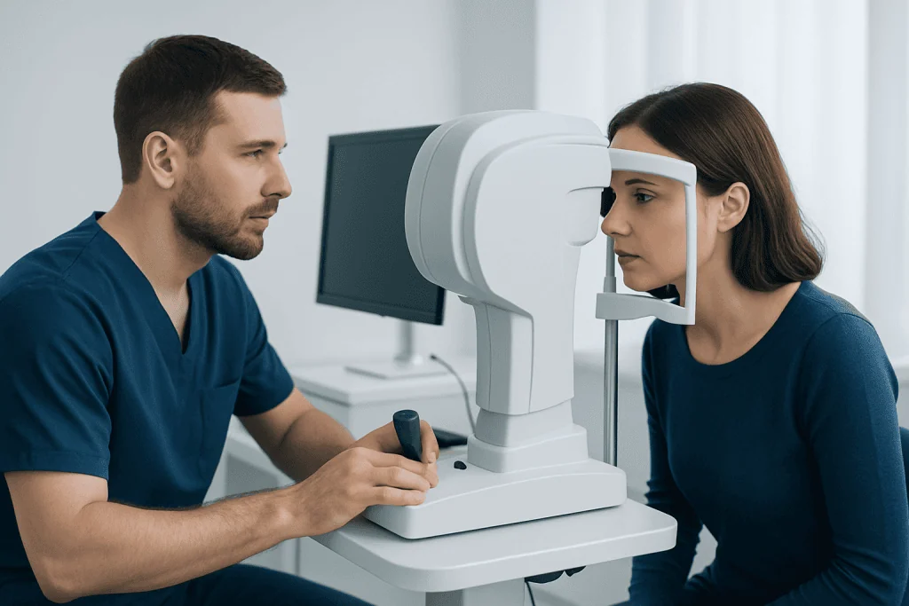
Light enters the eye through the cornea and lens, which focus it onto the retina. The retinal cells then transform light into nerve impulses. These impulses pass through several layers of retinal tissue before traveling along the optic nerve to visual centers in the brain. This process happens quickly and continuously.
The macula is the most detail-sensitive part of the retina. In the center of the macula is the fovea, a tiny area packed with cones that allows very precise vision. When disease affects the macula, a person may notice distortion, difficulty reading, trouble seeing faces clearly, or a dark or blurry spot in the center of vision.
The outer parts of the retina are more involved in side vision and night vision. Problems in the peripheral retina may not affect reading at first, but they can cause floaters, flashes, or gaps in the visual field. This is one reason that retinal disorders may be present before a person realizes there is a problem.
The retina also relies on support from neighboring structures, including the vitreous gel, the retinal pigment epithelium, and blood vessels under and within the retina. Conditions affecting these structures can lead to retinal swelling, bleeding, tears, or detachment.
Symptoms That May Point to a Retinal Problem
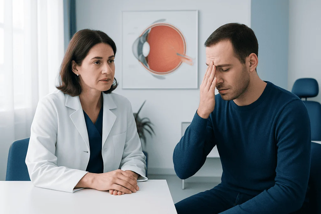
Retinal symptoms vary by cause and by which part of the retina is affected. Some changes are subtle and gradual, while others start suddenly. A person may notice symptoms in one eye only, which can make them easier to miss if the other eye still sees well.
Possible warning signs include:
- Blurred or reduced vision
- Straight lines appearing wavy or distorted
- New floaters, such as spots, threads, or cobweb-like shapes
- Flashes of light
- A shadow, curtain, or missing area in vision
- Difficulty seeing at night or in dim light
- Changes in color perception or central vision
Not every floater or brief visual disturbance means serious retinal disease. Floaters can become more noticeable with age as the vitreous gel changes. However, a sudden increase in floaters, especially when combined with flashes or loss of side vision, can signal a retinal tear or detachment and should be evaluated urgently.
Some retinal conditions, such as diabetic retinopathy or age-related macular degeneration, may cause few or no symptoms in the early stages. For that reason, routine dilated eye examinations remain important even when vision seems normal.
Common Retinal Conditions, Causes, and Risk Factors
Many different diseases can affect the retina. Some involve the macula, some involve blood vessels, and others affect the retinal tissue more broadly. Common examples include retinal detachment, retinal tears, diabetic retinopathy, age-related macular degeneration, macular holes, epiretinal membranes, retinal vein occlusion, retinal artery occlusion, inherited retinal disorders, and inflammatory or infectious retinitis.
Risk factors depend on the condition, but several are well recognized. Increasing age is a major factor for many retinal diseases. Diabetes can damage retinal blood vessels over time. High myopia, prior eye surgery, eye injury, and changes in the vitreous can increase the risk of a retinal tear or detachment. High blood pressure, smoking, family history, and certain inflammatory or autoimmune disorders may also contribute.
A common example is diabetic retinopathy, in which long-term high blood sugar damages small retinal blood vessels. Another is macular degeneration, which mainly affects central vision. These conditions are different, but both show why retinal disease is not a single diagnosis; it is a broad group of disorders that need accurate evaluation.
Retinal problems are not always preventable. Even so, early detection often improves the chance of preserving vision. People with diabetes, high myopia, a family history of retinal disease, or previous retinal problems usually benefit from regular follow-up with an eye specialist.
How Retinal Disorders Are Diagnosed
Diagnosis begins with a careful discussion of symptoms, medical history, and risk factors. An eye specialist will usually test visual acuity and examine the eye after dilation, which allows a better view of the retina. This examination helps identify bleeding, swelling, tears, detachments, or changes in the macula and blood vessels.
Imaging tests are often used to clarify the diagnosis and guide treatment. Optical coherence tomography creates detailed cross-sectional images of the retina and is especially useful for macular disease. Fundus photography can document retinal changes over time. Fluorescein angiography may be used to study blood flow and leakage in retinal vessels.
Ocular ultrasound can help when the view into the eye is blocked, for example by bleeding or a dense cataract. Visual field testing may be useful when a person reports missing areas in vision. Blood pressure measurement and laboratory tests may also be relevant when vascular, inflammatory, or systemic causes are suspected.
Because retinal conditions can look similar at first, self-diagnosis is not reliable. Prompt professional assessment is the safest way to determine whether symptoms reflect a benign change, a chronic retinal disease, or an urgent problem needing immediate treatment.
Treatment Options for Retinal Disease
Treatment depends entirely on the underlying diagnosis. Some retinal findings only need observation and regular follow-up. Others require medical or surgical treatment to reduce swelling, seal a tear, stop abnormal blood vessel growth, or repair a detached retina. The goal is usually to preserve vision, prevent further damage, and where possible improve sight.
Common treatment approaches include medicines injected into the eye for certain macular and vascular conditions, laser procedures for retinal tears or diabetic retinal disease, and surgery for more complex problems. Examples of specialist care may include eye injections, retinal laser treatment, and vitrectomy. A vitrectomy may be recommended when there is persistent bleeding, a macular hole, traction, or retinal detachment.
In some situations, treatment is urgent. A retinal tear may be treated quickly with laser or freezing therapy to reduce the chance of progression to detachment. A retinal detachment often requires surgical repair. For diabetic retinal disease, improving blood sugar, blood pressure, and cholesterol control is also an important part of care alongside eye treatment.
Vision recovery varies. Some people improve significantly after treatment, while others are treated mainly to prevent worsening rather than to restore normal sight. This depends on the disease, how early it is found, and whether central retinal structures such as the macula have already been damaged.
Prevention, Eye Protection, and Everyday Self-care
Not every retinal problem can be prevented, but healthy habits can lower risk and support long-term eye health. Managing diabetes carefully is one of the most important steps for protecting the retina. Controlling blood pressure and cholesterol, avoiding smoking, staying physically active, and attending routine medical care also matter.
Regular eye examinations are especially valuable for adults with diabetes, older adults, people with very nearsighted eyes, and anyone with a family history of serious eye disease. Dilated retinal exams may detect disease before symptoms begin. Early treatment is often simpler and more effective than treatment started after major vision loss.
Eye safety is another practical form of prevention. Protective eyewear during sports, construction work, or other activities with a risk of eye injury can reduce trauma-related retinal damage. People should also avoid ignoring sudden visual changes, even if symptoms seem to come and go.
At-home habits may support symptom monitoring. Some people with macular disease are asked to watch for distortion in straight lines or central blurring. However, home monitoring does not replace eye examinations. Any new or worsening symptoms should be discussed with a qualified clinician.
When to Seek Medical Care
A person should arrange medical assessment if they notice new blurred vision, distortion, increasing floaters, or reduced night vision, especially if symptoms affect daily activities. Even gradual changes deserve attention because some retinal diseases progress silently before causing obvious vision loss.
Urgent same-day eye care is important for sudden flashes of light, a rapid shower of new floaters, a curtain or shadow across vision, sudden loss of side vision, or sudden major reduction in sight. These symptoms can occur with a retinal tear or detachment, which needs prompt evaluation.
People with diabetes should not wait for symptoms before having retinal checks. Similarly, anyone with previous retinal disease or eye surgery should follow their specialist’s advice about review appointments. Quick action can help protect vision and may expand the range of treatment options.
For international patients who need coordinated eye care, Acibadem International’s multidisciplinary specialists in JCI-accredited hospitals diagnose and treat retinal conditions using modern imaging, medical retina therapies, and retinal surgery when appropriate.
Frequently asked questions
What does the retina do?
The retina detects light and converts it into electrical signals that travel to the brain. This process allows the brain to create the images a person sees. Without a healthy retina, vision cannot work properly even if other parts of the eye are normal.
Can retinal disease cause pain?
Many retinal disorders do not cause eye pain, especially in the early stages. A person may instead notice blurred vision, floaters, flashes, or distortion. That is why painless vision changes should still be taken seriously.
Are floaters always a sign of a serious retinal problem?
Not always. Floaters are common and often become more noticeable with age as the vitreous gel changes. However, a sudden increase in floaters, especially with flashes or a shadow in vision, can signal a retinal tear or detachment and needs urgent assessment.
Can retinal damage be reversed?
Some retinal conditions improve with treatment, while others cannot be fully reversed once damage has occurred. In many cases, treatment aims to slow progression and preserve the remaining vision. Early diagnosis generally offers the best chance of a good outcome.
Who is at higher risk for retinal problems?
Risk is higher in people with diabetes, older age, severe nearsightedness, high blood pressure, smoking history, prior eye injury, or a family history of retinal disease. Some conditions also develop after eye surgery or because of inherited factors. A doctor can help determine an individual's risk profile.
How often should the retina be checked?
The right schedule depends on age, symptoms, and medical history. People with diabetes or known retinal disease often need regular dilated eye exams, sometimes more than once a year. Those without risk factors should still follow routine eye examination advice from an eye care professional.
References
- American Academy of Ophthalmology
- National Eye Institute
- Centers for Disease Control and Prevention
- National Health Service
- World Health Organization
This article is for general information only and is not a substitute for professional medical advice. Please consult a qualified doctor about your individual situation.
Explore treatments in Turkey — costs, top hospitals & a free quote
JCI-accredited · board-certified surgeons · reply within 24h
Add us as a Preferred Source to see more of our trusted health content across Google Search, AI Overviews and Discover.