Cutaneous Horn — Explained by Medical Evidence, Not Myths

A cutaneous horn is a visible buildup of keratin, not a disease by itself. The most important part is the skin at the base, which can be benign, precancerous, or cancerous.
Key Takeaways
- A cutaneous horn is a visible buildup of keratin, not a disease by itself.
- The most important part is the skin at the base, which can be benign, precancerous, or cancerous.
- Medical examination and usually biopsy or removal are needed to confirm the cause.
- Treatment often involves complete removal and laboratory analysis of the lesion.
- Sun protection and regular skin checks can help lower the risk of related skin damage.
A cutaneous horn is a cone-shaped, hardened skin growth made of keratin, the same protein found in nails and hair. It is a descriptive term rather than a final diagnosis, because the skin at its base may be harmless, precancerous, or cancerous and usually needs medical evaluation.
What a Cutaneous Horn Really Is
A cutaneous horn is a firm, pointed or curved skin growth that looks like a small horn. It forms from compacted keratin, the tough protein that also makes up hair and nails. Although its appearance can be striking, the term cutaneous horn only describes how the lesion looks on the surface. It does not explain what condition is present underneath.
This distinction matters because the skin at the base of a cutaneous horn may be benign, such as a wart or seborrheic keratosis, or it may show precancerous changes, such as actinic keratosis, or skin cancer, especially squamous cell carcinoma. In other words, the horn itself is made of keratin, but the diagnosis depends on the tissue it grows from.
Cutaneous horns can develop on different parts of the body, but they are especially common on sun-exposed areas like the face, scalp, ears, forearms, and backs of the hands. They can occur in adults of different ages, though they are seen more often in older individuals because long-term sun exposure plays an important role in many cases.
How It Looks and Feels
The classic appearance is a raised, cone-like projection that may be white, yellow, tan, brown, or gray. Some cutaneous horns are short and stubby, while others are longer and more sharply pointed. The texture is usually hard, dry, and rough.
Many lesions cause no symptoms and are noticed because of their unusual look. Others may become tender if rubbed by clothing, shaving, or daily activities. The surrounding skin or the base of the horn may appear red, thickened, crusted, or inflamed.
Features that may increase concern include a broad base, pain, bleeding, rapid growth, ulceration, or a lesion on heavily sun-damaged skin. These signs do not prove cancer, but they do make prompt medical assessment more important.
- Hard, horn-like projection from the skin
- Rough or scaly surface
- Small or large size, sometimes increasing over time
- Possible tenderness, crusting, or bleeding at the base
- Most often found on sun-exposed areas
Why Cutaneous Horns Form

A cutaneous horn develops when excess keratin builds up over an underlying skin lesion. The horn is the visible result of this keratin production, but the reason it forms varies from person to person. This is why clinicians focus on identifying the lesion at the base rather than judging the horn by appearance alone.
Benign causes include viral warts, seborrheic keratosis, and other noncancerous growths. Precancerous causes include actinic keratosis, a sun-related change in the skin that can progress over time. Malignant causes can include squamous cell carcinoma and, less often, other forms of skin cancer such as basal cell carcinoma. Related concerns may overlap with conditions such as skin cancer.
Risk factors include long-term ultraviolet exposure, older age, fair skin, a history of sun-damaged skin, and previous precancerous or cancerous lesions. Chronic irritation and reduced immune function may also contribute in some people. Not every cutaneous horn is serious, but its presence signals the need for careful evaluation.
How Doctors Diagnose It
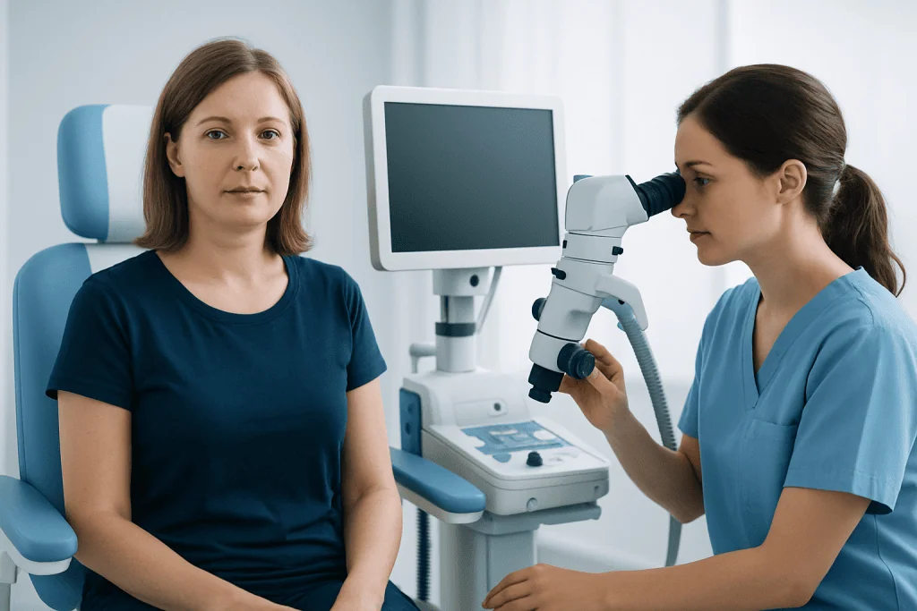
Diagnosis begins with a skin examination. A doctor will assess the size, shape, color, firmness, and location of the horn, along with the character of the skin at its base. Questions usually include how long it has been present, whether it has changed, and whether there is pain, bleeding, or prior skin cancer history.
Because the underlying cause cannot be confirmed by appearance alone, the most reliable way to diagnose a cutaneous horn is to remove part or all of the lesion and send it for laboratory analysis. This may be done through a biopsy or complete excision, depending on the lesion and the clinician’s judgment. If there is concern for suspicious sun-related change, the evaluation may also identify conditions such as actinic keratosis.
In many cases, complete removal is preferred because it both treats the visible lesion and allows the entire base to be examined under a microscope. This helps determine whether the lesion is benign, precancerous, or cancerous, and whether any further treatment is needed.
Treatment Options and What They Aim to Do
Treatment for a cutaneous horn depends on the diagnosis of the tissue at its base. In practice, many lesions are treated by complete removal so the area can be examined thoroughly. This approach is both diagnostic and therapeutic.
Common options include a simple in-office excision, shave removal in selected cases, or other dermatologic procedures depending on lesion size, location, and level of suspicion. If the pathology result shows a benign condition, no further treatment may be necessary beyond wound care and follow-up. If a precancerous lesion is found, the doctor may recommend monitoring and stronger sun-protection measures. If cancer is present, additional treatment may be planned based on the type and extent of disease.
For suspicious or confirmed malignancy, specialists may recommend skin cancer treatment tailored to the pathology report. In selected cases involving visible or sensitive areas, dermatology care can help guide removal, healing, and surveillance. The goal is not only to remove the horn but also to address the skin change that caused it.
Patients should avoid trying to cut, scrape, or treat the horn at home. Self-removal can cause bleeding, infection, scarring, and delayed diagnosis of a potentially important skin condition.
Prevention and Self-Care After Diagnosis
Because many cutaneous horns are linked to chronic sun damage, prevention focuses largely on protecting the skin from ultraviolet exposure. This includes using broad-spectrum sunscreen, wearing hats and protective clothing, seeking shade when possible, and avoiding tanning devices. These steps do not prevent every lesion, but they can reduce cumulative skin damage over time.
Regular skin self-checks are also helpful, especially for people with fair skin, a history of actinic keratoses, previous skin cancer, or significant lifetime sun exposure. A person should look for new rough growths, lesions that bleed or crust, and any spot that changes in size, shape, or color. Taking photos can help track change over time.
After a cutaneous horn is removed, follow the doctor’s wound-care instructions carefully. The area should be kept clean, monitored for signs of infection, and protected from further trauma and sun exposure as it heals. Follow-up visits are important if the pathology report shows precancerous or cancerous change.
When to Seek Medical Care
Any new horn-like skin growth should be assessed by a qualified doctor, ideally a dermatologist or another clinician experienced in skin lesions. Although many underlying causes are benign, there is no safe way to tell from appearance alone whether a cutaneous horn is linked to a precancerous or cancerous process.
Prompt medical care is especially important if the lesion grows quickly, becomes painful, bleeds, develops a sore at the base, or appears on heavily sun-damaged skin. People with a personal history of skin cancer, weakened immune function, or multiple unusual skin lesions should not delay evaluation.
Acibadem International’s multidisciplinary specialists and JCI-accredited hospitals diagnose and treat skin lesions for international patients, including assessment with dermatology services when appropriate. A timely examination can clarify the cause and guide the next steps with confidence.
Frequently asked questions
Is a cutaneous horn always cancer?
No. A cutaneous horn can arise from a benign, precancerous, or cancerous skin lesion. The only reliable way to know the cause is to examine the tissue, usually with biopsy or complete removal.
Can a cutaneous horn fall off on its own?
The outer keratin may break or come off, but that does not remove the underlying lesion. Even if the horn seems smaller, the skin at the base still needs medical evaluation because the important diagnosis is underneath.
What doctor should evaluate a cutaneous horn?
A dermatologist is often the most appropriate specialist for diagnosis and treatment. In some settings, a general practitioner, surgeon, or another skin-focused clinician may begin the evaluation and arrange biopsy or removal.
Is removing a cutaneous horn painful?
Removal is usually done with local anesthesia, so the area is numbed during the procedure. Mild soreness afterward is possible, but many people recover with simple wound care and follow-up instructions from their doctor.
Can a cutaneous horn come back after treatment?
It can recur if the underlying lesion is not fully treated or if a new lesion develops nearby. Follow-up depends on the pathology result, and ongoing skin checks are especially important when sun damage or precancerous changes are present.
Are cutaneous horns caused by poor hygiene?
No. Cutaneous horns are not caused by poor hygiene. They result from excess keratin building up over an underlying skin lesion, often related to sun damage, benign growths, or less commonly skin cancer.
This article is for general information only and is not a substitute for professional medical advice. Please consult a qualified doctor about your individual situation.
Hair transplant in Turkey — costs, graft counts & a free assessment
JCI-accredited · board-certified surgeons · reply within 24h
Add us as a Preferred Source to see more of our trusted health content across Google Search, AI Overviews and Discover.

More from the Health Library

What are neutrophils? A Doctor-Reviewed Answer

Fecal Elastase: A Complete Medical Overview

Runners Waist: A Complete Medical Overview

Extravasation: An Evidence-Based Guide for Patients

Ulnar Styloid Fracture: A Complete Medical Overview