Myelogram: What Patients Need to Know

A myelogram uses contrast dye in the spinal canal to highlight the spinal cord and nerve roots. It is commonly used to evaluate back pain, leg pain, numbness, weakness, or spinal canal narrowing.
Key Takeaways
- A myelogram uses contrast dye in the spinal canal to highlight the spinal cord and nerve roots.
- It is commonly used to evaluate back pain, leg pain, numbness, weakness, or spinal canal narrowing.
- The test usually includes a lumbar puncture followed by X-rays or a CT scan.
- Most people go home the same day, but temporary headache or soreness can occur.
- Patients should tell their doctor about allergies, pregnancy, blood thinners, and previous reactions to contrast dye.
A myelogram is a diagnostic imaging test that helps doctors see the spinal cord, nerve roots, and spinal canal more clearly by using contrast dye and X-ray or CT imaging. It is often recommended when symptoms suggest a spine or nerve problem and other imaging tests do not provide enough detail.
Overview: what a myelogram shows
A myelogram is a specialized imaging test used to look at the spinal canal, spinal cord, and nerve roots. During the test, a doctor places contrast dye into the fluid-filled space around the spinal cord using a thin needle, usually in the lower back. X-ray images and often a CT scan are then taken to show how the dye outlines the structures of the spine.
In simple terms, the test helps doctors see whether something is pressing on the nerves or narrowing the spinal canal. It can be useful for evaluating symptoms such as back pain, leg pain, numbness, tingling, or weakness when the cause is not fully clear on standard scans.
A myelogram does not replace every other imaging test. Instead, it is usually chosen when a doctor needs more detail than a standard X-ray can provide, or when an MRI is not possible or does not answer the clinical question. This makes it a focused test for specific spine and nerve concerns rather than a routine screening exam.
Why a doctor may recommend a myelogram
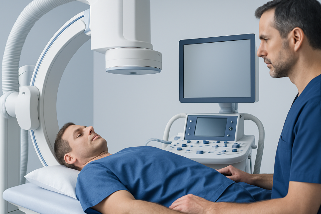
Doctors most often recommend a myelogram when they need a clearer view of the spaces around the spinal cord and nerve roots. This can help identify whether nerves are being compressed by a disc problem, bone spur, thickened ligaments, or other changes in the spine. It is especially useful when symptoms and physical examination suggest a nerve-related problem but the diagnosis remains uncertain.
The test may be used to help evaluate conditions such as spinal stenosis, herniated discs, nerve root compression, certain spinal injuries, or complications after spine surgery. In some cases, the doctor may compare the findings with symptoms on one side of the body to understand which nerve level is involved. Related spinal disorders may overlap with herniated disc or spinal stenosis findings, and a myelogram can help clarify the anatomy.
A myelogram may also be considered when a person cannot have an MRI, for example because of certain implanted devices, severe claustrophobia, or other limitations. When combined with CT, the test can give highly detailed information about the bony spinal canal and nerve root sleeves. This can be important for planning further care, including whether spine surgery might be appropriate.
How to prepare for the test

Preparation instructions can vary slightly between hospitals, so patients should follow the guidance given by their care team. In general, the doctor will review current symptoms, medical history, allergies, kidney problems, previous reactions to contrast dye, and any medicines that may increase bleeding risk. It is especially important to mention blood thinners, aspirin-containing products, or a history of seizures.
Patients may be asked to stop certain medications for a period of time before the test, but this should only be done under medical advice. They should also tell the team if they are pregnant or might be pregnant. Some centers ask patients to drink fluids before the procedure, while others may request limited food or drink for a few hours in advance.
Comfortable clothing is usually recommended, and jewelry or metal objects may need to be removed. Many people are advised to arrange for someone to drive them home afterward, particularly if they receive a sedative or feel uncomfortable after the procedure. Asking questions before the appointment often helps reduce anxiety and makes the process easier to understand.
What happens during a myelogram
A myelogram is usually performed in a radiology department by a doctor trained in image-guided procedures. The patient is positioned on an examination table, and the skin on the lower back is cleaned carefully. A local anesthetic is used to numb the area, and then a thin needle is guided into the spinal canal, most often in the lumbar region below the end of the spinal cord.
Once the needle is in the correct position, contrast dye is injected into the cerebrospinal fluid space. The table may be tilted or the patient may be asked to change position so the dye spreads to the area being examined. X-ray images are taken, and many patients then have a CT scan to produce more detailed cross-sectional images. This combined approach is sometimes called CT myelography.
Patients may feel pressure during the needle placement, but the test should not cause severe pain. It is important to stay as still as possible and follow instructions during imaging. The whole visit often takes longer than the injection itself because preparation, imaging, and post-procedure observation are all part of the process.
- Local anesthetic is used to numb the lower back.
- Contrast dye is placed into the spinal fluid space.
- X-rays and often CT images are taken afterward.
- Observation may be needed for a short time before going home.
Risks, side effects, and recovery
A myelogram is generally considered safe when performed by an experienced team, but like any invasive procedure it has some risks. The most common short-term side effect is a headache, especially when sitting or standing, because the procedure involves entering the fluid space around the spine. Mild soreness at the injection site, nausea, or temporary dizziness can also happen.
Less common risks include bleeding, infection, allergic reaction to the contrast dye, or irritation of the nerves. Serious complications are uncommon, but patients should know the warning signs and contact their doctor if they develop severe headache that does not improve, fever, neck stiffness, increasing back pain, weakness, or trouble with bladder or bowel control.
After the test, patients are often advised to rest for a period and drink fluids unless they have been told otherwise. Many can return to light activities within a day, but heavy lifting and strenuous exercise may need to be avoided briefly. If the myelogram findings show a structural problem affecting the spine or nerves, the next step may include referral for neurosurgical evaluation or another specialist review depending on the cause.
Understanding the results and treatment options
The images from a myelogram are interpreted by a radiologist and reviewed together with the patient’s symptoms, examination findings, and any previous scans. The test can show narrowing of the spinal canal, pressure on a nerve root, displacement of the spinal cord, or changes related to previous surgery. By itself, the result does not always provide a final diagnosis, but it can offer important evidence that guides the next step.
Treatment depends on what the test shows. Some people need only conservative care such as rest, physical therapy, posture changes, pain management, or medications recommended by their doctor. Others may need targeted procedures or surgery if there is significant nerve compression, spinal instability, or worsening neurological symptoms.
If the test suggests a problem involving the spinal cord, nerves, or surrounding structures, the doctor may recommend additional imaging or specialist assessment. In selected cases, further evaluation can overlap with services such as brain and nerve surgery when symptoms point to complex neurological involvement. Near the end of the care pathway, Acibadem International’s multidisciplinary specialists and JCI-accredited hospitals also evaluate and treat spinal and nerve-related conditions for international patients.
When to seek medical care
Medical advice should be sought if symptoms such as back pain, shooting leg pain, numbness, weakness, or walking difficulty are persistent, worsening, or affecting daily activities. These symptoms do not always mean a serious condition, but they do deserve proper evaluation, especially when they continue despite rest or basic treatment.
Urgent medical attention is important if there is sudden severe weakness, loss of sensation in the legs, loss of bladder or bowel control, fever with back pain, or severe pain after an injury. After a myelogram, prompt review is also recommended for severe persistent headache, signs of infection, confusion, or new neurological symptoms.
Patients should not try to interpret symptoms or imaging findings alone. A qualified doctor can decide whether a myelogram is needed, whether a different test would be better, and what treatment makes sense based on the full clinical picture.
Frequently asked questions
Is a myelogram painful?
Most people feel pressure or brief discomfort rather than severe pain. The skin is numbed with local anesthetic before the needle is inserted. Mild soreness afterward can happen, but it is usually short-lived.
How long does a myelogram take?
The injection part is relatively short, but the full appointment often takes longer because of preparation, imaging, and observation afterward. In many cases, patients should expect to spend a few hours at the hospital or imaging center. The exact timing depends on whether a CT scan is included.
Why would someone have a myelogram instead of an MRI?
A myelogram may be used when MRI cannot be performed or when MRI findings do not fully explain the symptoms. It can also provide excellent detail about the spinal canal and nerve root sleeves, especially when combined with CT. The choice depends on the clinical question and the patient’s situation.
What are the side effects after a myelogram?
The most common side effect is a headache, especially when upright. Some people also notice mild back soreness, nausea, or tiredness for a short time. Serious complications are uncommon, but new weakness, fever, or severe persistent headache should be reported promptly.
Can a myelogram diagnose a herniated disc?
A myelogram can show whether a disc is pressing on nearby nerves or narrowing the spinal canal. It does not look at soft tissues in exactly the same way as MRI, but it can still be very helpful, especially with CT. Doctors use the findings together with symptoms and examination results.
Do patients need someone to drive them home?
Many centers recommend arranging a ride home, especially if a sedative is used or if the patient may feel uncomfortable afterward. Even when sedation is not given, resting after the procedure is often advised. The care team will provide specific discharge instructions.
This article is for general information only and is not a substitute for professional medical advice. Please consult a qualified doctor about your individual situation.
Joint and spine care in Turkey — expert assessment & treatment
JCI-accredited · board-certified surgeons · reply within 24h
Add us as a Preferred Source to see more of our trusted health content across Google Search, AI Overviews and Discover.

More from the Health Library

Xy Dysgenesis: A Complete Medical Overview

Tongue Thrust: A Complete Medical Overview

Pinworms: An Evidence-Based Guide for Patients

Maitake Grifola: What Patients Need to Know

Psoriasis on Hands: An Evidence-Based Guide for Patients