Horseshoe Kidney: A Complete Medical Overview

Horseshoe kidney is present from birth and means the kidneys are fused, most often at the lower poles. Many people never notice symptoms and learn about it during imaging done for another reason.
Key Takeaways
- Horseshoe kidney is present from birth and means the kidneys are fused, most often at the lower poles.
- Many people never notice symptoms and learn about it during imaging done for another reason.
- Possible complications include urinary tract infections, kidney stones, urine flow blockage, and sometimes reduced kidney function.
- Diagnosis usually involves ultrasound or CT imaging, along with urine and blood tests when needed.
- Treatment depends on symptoms and complications rather than the kidney shape itself.
- Medical review is important for pain, repeated infections, blood in the urine, fever, or trouble passing urine.
Horseshoe kidney is a birth difference in which the two kidneys are connected, usually at their lower ends, forming a U-shape. Many people live well without symptoms, but some may need medical follow-up for urinary tract infections, kidney stones, drainage problems, or other associated conditions.
Overview
Horseshoe kidney is a congenital condition in which the two kidneys are joined together, usually at their lower ends, by a bridge of tissue called an isthmus. This creates a curved shape that resembles a horseshoe. It develops before birth, and in many people it does not cause obvious symptoms or serious health problems.
The kidneys in a horseshoe kidney are often located lower in the abdomen than usual and may not rotate into their typical position during fetal development. Because of this difference in shape and position, the drainage of urine can sometimes be less efficient. That can raise the chance of problems such as urinary tract infections, kidney stones, or blockage where urine leaves the kidney.
An important point for patients and families is that horseshoe kidney is not the same as kidney failure. Many people with this condition have normal kidney function throughout life. The need for treatment depends on whether complications are present, not simply on the appearance of the kidneys.
Horseshoe kidney may be found in children or adults. Sometimes it is discovered during evaluation for abdominal pain, recurrent infections, or stones. In other cases, it appears as an incidental finding during an ultrasound, CT scan, or other imaging test done for a different reason.
How Horseshoe Kidney Affects the Body
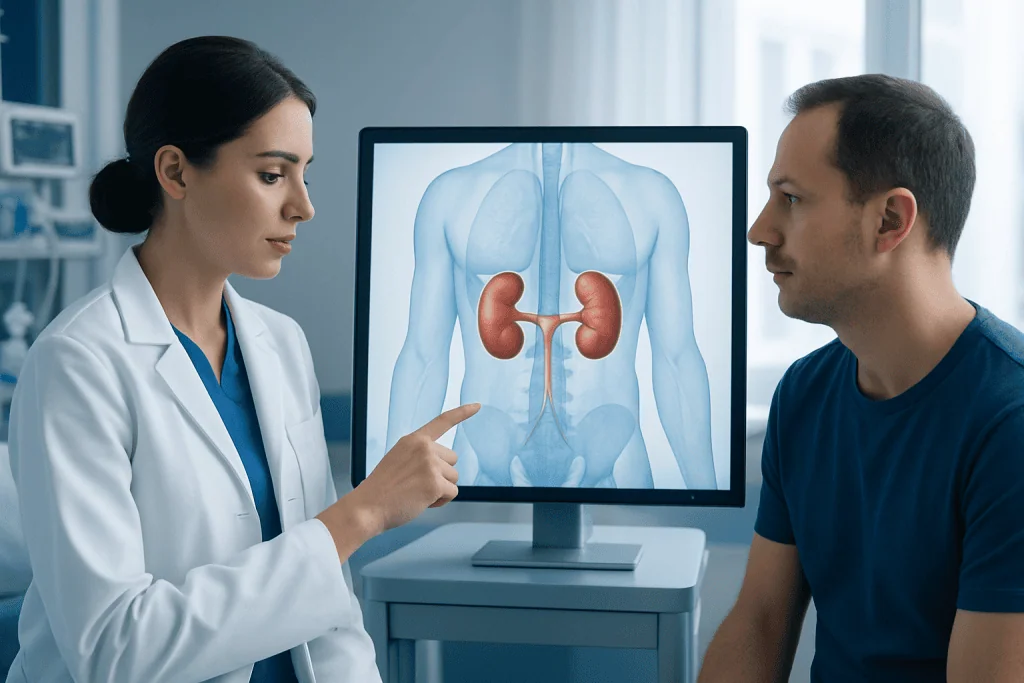
The kidneys filter waste products, balance fluids and salts, and help regulate blood pressure. In horseshoe kidney, these basic functions may remain entirely normal. The main difference is structural: the kidneys are connected and may sit lower than expected, which can influence urine flow and nearby anatomy.
Because the fused kidney can lie close to major blood vessels and the spine, it may be more vulnerable to injury from significant abdominal trauma. Also, the ureters, which carry urine from the kidneys to the bladder, may pass over the fused tissue in a way that makes drainage less smooth. This can increase the risk of swelling of the kidney, called hydronephrosis, if urine backs up.
Horseshoe kidney can also occur alongside other congenital differences, including some affecting the urinary tract, heart, skeleton, or chromosomes. Not every patient has associated conditions, but doctors may consider them, especially in infants and children. If there are signs of poor drainage or kidney swelling, evaluation for hydronephrosis may be helpful.
For many adults, the condition only becomes relevant when a complication develops. Understanding the kidney’s unique anatomy is important because it can affect how imaging is interpreted, how procedures are planned, and how symptoms are investigated over time.
Symptoms and Possible Complications

Many people with horseshoe kidney have no symptoms at all. When symptoms do occur, they are usually related to problems with urine drainage, infection, or stone formation rather than the fusion itself. Symptoms can begin in childhood or appear later in adult life.
Possible symptoms and complications include:
- Pain in the abdomen, side, or lower back
- Repeated urinary tract infections
- Burning or pain with urination
- Blood in the urine
- Kidney stones
- Nausea or vomiting during episodes of blockage or severe pain
- Fever if an infection is present
- Swelling of the kidney due to poor urine drainage
Urinary tract infections may be more common if urine does not drain fully. Symptoms can include frequent urination, urgency, pelvic discomfort, cloudy urine, or fever. Kidney stones may cause severe side pain, blood in the urine, and nausea. In some cases, stones are small and pass on their own; in other cases, they may need specialist treatment.
Although uncommon, some people may develop reduced kidney function over time, especially if recurrent obstruction or infection is not addressed. There may also be a slightly higher risk of certain kidney-related tumors or rare childhood cancers in specific settings, which is one reason regular medical follow-up may be advised when complications or associated abnormalities are present.
Causes and Risk Factors
Horseshoe kidney forms during fetal development. Early in pregnancy, the developing kidneys start in the lower pelvis and normally move upward while rotating into place. In horseshoe kidney, the lower parts of the kidneys join before this movement is complete, limiting how high they can rise and affecting their final position.
The exact reason this fusion happens is not always known. In most cases, it is not caused by anything a parent did or did not do during pregnancy. It is considered a developmental difference rather than an acquired disease. Families often want reassurance on this point, and in most cases there is no clear preventable cause.
Some people with horseshoe kidney also have other congenital or genetic conditions. It may be seen more often in association with certain chromosomal syndromes or structural differences in the urinary tract. The presence of other anomalies does not mean they will definitely cause health problems, but it can influence how thoroughly a child is assessed.
There are no typical lifestyle risk factors that cause horseshoe kidney after birth. However, factors such as dehydration, repeated infections, and a history of stone disease can increase the likelihood of complications in someone who already has this kidney anatomy.
Diagnosis and Monitoring
Horseshoe kidney is usually diagnosed with imaging. An ultrasound is often the first test because it is noninvasive and does not use radiation. It can show the location and shape of the kidneys and may also detect swelling, stones, or poor drainage. In some cases, a CT scan or MRI gives a clearer view of the anatomy and surrounding structures.
Doctors may also order urine and blood tests. A urine test can look for infection, blood, or other abnormalities. Blood tests help assess kidney function by measuring markers such as creatinine and estimating how well the kidneys are filtering. If infections are recurrent, a urine culture may be needed to identify the cause.
When there are signs of obstruction, specialists may use further imaging to study urine flow. Depending on the situation, this may include nuclear medicine scans or other functional tests. If stones are suspected, imaging helps determine their size, location, and whether they are likely to pass without intervention. Some patients may also be evaluated in relation to kidney stones if symptoms suggest this complication.
Monitoring depends on age, symptoms, and whether kidney function is normal. People without symptoms may only need periodic follow-up. Those with recurrent infections, stones, hydronephrosis, or reduced kidney function may need more regular reviews with a urologist or nephrologist.
Treatment Options
There is no need to separate or surgically change a horseshoe kidney simply because it is present. Treatment is directed at symptoms and complications. If a person has normal kidney function and no urinary problems, observation and routine follow-up may be all that is needed.
When infections occur, treatment usually involves antibiotics chosen by a doctor based on symptoms and, when appropriate, urine culture results. Recurrent infections may prompt further testing to check for reflux, blockage, or retained urine. If pain, fever, or signs of a kidney infection are present, prompt medical assessment is important.
Kidney stones may be managed with hydration, pain control, and observation when small and likely to pass. Larger stones or stones causing obstruction may require procedures such as kidney stone treatment. If there is a narrowing or blockage where urine exits the kidney, surgery may sometimes be recommended to improve drainage, including pyeloplasty in selected cases. A personalized approach matters because anatomy in horseshoe kidney can vary.
Care may involve more than one specialty, especially when imaging, surgery, and kidney function monitoring are all relevant. Near the end of the care pathway, some international patients choose centers with coordinated evaluation; Acibadem International’s multidisciplinary specialists and JCI-accredited hospitals diagnose and treat horseshoe kidney and related urinary conditions when advanced assessment is needed.
Prevention, Self-care, and Living With Horseshoe Kidney
Because horseshoe kidney is present from birth, it cannot be prevented after conception. What can often be reduced, however, is the risk of complications. Good hydration is one of the most practical steps, as it may help support healthy urine flow and lower the chance of some stones and infections.
Patients are often advised to seek prompt care for symptoms of urinary tract infection, such as burning with urination, frequent urination, fever, or cloudy urine. Delaying treatment can allow infection to spread or worsen kidney irritation. For people who form stones, doctors may recommend dietary changes based on the stone type and overall health.
General self-care may include:
- Drinking enough fluids unless a doctor has advised fluid restriction
- Not ignoring symptoms of infection or severe flank pain
- Attending follow-up appointments if kidney function or drainage needs monitoring
- Discussing contact sports or injury risk with a doctor if the kidney is low-lying and more exposed
- Following guidance on stone prevention if there is a history of stones
Living with horseshoe kidney is often very manageable. Many people work, exercise, travel, and live normal lives without major limitations. The key is knowing the diagnosis, understanding possible warning signs, and staying connected with appropriate medical follow-up if complications have occurred before.
When to Seek Medical Care
Medical advice should be sought if there is repeated urinary tract infection, ongoing abdominal or side pain, blood in the urine, or symptoms that suggest a stone. These concerns do not always mean a serious problem is present, but they do deserve proper assessment, especially in someone known to have horseshoe kidney.
Urgent medical evaluation is important for fever with urinary symptoms, severe or worsening flank pain, vomiting with suspected blockage, difficulty passing urine, or signs of dehydration. These symptoms can point to infection or obstruction, both of which may need prompt treatment to protect kidney function.
Children with horseshoe kidney may also need review if they have poor growth, recurrent fever without a clear source, bedwetting after being dry, or repeated abdominal complaints. Adults who are newly diagnosed should ask whether additional tests or follow-up imaging are appropriate based on their symptoms and kidney function.
If symptoms are persistent or the anatomy is complex, a specialist may recommend further imaging or procedural evaluation, including urology care. Timely assessment can help identify complications early and guide the most suitable treatment plan.
Frequently asked questions
Is horseshoe kidney serious?
Horseshoe kidney is not automatically serious, and many people have normal kidney function and no symptoms. It becomes medically important when it leads to infections, stones, blockage, or other urinary problems.
Can a person live a normal life with horseshoe kidney?
Yes, many people with horseshoe kidney live normal, healthy lives. Regular follow-up may be advised if there are symptoms, recurrent infections, stones, or concerns about drainage or kidney function.
Does horseshoe kidney need surgery?
Usually, no surgery is needed just because the kidneys are fused. Surgery or another procedure is considered only if a complication develops, such as a blockage, significant reflux, or a stone that does not pass.
Is horseshoe kidney hereditary?
Most cases are sporadic, meaning they happen without a clear inherited pattern. In some people, horseshoe kidney may occur along with genetic or chromosomal conditions, so the broader medical context matters.
Can horseshoe kidney cause pain?
It can, but pain is usually related to a complication rather than the fused shape alone. Common causes of pain include kidney stones, urinary blockage, swelling of the kidney, or infection.
How is horseshoe kidney found?
It is often found on imaging such as ultrasound or CT scan. Some people are diagnosed after symptoms develop, while others learn about it incidentally during testing for another reason.
This article is for general information only and is not a substitute for professional medical advice. Please consult a qualified doctor about your individual situation.
Explore treatments in Turkey — costs, top hospitals & a free quote
JCI-accredited · board-certified surgeons · reply within 24h
Add us as a Preferred Source to see more of our trusted health content across Google Search, AI Overviews and Discover.
Check your numbers in seconds
BMI, calories, due date, blood pressure and 30+ more clinical calculators — free, instant, doctor-reviewed ranges.

More from the Health Library

Aphrodisiac: A Complete Medical Overview

Pulled Hair Follicle Out — Explained by Medical Evidence, Not Myths

Best Multivitamin for Kids — Explained by Medical Evidence, Not Myths

Hemodynamic Instability: An Evidence-Based Guide for Patients

Hair Mask — Explained by Medical Evidence, Not Myths