Usg Pelvis Sonography: What Patients Need to Know

USG pelvis sonography is a safe, non-radiation imaging test for pelvic organs. It may be performed through the abdomen, through the vagina, or sometimes both for clearer detail.
Key Takeaways
- USG pelvis sonography is a safe, non-radiation imaging test for pelvic organs.
- It may be performed through the abdomen, through the vagina, or sometimes both for clearer detail.
- The test helps doctors assess symptoms such as pelvic pain, abnormal bleeding, urinary problems, or suspected cysts and fibroids.
- Preparation depends on the type of scan and may include arriving with a full bladder.
- Results must be interpreted together with symptoms, examination findings, and sometimes other tests.
USG pelvis sonography is a pelvic ultrasound that uses sound waves to create images of organs in the lower abdomen and pelvis. It is commonly used to investigate pain, bleeding, urinary or reproductive symptoms, and to assess pelvic structures without exposing the patient to radiation.
Overview: what USG pelvis sonography means
USG pelvis sonography is another name for pelvic ultrasound. It is an imaging test that uses high-frequency sound waves to create pictures of organs and structures in the pelvis. Depending on the patient and the clinical question, it may look at the uterus, ovaries, cervix, fallopian tube region, bladder, prostate, seminal vesicles, and nearby soft tissues.
This scan is widely used because it is noninvasive, does not use ionizing radiation, and can usually be completed quickly. In many cases, it is one of the first tests a doctor requests when a person has pelvic pain, abnormal bleeding, urinary symptoms, or concerns related to fertility or pregnancy.
Pelvic ultrasound can be performed in different ways. A transabdominal scan places the probe on the lower abdomen. A transvaginal scan places a slim, covered probe inside the vagina for a closer view of female pelvic organs. In some situations, both methods are used together because they provide complementary information.
Rather than being a single disease-specific test, USG pelvis sonography is a practical tool that helps guide diagnosis. It can identify normal anatomy, show structural changes, and help monitor known conditions over time.
Why doctors recommend this scan
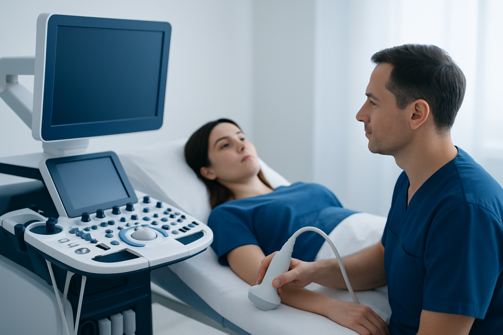
Doctors recommend USG pelvis sonography when symptoms suggest a problem in the pelvic organs or when there is a need to monitor an already known condition. It can be useful in women, men, and children, although the exact organs examined will differ. The test often helps narrow down the cause of symptoms before more invasive procedures are considered.
Common reasons for ordering a pelvic ultrasound include:
- Pelvic or lower abdominal pain
- Abnormal uterine bleeding or bleeding after menopause
- Missed periods, pregnancy assessment, or early pregnancy symptoms
- Suspected ovarian cysts, uterine fibroids, or pelvic masses
- Urinary retention, bladder symptoms, or difficulty emptying the bladder
- Fertility evaluation and follow-up
- Assessment of the prostate or residual urine in men
In women, the scan can help assess conditions such as ovarian cysts or uterine growths and can support further evaluation for endometriosis when symptoms and exam findings suggest it. In men, it may contribute to the assessment of prostate enlargement, bladder outlet issues, or other pelvic concerns.
Pelvic sonography can also be used during treatment planning or follow-up. For example, it may help guide decisions about whether a patient needs observation, medication, a repeat scan, or referral for additional imaging such as MRI.
Types of pelvic ultrasound and what each shows
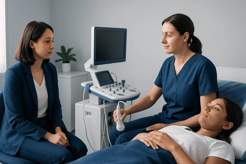
The type of USG pelvis sonography used depends on the patient’s anatomy, symptoms, age, and what the doctor needs to see. Transabdominal ultrasound is done over the lower abdomen with gel on the skin. It provides a broader overview of the pelvis and is often used first, especially in children, in pregnancy, or when a transvaginal scan is not suitable.
Transvaginal ultrasound offers a closer and often more detailed view of the uterus, endometrium, ovaries, and surrounding structures. Because the probe is closer to the organs, it may detect small cysts, fibroids, or changes in the lining of the uterus more clearly than an abdominal scan alone. Patients usually empty their bladder before this part of the exam.
Sometimes Doppler ultrasound is added. This measures blood flow and can help assess the circulation within a mass, the ovaries, or other pelvic tissues. It may be useful when doctors need more information about ovarian torsion, vascular patterns, or pregnancy-related concerns.
In selected cases, ultrasound findings may lead to further tests or procedures. If a structural issue is found and treatment is needed, a doctor may discuss options such as hysteroscopy for problems inside the uterus or laparoscopy when direct pelvic evaluation is necessary.
How to prepare and what happens during the exam
Preparation for USG pelvis sonography depends on the type of scan. For a transabdominal pelvic ultrasound, the patient is often asked to drink water beforehand and arrive with a comfortably full bladder. A full bladder can improve the view by moving bowel loops away from the pelvic organs. For a transvaginal scan, the opposite is usually true: the bladder is emptied before the examination.
During a transabdominal scan, the patient lies on an exam table while a sonographer places gel on the lower abdomen and moves a handheld probe over the skin. Pressure may be felt, especially if the bladder is full, but the test is usually well tolerated. During a transvaginal scan, a covered, lubricated probe is gently inserted into the vagina to obtain close-up images.
The examination typically takes a short time, although this can vary depending on the reason for the study and whether both scanning methods are used. Patients can usually return to normal activities right away. There is no recovery period after a standard diagnostic pelvic ultrasound.
Many patients feel anxious before imaging tests, especially if they are worried about what the scan might show. It can help to know that the test itself is considered safe and routine. Asking beforehand which type of scan is planned and how to prepare can make the experience more comfortable.
What the scan can detect and its limits
USG pelvis sonography can show organ size, shape, position, and many structural changes. It may detect ovarian cysts, uterine fibroids, thickening of the uterine lining, fluid collections, enlarged prostate, bladder distention, and some pelvic masses. In pregnancy, it can help confirm location, estimate gestational age, and evaluate certain symptoms in early pregnancy.
Ultrasound is especially useful because it can often distinguish between fluid-filled and solid structures. This helps doctors decide whether a finding is more likely to be a simple cyst, a fibroid, or another type of lesion that needs further evaluation. It may also support assessment when a person has symptoms of uterine fibroids or other common pelvic conditions.
At the same time, no imaging test answers every question. Ultrasound quality may be affected by bowel gas, body habitus, scar tissue, or the position of pelvic organs. Some conditions, including subtle endometriosis or very small abnormalities, may not be fully visible on ultrasound alone.
For this reason, results are interpreted together with a patient’s symptoms, physical examination, blood tests, and sometimes other imaging. If the findings are unclear, the doctor may recommend repeat ultrasound, MRI, CT in selected cases, or a procedure for direct visualization and sampling.
Understanding results and possible next steps
After the scan, a radiologist or another qualified doctor reviews the images and prepares a report. The report may describe normal findings or note changes such as cysts, fibroids, enlarged organs, fluid, or thickening of tissues. Some results are straightforward, while others need correlation with symptoms and follow-up over time.
A report may include terms that sound technical, such as simple cyst, complex cyst, echogenic lesion, endometrial thickness, or residual urine volume. These terms describe imaging features, not a final diagnosis by themselves. A doctor explains what the findings mean in the context of the patient’s age, symptoms, menstrual status, pregnancy status, and medical history.
Next steps vary. Some patients only need reassurance and routine observation. Others may need repeat imaging, laboratory tests, gynecologic or urologic review, or treatment planning. When a procedure is appropriate, options may include myomectomy for selected fibroids or other targeted care depending on the diagnosis.
Near the end of the care pathway, patients often benefit from coordinated specialist input. Acibadem International’s multidisciplinary specialists and JCI-accredited hospitals evaluate pelvic ultrasound findings and treat related conditions for international patients when further assessment or management is needed.
Safety, self-care, and when to seek medical care
USG pelvis sonography is generally considered safe because it uses sound waves rather than radiation. Most patients do not need any special aftercare. Drinking water, emptying the bladder after the test if needed, and returning to normal meals and activities are usually sufficient. Mild temporary discomfort can happen from probe pressure or a full bladder, but lasting side effects are not expected after a routine scan.
Self-care depends more on the symptoms that led to the scan than on the scan itself. Keeping a record of pelvic pain, menstrual changes, urinary symptoms, pregnancy-related symptoms, or fever can help the doctor interpret results more accurately. It is also helpful to bring prior imaging reports, medication lists, and any relevant laboratory results to follow-up appointments.
Medical care should be sought promptly if there is severe or worsening pelvic pain, heavy vaginal bleeding, fainting, fever, vomiting, painful urination with inability to pass urine, or symptoms during pregnancy such as one-sided pain or bleeding. These symptoms do not always mean an emergency, but they do need timely medical review.
Even if symptoms are mild, a patient should arrange a medical visit if pelvic pain persists, menstrual bleeding changes without explanation, abdominal swelling develops, or urinary symptoms continue. Early assessment can help identify common and treatable causes before they become more disruptive.
Frequently asked questions
Is USG pelvis sonography the same as a pelvic ultrasound?
Yes. USG stands for ultrasonography, so USG pelvis sonography refers to a pelvic ultrasound scan. It uses sound waves to create images of the pelvic organs.
Does pelvic sonography hurt?
Most patients find the test tolerable. A transabdominal scan may feel uncomfortable if the bladder is very full, and a transvaginal scan may cause mild pressure, but significant pain is not typical.
Do patients need a full bladder for the test?
Often yes for a transabdominal pelvic ultrasound, because a full bladder can improve image quality. For a transvaginal ultrasound, patients are usually asked to empty the bladder first.
Can USG pelvis sonography detect fibroids or ovarian cysts?
Yes, pelvic ultrasound commonly helps identify uterine fibroids and ovarian cysts. It can also show their size, location, and some imaging features, although further tests may sometimes be needed.
Is pelvic ultrasound safe during pregnancy?
Pelvic ultrasound is widely used in pregnancy and is generally considered safe when performed appropriately. It does not use ionizing radiation and is commonly used to assess early pregnancy and pregnancy-related symptoms.
What if the ultrasound result is normal but symptoms continue?
A normal scan can be reassuring, but it does not rule out every possible cause of symptoms. If symptoms persist, a doctor may recommend follow-up, repeat imaging, laboratory tests, or another type of evaluation.
References
- World Health Organization
- American College of Radiology
- American College of Obstetricians and Gynecologists
- Radiological Society of North America
- National Institute for Health and Care Excellence
This article is for general information only and is not a substitute for professional medical advice. Please consult a qualified doctor about your individual situation.
Explore treatments in Turkey — costs, top hospitals & a free quote
JCI-accredited · board-certified surgeons · reply within 24h
Add us as a Preferred Source to see more of our trusted health content across Google Search, AI Overviews and Discover.

More from the Health Library

Tai Chi: An Evidence-Based Guide for Patients

Pseudoseizure: A Complete Medical Overview

Spondylitt: What Patients Need to Know

Inhaler: What Patients Need to Know

Siadh: An Evidence-Based Guide for Patients