Ventricles: A Complete Medical Overview

The term ventricles usually refers to structures in both the brain and the heart. Brain ventricles hold and circulate cerebrospinal fluid, which cushions and supports the central nervous system.
Key Takeaways
- The term ventricles usually refers to structures in both the brain and the heart.
- Brain ventricles hold and circulate cerebrospinal fluid, which cushions and supports the central nervous system.
- Heart ventricles are the main pumping chambers that send blood to the lungs and the rest of the body.
- Ventricular problems can involve structure, pressure, rhythm, pumping strength, or fluid flow.
- Symptoms depend on whether the issue affects the brain or the heart and can range from headaches to shortness of breath or fainting.
- Early evaluation helps identify serious causes and guide the most appropriate treatment.
Ventricles are important hollow structures in the body, most commonly referring to the four fluid-filled spaces in the brain and the two lower pumping chambers of the heart. Understanding what ventricles do helps explain how certain brain and heart conditions are diagnosed, monitored, and treated.
Overview: what ventricles are
Ventricles are hollow chambers inside organs. In everyday medical use, the word usually refers to either the four ventricles of the brain or the two ventricles of the heart. Although they are very different structures, both sets of ventricles are essential because they help move vital fluids through the body.
In the brain, the ventricles contain cerebrospinal fluid, often called CSF. This clear fluid cushions the brain and spinal cord, helps remove waste, and supports a stable environment for the nervous system. The brain’s ventricular system includes two lateral ventricles, the third ventricle, and the fourth ventricle, all connected by narrow channels that allow fluid to circulate.
In the heart, the right and left ventricles are the lower chambers that do most of the pumping work. The right ventricle sends blood to the lungs to pick up oxygen, while the left ventricle pumps oxygen-rich blood to the rest of the body. Because the left ventricle must generate higher pressure, its muscular wall is thicker than the right.
When people search for information about ventricles, they may be trying to understand an imaging report, a diagnosis, or symptoms linked to the brain or heart. The meaning depends on context, so a clear explanation of both is often the most helpful starting point.
How the brain ventricles work
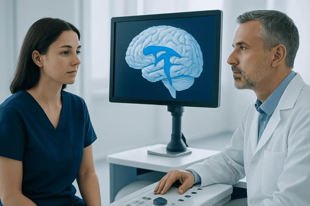
The brain’s ventricles are part of a continuous system that produces and circulates cerebrospinal fluid. Much of this fluid is made by specialized tissue called the choroid plexus. After it is produced, CSF flows through the ventricles and around the brain and spinal cord before being reabsorbed into the bloodstream.
This system helps protect delicate nervous tissue from sudden movement or minor trauma. It also plays a role in delivering nutrients, clearing metabolic waste, and maintaining pressure balance within the skull. If the flow of CSF becomes blocked or absorption is reduced, fluid can build up and enlarge the ventricles.
Doctors often examine the brain ventricles on ultrasound, CT, or MRI scans. Enlarged ventricles can be a sign of several conditions, including hydrocephalus, age-related brain changes, prior injury, bleeding, inflammation, or a mass that obstructs normal fluid pathways. The size of the ventricles alone does not always determine whether a problem is present; symptoms and the full clinical picture matter.
Depending on the cause, ventricular enlargement may develop gradually or more suddenly. This is why neurologic symptoms such as persistent headache, vomiting, balance problems, changes in memory, or altered alertness should be assessed by a qualified clinician.
How the heart ventricles work

The heart ventricles receive blood from the upper chambers of the heart, called the atria. After filling, they contract in a coordinated way to move blood forward. The right ventricle pushes blood into the pulmonary arteries and toward the lungs. The left ventricle then sends oxygenated blood into the aorta and throughout the body.
Normal ventricular function depends on healthy heart muscle, properly working valves, a reliable electrical conduction system, and blood vessels that are not excessively narrowed or stiff. If any of these are affected, the ventricles may not fill or pump efficiently. This can reduce oxygen delivery to tissues and lead to fatigue, breathlessness, chest discomfort, or swelling.
Heart ventricles are commonly evaluated with electrocardiography, echocardiography, cardiac CT, cardiac MRI, and sometimes stress testing or catheter-based studies. These tests help doctors look at chamber size, wall thickness, pumping strength, valve function, and abnormal rhythms.
Ventricular issues in the heart include weakening of the left ventricle in heart failure, enlargement from long-standing high blood pressure, scarring after a heart attack, and rhythm problems such as ventricular tachycardia. The type of ventricular problem strongly influences treatment choices.
Symptoms linked to ventricular problems
Symptoms vary depending on whether the ventricles in the brain or the heart are affected. Some ventricular changes cause no symptoms and are discovered incidentally on imaging. Others lead to noticeable problems that require follow-up or urgent care.
Possible symptoms of brain ventricular disorders include:
- Persistent or worsening headache
- Nausea or vomiting
- Blurred or double vision
- Difficulty with balance or walking
- Memory or concentration changes
- Drowsiness, confusion, or reduced alertness
- In infants, a rapidly enlarging head or bulging soft spot
Possible symptoms of heart ventricular disorders include:
- Shortness of breath, especially with activity or when lying flat
- Chest pain or pressure
- Palpitations or an unusually fast heartbeat
- Dizziness or fainting
- Fatigue or reduced exercise tolerance
- Swelling in the legs, ankles, or abdomen
Because these symptoms can overlap with many other conditions, they do not confirm a ventricular disorder by themselves. However, they are important clues that help doctors decide which tests are needed and how quickly evaluation should occur.
Causes and risk factors
Brain ventricular problems may result from blockage of cerebrospinal fluid flow, reduced absorption of fluid, congenital differences present at birth, bleeding, infection, trauma, tumors, or degenerative brain changes. In older adults, some people develop a pattern of enlarged ventricles associated with walking difficulties, urinary urgency, and cognitive changes, sometimes described as <a href="https://acibademinternational.com/diseases/normal-pressure-hydrocephalus/”>normal pressure hydrocephalus.
Heart ventricular problems can arise from coronary artery disease, high blood pressure, heart valve disease, cardiomyopathy, congenital heart defects, inflammation of the heart muscle, electrolyte disturbances, sleep apnea, or inherited rhythm conditions. Some ventricular rhythm disorders occur in otherwise healthy people, while others reflect underlying structural heart disease.
Risk factors differ by condition but may include older age, family history, diabetes, smoking, high cholesterol, obesity, uncontrolled blood pressure, prior stroke, prior head injury, or a history of infection affecting the brain or heart. In newborns and children, congenital structural differences can be especially important.
In many cases, prompt recognition of the underlying cause matters more than the size or appearance of the ventricles alone. A careful medical history, examination, and targeted testing help determine whether a finding is harmless, requires monitoring, or needs treatment.
How doctors diagnose ventricular conditions
Diagnosis starts with a history of symptoms and a physical examination. For suspected brain ventricular disorders, the clinician may assess vision, eye movements, strength, sensation, coordination, and mental status. For suspected heart problems, attention is given to pulse, blood pressure, breathing, heart sounds, fluid retention, and signs of poor circulation.
Imaging and functional tests are often central to diagnosis. Brain imaging may include ultrasound in infants, CT for urgent situations, or MRI for more detailed views of ventricular size and fluid pathways. Heart evaluation often includes an ECG to assess rhythm and an echocardiogram to examine ventricular size and pumping ability. Depending on the case, doctors may also use Holter monitoring, stress testing, cardiac MRI, or blood tests that look for heart strain or damage.
If a structural problem is suspected, specialized procedures may be considered. For example, doctors may use advanced neuroimaging to understand obstruction in the brain’s fluid pathways or a catheter-based study to clarify pressure and blood flow in the heart. Some people are referred to neurology, neurosurgery, cardiology, or electrophysiology depending on the likely cause.
The goal of diagnosis is not only to identify a ventricular abnormality but also to understand whether it is affecting function. This distinction helps avoid unnecessary treatment while ensuring that serious conditions are managed appropriately.
Treatment options and ongoing care
Treatment depends entirely on the type of ventricular problem, its cause, and how severe the symptoms are. Some findings only need observation and follow-up imaging. Others require medication, a procedure, or surgery. A doctor will tailor care to the individual rather than treating the word “ventricles” as a diagnosis by itself.
For brain-related ventricular disorders, treatment may focus on restoring normal cerebrospinal fluid flow or relieving pressure. In selected cases, this can involve a shunt procedure or endoscopic third ventriculostomy to create an alternative pathway for CSF. If a tumor, bleeding, or infection is responsible, treatment targets that specific cause as well. People with concerning neurologic symptoms may need input from neurosurgery specialists.
For heart-related ventricular disorders, treatment may include blood pressure control, medicines that support heart function, treatment of coronary artery disease, management of valve disease, or rhythm-directed therapies. Some patients benefit from procedures such as cardiac ablation for certain ventricular rhythm disorders, while others may need devices or advanced heart failure care.
Follow-up is often important even after treatment begins. Repeat imaging, heart function testing, rhythm monitoring, and symptom review help doctors see whether the ventricles are recovering, remaining stable, or showing signs that the treatment plan should change.
Self-care, prevention, and when to seek medical care
Not every ventricular problem can be prevented, especially those related to congenital anatomy or inherited conditions. Still, general health measures can reduce the risk of many heart-related ventricular disorders and support overall brain and vascular health. Helpful steps include controlling blood pressure, managing diabetes and cholesterol, avoiding smoking, staying physically active, maintaining a healthy weight, getting enough sleep, and taking prescribed medicines as directed.
People living with a known ventricular condition should keep regular follow-up appointments and report new symptoms promptly. It is also helpful to understand what type of ventricular issue is present, what tests are being monitored, and which warning signs require urgent attention. Clear communication with the medical team can make ongoing care safer and less stressful.
Medical care should be sought promptly for severe headache with vomiting, sudden confusion, new weakness, seizures, fainting, chest pain, severe shortness of breath, or a sustained rapid heartbeat. These symptoms do not always mean a ventricular emergency, but they should not be ignored.
For international patients who need further evaluation, Acibadem International’s multidisciplinary specialists and JCI-accredited hospitals diagnose and treat complex brain and heart conditions related to the ventricles using coordinated imaging, medical, and surgical care.
Frequently asked questions
What are ventricles in the body?
Ventricles are chambers inside organs. In medicine, the term most often refers to the four fluid-filled spaces in the brain and the two lower pumping chambers of the heart. Both are essential for normal body function.
Are brain ventricles and heart ventricles the same thing?
No. Brain ventricles hold and circulate cerebrospinal fluid, while heart ventricles pump blood. They share the same name because both are chamber-like structures, but they serve very different roles.
What does enlarged ventricles mean on a scan?
Enlarged ventricles means the chambers appear larger than expected on imaging. In the brain, this may reflect extra cerebrospinal fluid, blockage, or age-related changes. In the heart, it may suggest the muscle is stretched or working harder than normal, but the meaning depends on symptoms and other test results.
Can ventricular problems be serious?
Yes, some can be serious, especially if they affect pressure in the brain, heart pumping strength, or heart rhythm. However, not every ventricular finding is dangerous. A doctor interprets the result based on symptoms, examination, and imaging or heart tests.
How are ventricular disorders treated?
Treatment depends on the cause. Brain ventricular disorders may require monitoring, treatment of the underlying cause, or procedures that improve cerebrospinal fluid flow. Heart ventricular disorders may be managed with lifestyle measures, medications, rhythm treatments, or procedures for structural heart disease.
When should someone see a doctor about ventricles?
A person should seek medical advice if they have unexplained headaches, balance changes, memory changes, shortness of breath, palpitations, chest discomfort, or fainting. Urgent care is important for severe neurologic symptoms, chest pain, significant breathing difficulty, or loss of consciousness.
References
- National Institute of Neurological Disorders and Stroke
- American Heart Association
- National Heart, Lung, and Blood Institute
- Mayo Clinic
- MedlinePlus
This article is for general information only and is not a substitute for professional medical advice. Please consult a qualified doctor about your individual situation.
Explore treatments in Turkey — costs, top hospitals & a free quote
JCI-accredited · board-certified surgeons · reply within 24h
Add us as a Preferred Source to see more of our trusted health content across Google Search, AI Overviews and Discover.
Check your numbers in seconds
BMI, calories, due date, blood pressure and 30+ more clinical calculators — free, instant, doctor-reviewed ranges.

More from the Health Library

Paralyzed: A Complete Medical Overview

Farxiga Cost — Explained by Medical Evidence, Not Myths

IUD Strings: A Complete Medical Overview

Sodium Citrate: An Evidence-Based Guide for Patients

Cooing — Explained by Medical Evidence, Not Myths