Retroperitoneal Lymph Nodes: An Evidence-Based Guide for Patients
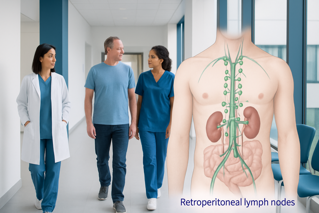
Retroperitoneal lymph nodes are part of the body's immune system and lie deep behind the abdominal lining. Enlargement can happen for several reasons, including infections, inflammatory conditions, and cancers.
Key Takeaways
- Retroperitoneal lymph nodes are part of the body's immune system and lie deep behind the abdominal lining.
- Enlargement can happen for several reasons, including infections, inflammatory conditions, and cancers.
- Imaging tests such as ultrasound, CT, MRI, or PET-CT often help identify the cause and guide next steps.
- Not all enlarged lymph nodes are cancerous, but persistent or unexplained enlargement should be evaluated by a doctor.
- Treatment depends on the underlying cause and may involve monitoring, medicines, or procedures such as biopsy.
Retroperitoneal lymph nodes are lymph nodes located deep in the back part of the abdomen, near major blood vessels and organs. They are not usually felt from the outside, so changes are often found on imaging and then assessed to determine whether they are related to infection, inflammation, or cancer.
Overview: what retroperitoneal lymph nodes are
Retroperitoneal lymph nodes are lymph nodes located in the retroperitoneum, the space deep in the abdomen behind the lining called the peritoneum. This area contains important structures such as the kidneys, adrenal glands, pancreas, major blood vessels, parts of the intestines, and the ureters. Lymph nodes in this region help filter lymph fluid and support the body’s immune defenses.
Because these nodes sit deep inside the body, they usually cannot be seen or felt during a routine self-exam. Many people only learn about retroperitoneal lymph nodes after a scan done for abdominal pain, back pain, weight loss, fever, follow-up of a known condition, or cancer staging. In some cases, enlarged nodes are found incidentally and do not cause symptoms.
The term retroperitoneal lymphadenopathy means that one or more of these nodes are enlarged or appear abnormal on imaging. Enlargement alone does not give a diagnosis. Doctors interpret the size, shape, number, and location of nodes together with symptoms, blood tests, and imaging findings to understand whether the cause is more likely to be reactive, inflammatory, infectious, or malignant.
Symptoms and how enlarged nodes may be found
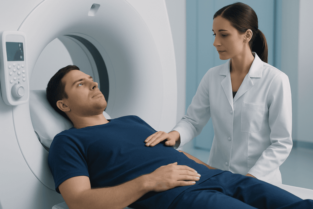
Retroperitoneal lymph nodes themselves do not always cause symptoms, especially when they are only mildly enlarged. When symptoms do occur, they are often related to pressure on nearby organs or to the underlying illness rather than to the nodes alone. This is one reason why imaging plays such an important role in evaluation.
Possible symptoms can include dull abdominal discomfort, back pain, a feeling of fullness, nausea, changes in appetite, or unexplained weight loss. If the enlarged nodes press on the ureters, they may affect urine flow and lead to flank pain or kidney swelling. Some people also have fever, night sweats, tiredness, or generalized unwellness, depending on the cause.
Retroperitoneal lymph nodes are commonly detected during tests such as abdominal ultrasound, computed tomography, or magnetic resonance imaging. In people with a known cancer, they may be seen during staging or follow-up scans. In others, the finding may prompt further review for conditions such as lymphoma or infection.
- Some people have no symptoms at all.
- Others have symptoms linked to pressure on nearby structures.
- General symptoms such as fever or weight loss may point to a broader medical cause.
Common causes and risk factors

There is no single cause of enlarged retroperitoneal lymph nodes. One broad group of causes is infection. Certain viral, bacterial, fungal, and mycobacterial infections can trigger the immune system and make lymph nodes enlarge. Inflammatory and autoimmune conditions can do the same, especially when there is ongoing immune activation in the body.
Another important group of causes is cancer. Some cancers start in the lymphatic system itself, while others spread to retroperitoneal lymph nodes from nearby or distant organs. Examples can include testicular cancer, kidney cancer, gynecologic cancers, gastrointestinal cancers, and colon cancer. In this setting, enlarged nodes may be part of cancer staging, which helps doctors understand how far a disease has spread and which treatments may be appropriate.
Less commonly, nodes may appear enlarged because of benign reactive changes, rare immune disorders, or treatment-related effects. Risk factors depend on the underlying cause. For example, age, smoking history, certain infections, personal or family history of cancer, immune suppression, and previous cancer diagnosis may all influence the likelihood of a particular explanation.
Doctors do not rely on one scan feature alone to decide whether enlarged nodes are serious. A node may be enlarged from a harmless temporary reaction, while a normal-sized node can occasionally still be involved by disease. This is why the overall clinical picture matters.
How doctors diagnose the cause
Evaluation usually begins with a detailed medical history and physical examination. A doctor may ask about the timing of symptoms, fever, night sweats, weight loss, travel history, infections, medications, previous cancers, and family history. Although retroperitoneal nodes are deep and difficult to examine directly, other lymph node areas and abdominal findings may provide useful clues.
Blood tests can help assess inflammation, infection, blood counts, liver and kidney function, and tumor-related concerns when relevant. Imaging is central to diagnosis. A CT scan often gives a clear view of the retroperitoneal area and can show the size, shape, and distribution of nodes. MRI may be helpful in selected cases, and PET-CT can provide additional information when cancer or inflammatory disease is suspected. In some situations, doctors may use PET-CT imaging to assess metabolic activity in enlarged nodes.
If imaging and laboratory findings do not clearly explain the enlargement, a biopsy may be recommended. This means taking a small tissue sample so it can be examined under a microscope. Depending on the location, biopsy may be performed with image guidance or, less commonly, through surgery. Tissue diagnosis is often the most reliable way to distinguish between infection, inflammation, lymphoma, and metastatic disease.
In some patients, careful follow-up is appropriate rather than immediate biopsy. This may be considered when the nodes are only mildly enlarged, symptoms are limited, and there are signs that a temporary infection or inflammatory process could be responsible. Follow-up imaging helps confirm whether the nodes shrink, remain stable, or grow.
Treatment options and what they depend on
Treatment for retroperitoneal lymph nodes is directed at the cause, not simply at the enlargement itself. If the nodes are reacting to an infection, treatment may involve targeted antimicrobial therapy or supportive care, depending on the organism and the severity of illness. If an inflammatory or autoimmune condition is responsible, management may include medications that calm the immune response.
When cancer is the cause, treatment is based on the cancer type, stage, and the person’s overall health. Options may include surgery, chemotherapy, radiation therapy, immunotherapy, or combinations of these. For some patients, an enlarged retroperitoneal node is one part of a larger treatment plan coordinated across specialties. If there is concern for a kidney-related source, evaluation may connect with care for kidney cancer or other urinary tract conditions.
Procedures may also be needed when enlarged nodes cause complications. For example, if pressure on a ureter blocks urine flow, doctors may need to relieve the obstruction while treating the underlying problem. If tissue diagnosis is needed, a guided biopsy can be part of the management pathway. Cross-sectional imaging such as MRI or CT scanning can also help plan treatment and monitor response over time.
Near the end of the care pathway, some patients benefit from review in a multidisciplinary center. Acibadem International’s multidisciplinary specialists and JCI-accredited hospitals evaluate and treat retroperitoneal lymph node conditions for international patients when advanced imaging, biopsy, or oncology input is needed.
Prevention, self-care, and living with uncertainty
There is no specific way to prevent enlarged retroperitoneal lymph nodes because they are a sign rather than a disease on their own. Prevention focuses on reducing risk factors for the underlying conditions where possible. This may include avoiding tobacco, keeping up with recommended vaccinations, seeking timely care for infections, and following screening advice for cancers based on age, sex, and personal risk.
Self-care after the finding of enlarged nodes often centers on staying engaged with follow-up. It can be stressful to hear that deep abdominal lymph nodes are enlarged, especially when the cause is not immediately clear. Keeping copies of imaging reports, attending recommended appointments, and asking what changes doctors are looking for on follow-up can help make the process easier to understand.
People should avoid assuming the cause from internet searches alone. Not all enlarged nodes mean cancer, and not all cases need urgent invasive treatment. At the same time, unexplained or persistent enlargement should not be ignored. A calm, stepwise evaluation usually provides the safest path to diagnosis and care.
- Follow imaging and blood test plans as advised.
- Report new symptoms such as fever, worsening pain, or urinary changes.
- Maintain general health habits, including nutrition, sleep, and physical activity as tolerated.
When to seek medical care
Medical review is appropriate if retroperitoneal lymph nodes are found on a scan and there is no clear explanation, or if symptoms such as ongoing abdominal pain, back pain, fever, night sweats, tiredness, or weight loss are present. Evaluation is also important for anyone with a personal history of cancer, immune suppression, or a recent serious infection.
More urgent medical attention may be needed if there is severe abdominal or flank pain, vomiting that prevents fluid intake, reduced urine output, swelling related to possible urinary blockage, or rapidly worsening general condition. These symptoms do not necessarily mean a dangerous cause, but they should be assessed promptly.
Patients do not need to decide on their own whether enlarged retroperitoneal lymph nodes are benign or serious. A qualified doctor can interpret the imaging findings in context, arrange the right tests, and explain whether monitoring, biopsy, or treatment is the most appropriate next step.
Frequently asked questions
Are retroperitoneal lymph nodes normal?
Yes. Retroperitoneal lymph nodes are a normal part of the lymphatic and immune systems. The concern usually arises when they are enlarged, unusually shaped, or associated with other symptoms or abnormal test results.
Do enlarged retroperitoneal lymph nodes always mean cancer?
No. They can enlarge because of infection, inflammation, immune conditions, or other reactive causes. Cancer is one possible explanation, but imaging and sometimes biopsy are needed to determine the cause.
Can retroperitoneal lymph nodes cause back pain?
Yes, they can in some cases, especially if enlarged nodes press on nearby tissues or structures. However, back pain is common and has many causes, so imaging and clinical assessment are needed to know whether the nodes are related.
How are retroperitoneal lymph nodes checked?
They are usually evaluated with imaging such as CT, MRI, ultrasound, or PET-CT, depending on the clinical situation. Blood tests and, in some cases, a biopsy are used to find the underlying cause.
Can enlarged retroperitoneal lymph nodes go away on their own?
Sometimes they can, especially if they are reacting to a temporary infection or inflammation. Doctors may recommend repeat imaging after a period of observation to confirm that the nodes are shrinking or staying stable.
When is a biopsy needed?
A biopsy is usually considered when imaging and blood tests do not clearly explain the enlarged nodes, when cancer is suspected, or when the diagnosis will change treatment decisions. The exact approach depends on where the nodes are located and how safely they can be reached.
This article is for general information only and is not a substitute for professional medical advice. Please consult a qualified doctor about your individual situation.
Explore treatments in Turkey — costs, top hospitals & a free quote
JCI-accredited · board-certified surgeons · reply within 24h
Add us as a Preferred Source to see more of our trusted health content across Google Search, AI Overviews and Discover.

More from the Health Library

Allulose — Explained by Medical Evidence, Not Myths

Peptides for Weight Loss: A Complete Medical Overview

Misophonia: A Complete Medical Overview

Suicidal Ideation: What Patients Need to Know
Understanding Mammogram Results and Follow-Up Care