Venogram: An Evidence-Based Guide for Patients

A venogram is a diagnostic imaging test that makes veins visible using contrast dye and X-ray imaging. It may be used to evaluate suspected blood clots, narrowed veins, vein blockages, or anatomy before treatment.
Key Takeaways
- A venogram is a diagnostic imaging test that makes veins visible using contrast dye and X-ray imaging.
- It may be used to evaluate suspected blood clots, narrowed veins, vein blockages, or anatomy before treatment.
- Many people have a venogram as a planned outpatient procedure with only brief discomfort.
- Doctors often choose less invasive imaging first, such as ultrasound, and reserve venography for selected cases.
- The main risks relate to contrast dye, radiation exposure, bruising, and rarely bleeding, infection, or kidney strain.
A venogram is an imaging test used to view veins after a contrast dye is injected, helping doctors detect blood clots, blockages, and other vein-related problems. It is usually considered when more common tests do not fully explain symptoms or when detailed vein mapping is needed for treatment planning.
What is a venogram?
A venogram, also called venography, is an imaging test that shows how blood moves through the veins. During the test, a clinician injects a contrast dye into a vein so that the vein becomes visible on X-ray images. This helps identify areas where blood flow is slowed, blocked, narrowed, or redirected.
Patients often ask whether a venogram is the same as a routine scan. It is more specific than many standard imaging tests because it focuses on the venous system and provides a detailed map of vein anatomy. That level of detail can be helpful when symptoms suggest a vein problem but earlier tests are unclear.
Today, ultrasound, CT, or MRI often answer the question first. A venogram is usually used when a doctor needs more precise information, when treatment is being planned, or when there is concern about conditions such as deep vein thrombosis or complex venous obstruction.
Why a doctor may recommend a venogram
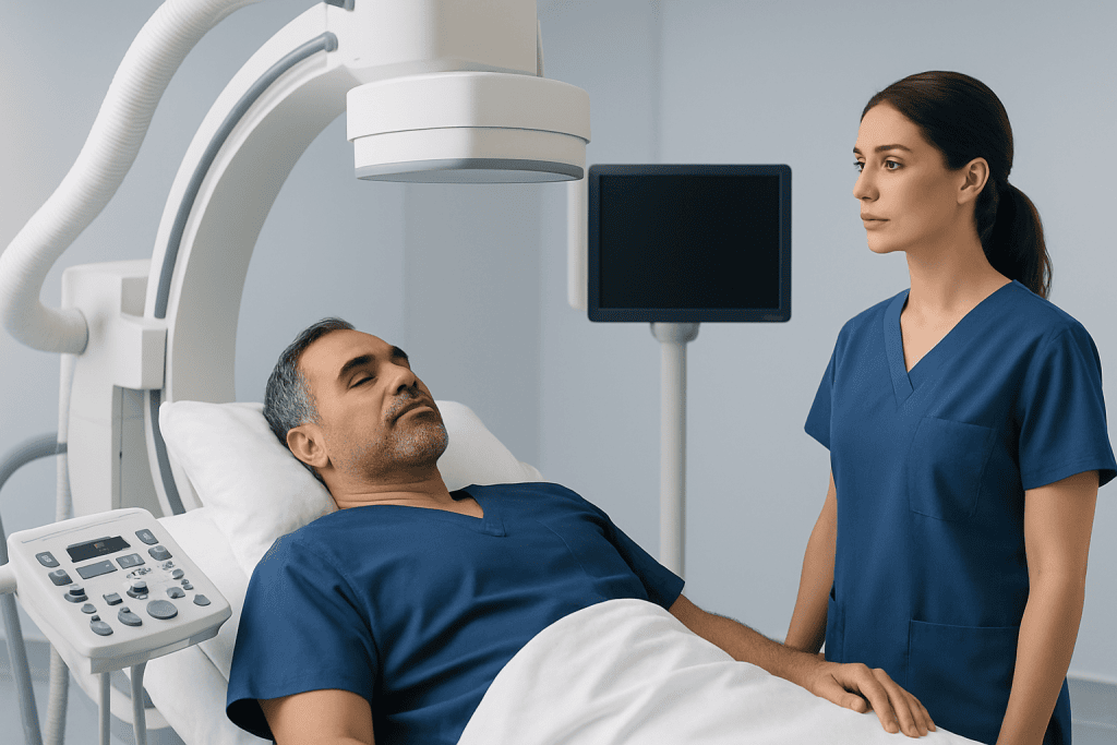
A venogram is recommended to clarify suspected problems in the veins. These may include a blood clot, chronic vein narrowing, compression of a major vein, congenital differences in vein structure, or complications related to prior surgery or long-term catheters. In some cases, it is also used before an intervention so the care team can plan the safest and most effective approach.
Symptoms that may lead to this test include persistent leg swelling, pain, skin color changes, unexplained heaviness, enlarged superficial veins, or signs that blood is not draining normally. In the pelvis, abdomen, or arms, symptoms may be less typical but can still point to venous disease.
Venography may also be part of preparation for procedures that treat blocked or narrowed veins. For example, a specialist may use the test before or during angioplasty and stent placement to define the exact location and extent of a blockage.
- To look for a suspected clot
- To evaluate chronic vein obstruction or narrowing
- To assess veins before a vascular procedure
- To investigate symptoms not fully explained by ultrasound
How the venogram procedure is performed
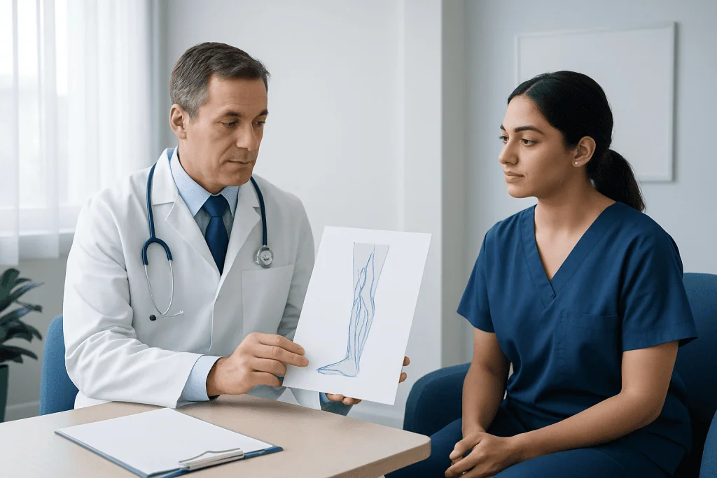
The exact method depends on which veins are being examined, but the general steps are similar. A small intravenous line or needle is placed into a vein, often in the foot, leg, arm, or groin depending on the area of interest. Contrast dye is then injected while a series of X-ray images is taken.
Patients are usually asked to lie still on an examination table. The skin may be cleaned carefully, and local anesthetic may be used if a catheter needs to be placed. Some people feel a brief sting from the needle, pressure at the access site, or a temporary warm sensation as the contrast moves through the vein.
In more advanced cases, venography may be performed as part of a catheter-based vascular evaluation. During the same session, doctors may also measure pressure differences in the veins or proceed to treatment if a clear problem is found and prior consent has been given. If this is planned, patients may be cared for by teams experienced in interventional radiology.
The test length varies. A straightforward venogram may take less than an hour, while a more detailed study combined with a planned intervention can take longer. Most patients go home the same day unless observation or treatment is needed.
How to prepare and what to expect afterward
Before the test, the medical team reviews symptoms, allergies, medications, and kidney health. This is important because contrast dye may not be suitable for everyone, especially people with severe kidney disease or a history of allergic reaction to contrast material. Patients should also mention blood thinners, diabetes medicines, pregnancy, or any implanted devices if other imaging is being considered.
Instructions vary by center. Some patients may be asked not to eat or drink for a few hours before the procedure, especially if sedation is planned. Comfortable clothing and arranging a ride home can be helpful if medicines that affect alertness will be used.
After the venogram, mild soreness or bruising at the injection site can occur. Drinking fluids may be advised to help clear the contrast, unless a doctor has restricted fluid intake for another health reason. Patients are usually told when they can resume normal activity and which symptoms should prompt a call, such as increasing pain, swelling, fever, or bleeding from the access site.
Benefits, limits, and possible risks
The main benefit of a venogram is detail. It can show the shape and openness of veins with a high degree of precision, which is especially useful when planning treatment or evaluating difficult cases. In selected situations, it remains one of the clearest ways to define a venous blockage or abnormal flow pattern.
At the same time, a venogram is more invasive than ultrasound because it requires contrast injection and X-ray exposure. For that reason, doctors do not usually start with venography unless there is a specific need. Noninvasive tests often provide enough information first.
Risks are generally low but important to understand. They may include bruising, discomfort, bleeding, infection at the needle or catheter site, contrast allergy, and kidney strain in vulnerable patients. Radiation exposure is usually limited, but clinicians still aim to keep it as low as reasonably possible. Rarely, the procedure can irritate a vein or contribute to clot-related complications.
- Benefits: detailed vein imaging, treatment planning, targeted diagnosis
- Limits: invasive compared with ultrasound, uses contrast and X-rays
- Possible risks: bruising, bleeding, infection, allergy, kidney effects, radiation exposure
How venogram results are used
The images are interpreted by a radiologist or vascular specialist, who looks for normal flow, narrowing, blockage, backflow, collateral veins, or structural differences. Results are then matched to the patient’s symptoms, physical examination, and other tests. A venogram is rarely interpreted in isolation; it is one part of a broader clinical picture.
If the venogram shows a blood clot, the doctor may recommend medicines to reduce the risk of clot growth or movement, along with follow-up care. If it shows a narrowed or compressed vein, treatment options may include observation, medication, or a procedure to open the vein. Some patients may be referred for varicose veins treatment or other vascular care depending on the source of symptoms.
When a venogram is normal, that can still be useful. It helps rule out important venous causes of swelling or pain and guides attention toward other explanations, such as lymphatic issues, joint disease, nerve-related symptoms, or arterial circulation problems. In some cases, the next step may be additional vascular testing or evaluation for related conditions such as peripheral artery disease.
When to seek medical care
Medical care should be sought promptly for sudden leg swelling, new one-sided pain, warmth or redness in a limb, chest pain, or shortness of breath. These symptoms do not always mean a blood clot is present, but they deserve urgent evaluation because some causes need timely treatment.
Patients should also contact a healthcare professional if they have persistent or worsening limb swelling, visible vein changes with pain, or symptoms that keep returning despite previous treatment. People with a history of clots, cancer treatment, recent surgery, prolonged immobility, or an indwelling catheter may need especially careful assessment.
After a venogram, urgent advice is appropriate if there is significant bleeding, rapidly increasing swelling, severe pain, fever, difficulty breathing, or signs of an allergic reaction. For international patients who need evaluation or treatment planning, Acibadem International’s multidisciplinary specialists and JCI-accredited hospitals provide diagnosis and care for vascular conditions.
Frequently asked questions
Is a venogram painful?
Most people feel only brief discomfort from the needle or IV placement. Some also notice a warm sensation or pressure when the contrast dye is injected. Significant pain is not expected, and the team can help if a patient is uncomfortable.
How is a venogram different from an ultrasound?
An ultrasound uses sound waves and does not require contrast dye or X-rays. A venogram is more invasive, but it can provide a more detailed view of vein anatomy and blood flow in selected cases. Doctors often use ultrasound first and choose venography when more information is needed.
Why would someone need a venogram if other imaging exists?
A venogram may be helpful when symptoms are difficult to explain, when ultrasound findings are unclear, or when treatment is being planned. It can also help define the exact location and severity of a blockage or narrowing. In some cases, it is performed during a procedure so diagnosis and treatment can be closely coordinated.
Are there risks from the contrast dye?
Yes, but serious problems are uncommon. Some people can have an allergic reaction, and people with reduced kidney function may need extra precautions or a different test. This is why doctors review allergies, kidney health, and medications before the procedure.
How long does it take to recover after a venogram?
Recovery is often quick, especially when the test is done as a simple outpatient study. Mild bruising or soreness at the injection site can happen for a short time. If a catheter-based treatment is also performed, recovery instructions may be more specific and activity may be limited temporarily.
Can a venogram diagnose a blood clot?
Yes, a venogram can show whether a vein is blocked or narrowed in a way that suggests a clot. It may be used when the diagnosis remains uncertain after initial testing or when detailed imaging is needed before treatment. The final diagnosis is based on the imaging together with the patient’s symptoms and examination.
References
- American College of Radiology
- Society of Interventional Radiology
- Centers for Disease Control and Prevention
- National Heart, Lung, and Blood Institute
- Radiological Society of North America
This article is for general information only and is not a substitute for professional medical advice. Please consult a qualified doctor about your individual situation.
Explore treatments in Turkey — costs, top hospitals & a free quote
JCI-accredited · board-certified surgeons · reply within 24h
Add us as a Preferred Source to see more of our trusted health content across Google Search, AI Overviews and Discover.

More from the Health Library

Vellus Hair: What Patients Need to Know

Compazine: A Complete Medical Overview

Negative Reinforcement: A Complete Medical Overview

Adderall Half Life: An Evidence-Based Guide for Patients
Splenomegaly — Explained by Medical Evidence, Not Myths