Angiomas: A Complete Medical Overview
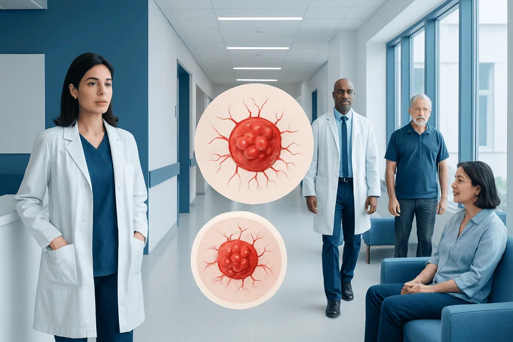
Most angiomas are noncancerous and do not cause serious health problems. Angiomas vary by type, including cherry angiomas, infantile hemangiomas, venous angiomas, and lymphangiomas.
Key Takeaways
- Most angiomas are noncancerous and do not cause serious health problems.
- Angiomas vary by type, including cherry angiomas, infantile hemangiomas, venous angiomas, and lymphangiomas.
- A doctor may recommend observation, imaging, laser therapy, medication, or surgery depending on the lesion.
- Any skin growth that changes quickly, bleeds repeatedly, causes pain, or is difficult to identify should be examined.
- Treatment decisions are based on symptoms, cosmetic concerns, location, age, and whether deeper structures are involved.
Angiomas are usually benign growths made up of blood vessels or lymphatic vessels. They can appear as red, purple, or blue marks on the skin or mucous membranes, and while many are harmless, some need medical assessment because of their size, location, symptoms, or uncertain diagnosis.
Overview: what angiomas are and why they appear
Angiomas are abnormal clusters or overgrowths of small blood vessels or lymphatic vessels. In everyday use, the term often refers to visible skin lesions such as cherry angiomas, but it can also include deeper vascular lesions in soft tissue or internal organs. Most angiomas are benign, meaning they are not cancer, and many never need treatment.
These lesions can be present at birth, appear in infancy, or develop later in adult life. Their appearance depends on the type and depth of the vessels involved. Some look like small bright-red dots, while others are bluish, raised, soft, or spongy. A few may grow quickly for a period of time and then stabilize or shrink.
Because the word “angioma” covers several different conditions, medical evaluation is sometimes important to confirm exactly what kind of lesion is present. Some vascular birthmarks and benign tumors can look similar at first glance. A careful diagnosis helps guide whether simple observation is enough or whether treatment is the better option.
Main types of angiomas
Doctors classify angiomas by the kind of vessels involved, when they appear, and how they behave over time. A common type in adults is the cherry angioma, a small red or purple spot caused by tiny blood vessel proliferation near the skin surface. These often increase with age and are usually harmless.
Infantile hemangiomas are another well-known vascular lesion. They often become visible in the first weeks of life, may grow during infancy, and then gradually shrink over several years. Some require monitoring only, while others need treatment if they interfere with vision, breathing, feeding, or skin integrity. Readers interested in related vascular skin lesions may also encounter hemangioma as a distinct but closely related diagnosis.
Other angiomas include venous angiomas, which may appear blue or compressible because they involve veins, and lymphangiomas, which arise from lymphatic vessels and can form soft, cystic, or clustered lesions. Some vascular malformations are present from birth and grow proportionally with the body rather than going through a growth-and-shrink cycle.
- Cherry angiomas: common, small, red skin spots in adults
- Infantile hemangiomas: vascular growths that often appear after birth
- Venous lesions: blue or purple, soft vascular abnormalities
- Lymphangiomas: lesions involving lymphatic channels
- Mixed vascular lesions: combinations of blood and lymphatic vessel changes
Symptoms and possible complications
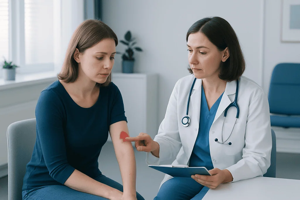
Many angiomas cause no symptoms at all and are discovered incidentally. A cherry angioma may simply appear as a tiny dome-shaped red papule on the trunk, arms, or shoulders. Larger or deeper lesions can produce swelling, a visible lump, skin discoloration, or asymmetry in the affected area.
Symptoms depend heavily on location. An angioma on the skin may bleed if rubbed by clothing or shaved. A lesion near the eye, nose, mouth, or airway can interfere with normal function. Deeper vascular lesions may cause discomfort, pressure, or cosmetic concerns, especially if they enlarge over time.
Complications are uncommon in many superficial lesions, but they can occur. Ulceration, repeated bleeding, infection of broken skin, and pain are possible in some angiomas. In infants and children, specialists pay special attention to lesions that affect vital functions or are associated with more extensive vascular abnormalities. If a vascular mark seems unusual, the diagnosis may need to be distinguished from other skin findings such as skin cancer or pigmented lesions.
Causes and risk factors
The exact cause of many angiomas is not fully understood. In general, they arise when blood vessels or lymphatic vessels develop or multiply in an unusual way. This process may be influenced by genetic factors, developmental changes before birth, local tissue signals, and age-related changes in the skin.
Some angiomas are congenital, meaning they are present at birth or result from vessel development during pregnancy. Others appear later in life without a clear trigger. Cherry angiomas tend to become more common with age. Family history may play a role in certain people, though many patients have no known inherited risk.
Hormonal influences, skin aging, and underlying vascular biology may contribute to some lesions, but a single cause usually cannot be identified. Importantly, most angiomas are not related to poor hygiene, infection, or anything a person did wrong. A doctor’s assessment is most helpful when a lesion is new, changing, symptomatic, or difficult to classify by appearance alone.
How angiomas are diagnosed
Diagnosis often starts with a physical examination and a review of when the lesion appeared, whether it has changed, and whether it causes bleeding, pain, or functional problems. Dermatologists, pediatricians, vascular specialists, or surgeons may all be involved depending on the lesion’s type and location. In many cases, the diagnosis can be made clinically.
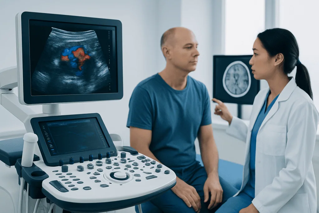
When a lesion is deeper, larger, or uncertain, imaging may be recommended. Ultrasound can help show blood flow and depth. Magnetic resonance imaging is useful for evaluating more complex or extensive vascular lesions. If a lesion has atypical features, a biopsy may occasionally be needed to confirm the diagnosis and rule out other conditions.
Accurate diagnosis matters because the term angioma may overlap with vascular tumors, vascular malformations, and other skin growths. Some patients are referred for advanced evaluation in centers that manage complex vascular anomalies. In selected cases, MRI imaging or dermatology assessment helps define the lesion and the safest treatment plan.
Treatment options and when treatment is needed
Not all angiomas need treatment. If a lesion is small, clearly benign, and not causing symptoms, observation may be the best approach. Doctors often recommend simple monitoring for stable cherry angiomas or small lesions that do not affect comfort, function, or appearance in a troubling way.
When treatment is appropriate, the choice depends on the type of angioma, the patient’s age, and the lesion’s size and location. Options may include laser therapy, medications, sclerotherapy, electrocautery, cryotherapy, or surgical removal. Infantile hemangiomas that threaten important functions may be treated with medicines under specialist supervision. Superficial lesions that bleed repeatedly or are frequently irritated may be removed for comfort or practical reasons.
Procedural treatment is also considered when there is diagnostic uncertainty, ulceration, pain, recurrent trauma, or significant cosmetic impact. In some cases, surgery is the clearest option, especially for localized lesions that can be removed safely. Depending on the clinical situation, doctors may discuss laser treatment, sclerotherapy, or vascular surgery as part of a personalized plan.
Near the end of the care pathway, patients with more complex vascular lesions may benefit from coordinated assessment by dermatology, radiology, pediatrics, surgery, and vascular specialists. Acibadem International’s multidisciplinary specialists and JCI-accredited hospitals diagnose and treat angiomas and related vascular conditions for international patients.
Self-care, monitoring, and prevention
There is no guaranteed way to prevent most angiomas, especially those linked to age or natural vessel development. However, practical self-care can reduce irritation and help patients notice meaningful changes early. For example, lesions in areas exposed to shaving, jewelry, waistbands, or friction may need gentle protection.
Patients are often advised not to pick, scratch, or try to remove an angioma at home. Home removal can lead to bleeding, infection, scarring, and delayed diagnosis if the lesion is not actually an angioma. Photographing a lesion occasionally in the same lighting can be a helpful way to monitor whether it is changing in size, color, or shape.
General skin care also matters. Using sunscreen, protecting vulnerable skin from repeated trauma, and seeking medical advice for lesions that crack or bleed can be useful habits. For children with diagnosed vascular lesions, follow-up visits are important so doctors can assess growth, function, and any need for treatment over time.
When to seek medical care
Medical care should be sought if a suspected angioma appears suddenly and grows quickly, bleeds often, becomes painful, develops an ulcer, or shows signs of infection such as warmth, swelling, or drainage. Assessment is also important if the lesion interferes with seeing, breathing, feeding, hearing, movement, or normal daily activities.
Parents should arrange prompt evaluation for infants who have a vascular lesion near the eye, nose, lips, diaper area, or airway, or for marks that are large, rapidly enlarging, or multiple. Adults should also have any new or changing skin lesion checked if the diagnosis is uncertain. Although most angiomas are benign, some skin growths can resemble each other, and a proper medical opinion provides clarity and reassurance.
Urgent care is appropriate if bleeding does not stop with gentle pressure, if swelling rapidly increases, or if the person has significant pain or breathing difficulty. A qualified doctor can decide whether the lesion is a harmless angioma, a different vascular condition, or another skin problem that needs specific treatment.
Frequently asked questions
Are angiomas dangerous?
Most angiomas are not dangerous and are considered benign. They often cause little more than a visible skin mark, although some need treatment if they bleed, grow, or affect important functions.
What is the difference between an angioma and a hemangioma?
The word angioma is a broad term for benign lesions involving blood vessels or lymphatic vessels. Hemangioma is a more specific type of vascular growth, commonly seen in infancy, with its own typical pattern of growth and gradual regression.
Do cherry angiomas go away on their own?
Cherry angiomas usually do not disappear spontaneously. They often remain stable over time, though some may slowly enlarge or increase in number with age.
Can angiomas bleed?
Yes, some angiomas can bleed, especially if they are rubbed, scratched, shaved, or injured. Repeated bleeding is a good reason to seek medical advice because treatment may help prevent further irritation.
How do doctors remove an angioma?
Removal methods depend on the lesion type and location. Common approaches include laser therapy, electrocautery, cryotherapy, sclerotherapy, medication for selected lesions, or surgical excision.
Should a child with an angioma see a specialist?
A child should be assessed if the lesion is large, rapidly growing, ulcerated, or located near the eye, nose, mouth, or airway. Many childhood vascular lesions are harmless, but specialist review helps identify those that need closer monitoring or treatment.
References
- American Academy of Dermatology
- National Institute of Arthritis and Musculoskeletal and Skin Diseases
- MedlinePlus
- National Organization for Rare Disorders
- American Academy of Pediatrics
This article is for general information only and is not a substitute for professional medical advice. Please consult a qualified doctor about your individual situation.
Oncology care in Turkey — second opinion and treatment plan
JCI-accredited · board-certified surgeons · reply within 24h
Add us as a Preferred Source to see more of our trusted health content across Google Search, AI Overviews and Discover.