Bone Island: A Complete Medical Overview

A bone island, also called enostosis, is usually benign and often found by chance on X-ray, CT, or MRI. Most bone islands do not cause symptoms and do not need treatment.
Key Takeaways
- A bone island, also called enostosis, is usually benign and often found by chance on X-ray, CT, or MRI.
- Most bone islands do not cause symptoms and do not need treatment.
- Further evaluation may be needed if the lesion is large, painful, changing over time, or seen in a person with a history of cancer.
- Diagnosis relies on imaging appearance and sometimes follow-up imaging to confirm stability.
- A bone island is different from bone cancer and other bone lesions, although careful review is sometimes necessary.
A bone island is a small area of compact bone located within spongy bone. It is usually a benign incidental imaging finding, but a doctor may recommend follow-up tests if the spot is painful, growing, or difficult to distinguish from other bone conditions.
What Is a Bone Island?
A bone island is a small focus of dense, compact bone located inside the softer cancellous, or spongy, part of a bone. Doctors also call it enostosis. In most cases, it is discovered incidentally during imaging done for another reason, such as a sports injury, back pain, or routine evaluation after trauma.
For most people, a bone island is a harmless finding rather than a disease. It usually does not weaken the bone, spread to other parts of the body, or turn into cancer. Once its typical appearance is recognized, no treatment is often needed.
Bone islands can appear in many parts of the skeleton, including the pelvis, femur, ribs, spine, and other long bones. They may be tiny and barely noticeable or somewhat larger, but size alone does not always mean danger. What matters most is how the lesion looks on imaging and whether it stays stable over time.
Because the term can sound unfamiliar or worrying, many patients understandably ask whether a bone island is serious. In the great majority of cases, the answer is no. Still, doctors may look more closely when the imaging pattern is unusual or when the patient has symptoms or a medical history that raises concern.
How a Bone Island Is Usually Found
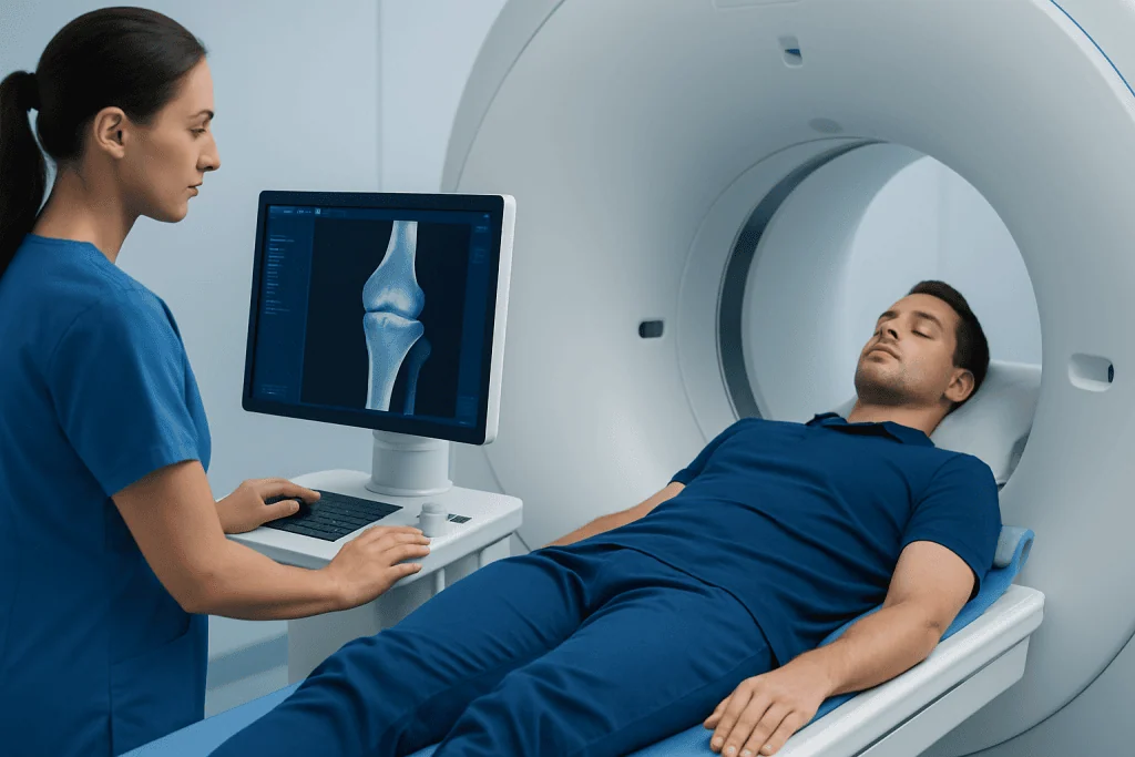
Bone islands are most often found by chance. A person may have an X-ray after a fall, a CT scan for abdominal pain, or an MRI for joint symptoms, and the radiologist notices a dense spot within a bone. Since these lesions usually do not cause problems, many people never know they have one until imaging reveals it.
On imaging, a bone island often has a very characteristic appearance. It is typically round, oval, or slightly irregular, with dense margins that blend into the surrounding bone through fine radiating lines. This appearance helps radiologists distinguish it from more concerning lesions.
Different imaging tests can show a bone island in different ways. An X-ray or CT usually shows it best because both display bone density clearly. MRI can also detect it, although the appearance may be less intuitive for patients reading reports. In selected cases, a doctor may compare prior images to confirm that the lesion has not changed.
When the imaging features are classic and the patient has no concerning symptoms, the finding is often simply documented and observed without any further steps. If the appearance is less typical, the doctor may recommend additional imaging through services such as diagnostic radiology evaluation to better define the lesion.
Symptoms and What They May Mean

Most bone islands cause no symptoms at all. They do not usually lead to swelling, fever, redness, or limitation of movement. In many cases, the pain that led to the imaging study comes from a different problem, while the bone island is an unrelated incidental finding.
Less commonly, a person may have pain in the same region where the bone island is seen. Even then, the lesion itself is not always the cause. Muscular strain, arthritis, tendon irritation, osteoarthritis, stress injury, or other bone and joint conditions may better explain the symptoms.
Doctors become more cautious when pain is persistent, worsening, or accompanied by other signs such as night pain, unexplained weight loss, or a known cancer history. These situations do not mean the lesion is dangerous, but they do make a closer review important.
Very large bone islands, sometimes called giant bone islands, may be discussed separately in imaging reports. Even these can still be benign, but they are more likely to prompt follow-up because their size can overlap with other conditions. The key point is that symptoms are assessed in the full clinical context, not based on the image alone.
Why Bone Islands Happen and Who Gets Them
The exact cause of a bone island is not always clear. It is generally considered a developmental or anatomical variation in which a small focus of compact bone remains within cancellous bone. It is not usually related to injury, infection, or poor lifestyle habits.
Bone islands can be seen in adults of many ages and are often found in otherwise healthy people. Because they are usually silent, the true number of people who have them is probably higher than the number actually diagnosed. They may be identified more often today simply because imaging is used more frequently.
There are no well-established day-to-day risk factors that a person can control, such as diet or exercise, that clearly cause bone islands. They are not typically linked with calcium intake or common overuse patterns. In that sense, they differ from fractures, osteoporosis, or joint degeneration.
What changes the medical approach is not usually the cause of the bone island, but the context in which it appears. For example, if a dense bone lesion is seen in someone with a history of cancer, clinicians may need to distinguish it from other bone findings. This is why medical history remains an important part of evaluation.
How Doctors Tell a Bone Island From Other Bone Lesions
The main challenge with a bone island is not that it is dangerous, but that it can resemble other dense bone lesions on imaging. Radiologists assess its location, shape, borders, internal density, and whether it has remained unchanged over time. A classic bone island has sharp, thorn-like margins that merge with surrounding trabecular bone.
Several other conditions may enter the discussion when a dense bone lesion is seen. These can include healing bone injury, osteoid osteoma, osteoblastic metastasis, or other benign and malignant bone processes. In some patients, doctors may also consider broader orthopedic conditions involving the bone structure, including problems reviewed under bone tumors, even though a bone island itself is usually not a tumor in the dangerous sense patients fear.
Additional tests are not always necessary, but they may be recommended if the lesion appears atypical. Follow-up X-rays, CT, MRI, or occasionally a bone scan can help show whether the area is stable. Stability over time strongly supports a benign diagnosis.
Rarely, a biopsy is considered when imaging cannot confidently exclude another diagnosis. This is uncommon and generally reserved for cases with unusual features, lesion growth, unexplained pain, or high-risk medical history. When tissue sampling is needed, the care plan is usually guided by orthopedic and radiology specialists together.
Treatment and Follow-Up
Most bone islands need no treatment. If the lesion has a typical benign appearance and the person has no concerning symptoms, reassurance is often the only management required. Many patients simply continue normal daily activities without restrictions.
When symptoms are present, treatment is directed at the actual cause of the pain rather than the bone island itself. For example, if imaging was done because of back pain and the real problem is a disc disorder or muscle strain, the doctor treats that condition. In some cases, a broader musculoskeletal assessment may be useful through orthopedics and traumatology.
If the imaging appearance is uncertain, the doctor may recommend interval imaging after several months or longer to confirm the lesion is unchanged. This kind of follow-up is common in medicine and does not necessarily mean something serious is suspected. It is simply a careful way to confirm a benign diagnosis.
Rarely, if a lesion remains unclear or causes diagnostic concern, advanced evaluation may be discussed. Multidisciplinary review can help decide whether observation, repeat imaging, or biopsy is the best next step. In centers experienced with complex bone conditions, supportive care may also involve specialist hematology or bone-focused assessment when the wider clinical picture suggests another disorder, though this is not typical for a simple bone island.
Self-Care, Outlook, and When to Seek Medical Care
Because a bone island is usually benign, no special home treatment is needed for the lesion itself. A person can generally stay active, follow regular bone-health habits, and manage any unrelated musculoskeletal discomfort according to medical advice. Healthy routines such as exercise, balanced nutrition, and avoiding smoking remain beneficial for overall skeletal health, even though they do not specifically change a bone island.
The outlook is excellent in most cases. Bone islands commonly remain stable for years and never lead to complications. Once the finding is clearly identified, many people do not need repeated follow-up unless new symptoms appear or a doctor advises surveillance based on individual circumstances.
Medical care should be sought if bone pain is persistent, worsening, or occurs at night; if there is swelling or reduced function near the area; or if the person has a history of cancer and a new bone lesion is found. Follow-up is also important if imaging reports say the lesion is atypical, enlarging, or indeterminate.
For patients who need further assessment, experienced teams can help interpret imaging and plan next steps. Acibadem International’s multidisciplinary specialists at JCI-accredited hospitals diagnose and treat bone and joint conditions for international patients when more detailed evaluation is needed.
Frequently asked questions
Is a bone island cancer?
A bone island is usually a benign finding and is not considered cancer. Doctors may investigate further only if the imaging appearance is unusual or the patient has symptoms or a medical history that raises concern.
Can a bone island cause pain?
Most bone islands do not cause pain. If pain is present, doctors look carefully to see whether another bone, joint, or soft-tissue problem is the true cause.
Does a bone island need to be removed?
In most cases, no removal is needed. Treatment is generally unnecessary unless the diagnosis is uncertain and further evaluation shows another condition that requires care.
How is a bone island diagnosed?
It is usually diagnosed based on its typical appearance on imaging such as X-ray or CT. Sometimes follow-up imaging is used to confirm that the lesion stays stable over time.
Can a bone island grow?
Many bone islands remain stable. If a lesion appears to enlarge, a doctor may recommend closer imaging review or additional tests to make sure it is truly a benign bone island.
What is the difference between a bone island and a bone tumor?
A bone island is a small area of dense bone within normal bone and is usually harmless. A bone tumor is a broader term that can include benign or malignant growths, so the imaging pattern and clinical context are important.
References
- American Academy of Orthopaedic Surgeons
- Radiological Society of North America
- American College of Radiology
- National Cancer Institute
This article is for general information only and is not a substitute for professional medical advice. Please consult a qualified doctor about your individual situation.
Joint and spine care in Turkey — expert assessment & treatment
JCI-accredited · board-certified surgeons · reply within 24h
Add us as a Preferred Source to see more of our trusted health content across Google Search, AI Overviews and Discover.