S4 Gallop: An Evidence-Based Guide for Patients

An S4 gallop, also called a fourth heart sound or atrial gallop, occurs late in the heart’s filling phase. It is commonly associated with reduced flexibility of the left ventricle, often related to high blood pressure, heart muscle thickening, or coronary artery disease.
Key Takeaways
- An S4 gallop, also called a fourth heart sound or atrial gallop, occurs late in the heart’s filling phase.
- It is commonly associated with reduced flexibility of the left ventricle, often related to high blood pressure, heart muscle thickening, or coronary artery disease.
- An S4 sound is interpreted together with symptoms, medical history, examination findings, and tests such as an electrocardiogram and echocardiogram.
- It may be absent in conditions with an irregular rhythm such as atrial fibrillation because coordinated atrial contraction is needed to produce it.
- New chest pain, severe shortness of breath, fainting, or sudden worsening symptoms require urgent medical assessment.
An S4 gallop is a low-pitched extra heart sound heard just before the normal first heart sound. It can occur when the upper heart chamber pushes blood into a stiff lower chamber, so it may be a clue to an underlying heart condition but is not a diagnosis on its own.
What Is an S4 Gallop?
An S4 gallop is an additional heart sound that a clinician may hear with a stethoscope. It happens immediately before the usual first heart sound, often described as occurring just before “lub.” The term gallop reflects the rhythm created when this extra sound is heard alongside the normal heartbeat.
The sound is produced near the end of diastole, the phase when the heart’s lower chambers, called ventricles, relax and fill with blood. At this point, the atria contract to give the ventricles a final small push of blood. If a ventricle is unusually stiff, that push can create vibrations audible as an S4 sound. For this reason, it is also called an atrial gallop or fourth heart sound.
An S4 gallop is a physical examination finding, not a disease by itself. Its importance depends on the person’s age, blood pressure, heart rhythm, symptoms, and other findings. In many adults, especially when it is newly detected, it prompts a clinician to consider whether the heart muscle is less able to relax and fill normally.
How the Heart Sound Fits Into a Normal Heartbeat
Clinicians commonly describe two normal heart sounds. S1 is made mainly when the mitral and tricuspid valves close as the ventricles begin to contract. S2 occurs when the aortic and pulmonary valves close as the ventricles finish pumping and begin to relax. These sounds are typically heard as “lub-dub.”
S4 occurs before S1, during the final portion of ventricular filling. It is usually low in pitch and may be difficult to hear without careful listening in a quiet setting. A clinician may listen at the apex of the heart, near the lower left side of the chest, using the bell of the stethoscope. The sound can be more noticeable in certain body positions or when the heart rate is faster.
S4 should not be confused with an S3 gallop. An S3 occurs shortly after S2, earlier in ventricular filling, and has different possible causes. While either sound can provide useful clinical information, neither can establish a diagnosis alone. The examination guides the next steps, which may include blood pressure measurement, heart rhythm assessment, or imaging.
What Can Cause an S4 Gallop?

An S4 gallop is most often linked with a ventricle that has become less compliant, meaning it does not relax or stretch as easily during filling. Long-standing high blood pressure is a frequent reason. Over time, increased pressure can cause the left ventricular muscle to thicken, making the chamber stiffer and increasing resistance to blood entering it.
Other possible causes include coronary artery disease or a prior heart attack, <a href="https://acibademinternational.com/diseases/hypertrophic-cardiomyopathy/”>hypertrophic cardiomyopathy, aortic valve narrowing, and conditions that lead to thickening or scarring of the heart muscle. In these situations, the heart may still pump blood effectively but have difficulty relaxing between beats, a pattern sometimes described as diastolic dysfunction.
An S4 can occasionally be heard in older adults without clear symptoms of heart disease, but it should not automatically be dismissed as a normal effect of aging. A clinician considers the whole clinical picture. The sound is generally not heard when atrial contraction is absent or uncoordinated, as in atrial fibrillation, because the atria need to contract effectively for an S4 to occur.
- Long-standing high blood pressure
- Left ventricular hypertrophy, or thickened heart muscle
- Coronary artery disease or previous heart muscle injury
- Aortic stenosis, meaning narrowing of the aortic valve
- Hypertrophic cardiomyopathy and some other heart muscle disorders
Symptoms and What They May Mean
An S4 gallop itself does not cause symptoms and cannot be felt by the person experiencing it. It is identified during a clinical examination. Some people have no symptoms, particularly when the underlying change is mild or has developed gradually.
When symptoms are present, they are usually related to the condition affecting the heart rather than to the extra sound. Possible symptoms include getting short of breath during activity, reduced exercise tolerance, fatigue, chest discomfort, awareness of a fast or irregular heartbeat, ankle swelling, or needing more pillows to sleep comfortably. These symptoms can also arise from non-heart conditions, so professional assessment is important.
A symptom pattern can help guide evaluation. For example, breathlessness that develops gradually in a person with high blood pressure may lead a clinician to assess heart structure and function. Chest pressure brought on by exertion may lead to assessment for reduced blood flow to the heart. However, symptoms alone cannot determine the cause, and an S4 gallop does not confirm heart failure, coronary artery disease, or any other single diagnosis.
How Clinicians Evaluate an S4 Gallop
Evaluation begins with a conversation about current symptoms, medical history, medicines, lifestyle, and family history of heart disease. A clinician will check blood pressure, pulse, oxygen level when appropriate, and signs such as swelling, lung sounds, or heart murmurs. They will also determine whether the rhythm is regular and whether other heart sounds are present.
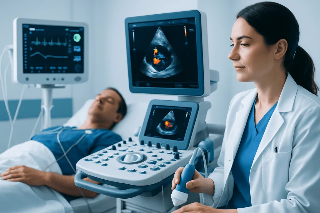
An electrocardiogram, also called an ECG or EKG, may be used to assess heart rhythm and look for evidence of heart muscle thickening, prior injury, or reduced blood flow. An echocardiogram uses ultrasound to show the heart chambers, valves, pumping strength, and how the ventricles relax and fill. It is often the most useful test when an extra heart sound raises concern about structural heart disease.
Depending on the findings, clinicians may recommend blood tests, chest imaging, exercise testing, ambulatory rhythm monitoring, or more specialized cardiac imaging. The goal is not simply to confirm an S4 sound; it is to identify whether a treatable condition is affecting heart function. Results should be discussed with a qualified doctor, who can explain what they mean in the context of the individual’s health.
Treatment and Everyday Heart Care
There is no treatment aimed specifically at eliminating an S4 gallop. Care focuses on the underlying cause. For example, controlling elevated blood pressure can reduce strain on the heart and may help prevent further thickening or stiffness of the heart muscle. If valve disease, coronary artery disease, or cardiomyopathy is found, management is tailored to that condition and to the person’s symptoms and overall health.
Depending on the diagnosis, treatment may include lifestyle measures, prescribed medicines, monitoring, or procedures. It is important not to start, stop, or change heart medicines without medical advice. People with high blood pressure, diabetes, high cholesterol, kidney disease, or a history of heart disease should attend regular follow-up appointments, even if they feel well.
Supportive heart-healthy habits can make a meaningful difference alongside medical care. These include avoiding tobacco, following a balanced eating pattern rich in vegetables, fruit, whole grains, and minimally processed foods, limiting excess salt when advised, staying physically active at a level approved by a clinician, maintaining a weight that supports health, and getting adequate sleep. Alcohol intake should be discussed with a healthcare professional, particularly for people with high blood pressure or heart disease.
Acibadem International’s multidisciplinary specialists and JCI-accredited hospitals assess and treat cardiovascular conditions for international patients, with care plans based on individual clinical findings.
When to Seek Medical Care
Anyone told they have an S4 gallop should arrange follow-up with a doctor, especially if they have high blood pressure, known heart disease, diabetes, kidney disease, or a family history of cardiomyopathy or early heart disease. A timely appointment is also appropriate for new breathlessness, declining exercise capacity, palpitations, leg swelling, or repeated dizziness.
Emergency medical care is needed for chest pain, pressure, tightness, or discomfort that is severe, persistent, or occurs with sweating, nausea, shortness of breath, or pain spreading to the arm, jaw, back, or shoulder. Sudden severe breathlessness, fainting, bluish lips, confusion, or a very rapid or irregular heartbeat with feeling unwell also need urgent evaluation.
Most people who have an extra heart sound do not need to panic. Nevertheless, prompt assessment is valuable because many contributing conditions can be managed effectively when identified early. Keeping a record of symptoms, blood pressure readings if available, medicines, and relevant family history can help make the clinical visit more useful.
Frequently asked questions
Is an S4 gallop dangerous?
An S4 gallop is not dangerous by itself; it is an examination finding rather than a diagnosis. It can indicate that a heart ventricle is stiff, so clinicians evaluate it alongside symptoms, blood pressure, heart rhythm, and testing. The level of concern depends on the underlying cause.
Can an S4 gallop be normal?
An S4 sound may occasionally be detected in older adults, but a new finding should still be interpreted carefully. It is more often associated with conditions that reduce the heart muscle’s ability to relax, such as long-standing high blood pressure. A doctor can determine whether further evaluation is needed.
What is the difference between S3 and S4 gallops?
S3 occurs shortly after the second heart sound, early in the filling phase of the ventricles. S4 occurs just before the first heart sound, when the atria contract to complete ventricular filling. They have different mechanisms and may point toward different clinical situations.
Can high blood pressure cause an S4 heart sound?
Yes. Persistently elevated blood pressure can make the left ventricular muscle thicker and less flexible over time. When the atrium pushes blood into this stiff ventricle, an S4 sound may be produced.
Does an S4 gallop mean heart failure?
No. An S4 gallop does not by itself mean that a person has heart failure. It can be associated with impaired relaxation of the heart muscle, which may occur in some forms of heart failure, but diagnosis requires a full clinical assessment and often an echocardiogram.
Can an S4 gallop occur with atrial fibrillation?
An S4 is generally absent in atrial fibrillation because the atria do not contract in an organized way. Coordinated atrial contraction is needed to create the extra sound. Other abnormal heart sounds or signs may still be present, depending on the person’s heart health.
References
- American Heart Association
- American College of Cardiology
- National Heart, Lung, and Blood Institute
- Merck Manual Professional Edition
- Mayo Clinic
This article is for general information only and is not a substitute for professional medical advice. Please consult a qualified doctor about your individual situation.
Explore treatments in Turkey — costs, top hospitals & a free quote
JCI-accredited · board-certified surgeons · reply within 24h
Add us as a Preferred Source to see more of our trusted health content across Google Search, AI Overviews and Discover.
Check your numbers in seconds
BMI, calories, due date, blood pressure and 30+ more clinical calculators — free, instant, doctor-reviewed ranges.

More from the Health Library



Related Specialists

Prof. Dr. Muhsin Eraslan
Ophthalmology
Dr. Ayça Sözen
Pediatrics
Prof. Dr. Javad Parvızı
Orthopedic Surgery & Traumatology
Dyt. Nur Ecem Baydı Ozman
Nutrition & Diet