Caliectasis: An Evidence-Based Guide for Patients

Caliectasis means widening of the kidney’s urine-collecting calyces, usually seen on ultrasound or CT imaging. It may result from a temporary or persistent problem with urine drainage, including kidney stones, narrowing, infection or pressure during pregnancy.
Key Takeaways
- Caliectasis means widening of the kidney’s urine-collecting calyces, usually seen on ultrasound or CT imaging.
- It may result from a temporary or persistent problem with urine drainage, including kidney stones, narrowing, infection or pressure during pregnancy.
- Some people have no symptoms, while others develop flank pain, urinary symptoms, fever or blood in the urine.
- Tests help determine whether there is obstruction, infection, reflux or reduced kidney function.
- Treatment focuses on the cause; urgent drainage may be needed when obstruction occurs with infection, severe pain or impaired kidney function.
- Prompt medical assessment is important for fever, severe pain, vomiting, reduced urine output or symptoms of a urinary infection.
Caliectasis describes enlargement of one or more calyces, the small cup-shaped parts of the kidney that collect urine before it flows into the ureter. It is an imaging finding rather than a diagnosis itself, and its importance depends on the underlying cause, symptoms, kidney function and degree of urine-flow obstruction.
Caliectasis: what it means
Caliectasis is the medical term for dilation, or widening, of the renal calyces. The calyces are small chambers inside each kidney that collect urine produced by the kidney tissue. Urine normally passes from the calyces into the renal pelvis, then through the ureter to the bladder. When urine does not drain as freely as expected, pressure can cause one or more calyces to expand.
Caliectasis is usually identified during an ultrasound, CT scan, MRI scan or another imaging test performed for pain, urinary symptoms, pregnancy monitoring or an unrelated concern. It may affect one kidney or both. The report may also describe the finding as calyceal dilatation, dilated calyces, or fullness of the collecting system.
Although caliectasis is sometimes discussed alongside hydronephrosis, the terms are not completely interchangeable. Hydronephrosis generally refers to dilation of the renal pelvis and often the calyces as a result of backed-up urine. Caliectasis may be an early, localized or milder collecting-system change. A clinician considers the imaging pattern, symptoms and test results rather than relying on one term alone.
How caliectasis may feel
Caliectasis itself does not always cause symptoms. Mild dilation may be found incidentally and may not be associated with ongoing obstruction or kidney damage. Symptoms, when present, usually arise from the condition affecting urine flow, such as a stone, infection or narrowing of the urinary tract.
A person may notice aching or sharp pain in the side or back below the ribs, often called flank pain. Pain from a kidney stone can be severe, may come in waves and can travel toward the lower abdomen or groin. Other possible symptoms include nausea, vomiting, burning with urination, a need to urinate more frequently, cloudy urine or visible blood in the urine.
Fever, chills, feeling unwell and flank pain may signal a urinary tract infection involving the kidney. This combination deserves prompt assessment, particularly if imaging has shown impaired drainage. In infants and children, signs can be less specific and may include fever, poor feeding, vomiting, irritability, abdominal discomfort or recurrent urinary infections.
- Many cases are painless and discovered on imaging.
- Flank pain may suggest a stone or other obstruction.
- Burning, urgency or cloudy urine can occur with a urinary infection.
- Fever with urinary symptoms needs urgent medical attention.
Why urine may back up in the kidney

The most common reason for caliectasis is partial or complete obstruction somewhere along the route urine takes out of the kidney. A kidney stone is a frequent cause because it can lodge in the ureter and reduce urine flow. The size and location of a stone, as well as the degree of obstruction, influence symptoms and treatment decisions.
Other possible causes include a narrowed ureter, sometimes related to scar tissue after surgery, inflammation or prior stone passage; congenital differences in urinary tract anatomy; and masses that press on or involve the urinary tract. An enlarged prostate can interfere with bladder emptying and, in more advanced cases, contribute to back pressure affecting both kidneys. Blood clots, rare urinary tract tumors and injury may also obstruct flow.
Urine can also move backward from the bladder toward the kidneys, a condition called vesicoureteral reflux. This is more commonly evaluated in children with recurrent urinary infections or prenatal urinary tract dilation. During pregnancy, hormonal changes and pressure from the growing uterus can cause temporary dilation of the collecting system, often more noticeable on one side. A medical team can distinguish expected pregnancy-related changes from a stone or infection when symptoms occur.
Not every imaging appearance of mild caliectasis reflects a fixed blockage. Hydration status, a full bladder and the timing of the scan can affect the appearance of the collecting system. Follow-up imaging or functional testing may be recommended when the clinical picture is unclear.
How doctors investigate caliectasis
Assessment begins with a review of symptoms, prior stones or urinary infections, medical history, medications and family history. A clinician may ask about urine output, pregnancy, previous urinary surgery and conditions that can affect the bladder. The physical examination may include checking for tenderness over the kidney area, fever, signs of dehydration and bladder fullness.
A urinalysis can identify blood, white blood cells, bacteria or protein in the urine. If infection is suspected, a urine culture can help identify the organism and guide antibiotic selection. Blood tests may measure creatinine and other markers of kidney function, especially when both kidneys may be affected, a person has one functioning kidney, or significant obstruction is possible.
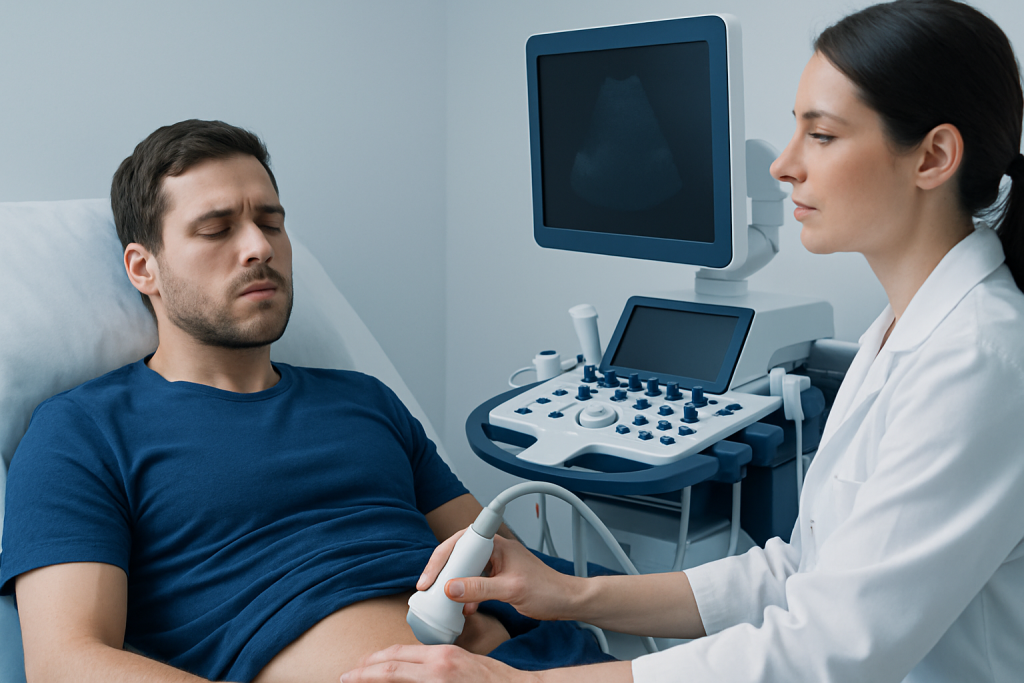
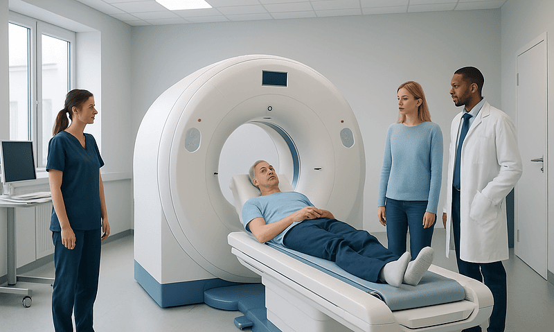
Ultrasound is commonly used because it does not involve radiation and can show collecting-system dilation, some stones, bladder emptying and kidney size. CT imaging can provide more detailed information about stones and other causes of obstruction. In selected situations, MRI, a nuclear medicine renal scan, or imaging of the bladder and ureters may help show how effectively urine drains and how well each kidney is functioning.
Imaging findings are interpreted in context. A report may note mild, moderate or severe dilation, but the next step is based on the likely cause and whether there are warning signs such as infection, worsening pain or reduced kidney function. Referral to a urologist or kidney specialist may be appropriate for persistent, unexplained or complex cases.
Treatment is guided by the cause
There is no single treatment for caliectasis because it is a sign of altered urine drainage, not a disease on its own. If imaging shows mild dilation without symptoms, infection or impaired kidney function, a clinician may recommend observation and repeat ultrasound. This approach can be appropriate when temporary dilation is likely and there is no evidence of significant obstruction.
When a kidney stone is responsible, treatment depends on its size, location, symptoms and effect on urine flow. Some stones pass with time and supportive care, while others require a procedure to break, remove or bypass the stone. Pain relief, anti-nausea medicines and hydration advice should be individualized by a healthcare professional, particularly for people with kidney disease, pregnancy or other medical conditions.
Infection requires prompt evaluation and appropriate antibiotics. An infected, obstructed kidney is a medical emergency because bacteria can become trapped behind a blockage. In this situation, doctors may need to drain urine urgently using a ureteral stent, a small tube placed inside the ureter, or a nephrostomy tube that drains the kidney through the skin. Definitive treatment of the obstruction is usually planned once the acute infection is controlled.
Procedures may also be considered for ureteral narrowing, reflux, congenital urinary tract differences or pressure from another condition. The aim is to protect kidney function, relieve symptoms and prevent repeated infection. Follow-up tests help confirm that drainage has improved and that the kidneys are functioning appropriately.
Protecting urinary and kidney health
Caliectasis cannot always be prevented, particularly when it is related to anatomy, pregnancy or an unexpected stone. However, early attention to urinary symptoms may reduce the risk of complications. People should seek medical advice for repeated urinary infections, recurrent stones, blood in the urine or persistent flank discomfort rather than assuming symptoms will resolve on their own.
For people who have had kidney stones, prevention is tailored to the stone type and individual risk factors. General measures may include drinking enough fluid to maintain regular pale-yellow urine, unless a clinician has advised fluid restriction; limiting excess salt; and following individualized dietary guidance. It is not advisable to make major dietary restrictions or use supplements marketed for “kidney cleansing” without medical advice.
Completing prescribed treatment for urinary infections and attending follow-up imaging are important when recommended. People with diabetes, neurologic bladder conditions, an enlarged prostate or a history of urinary tract surgery may benefit from regular monitoring because these factors can affect urine flow.
Acibadem International’s multidisciplinary specialists and JCI-accredited hospitals evaluate urinary tract conditions for international patients, using imaging, laboratory testing and urology care when needed.
When to seek medical care
Medical review should be arranged soon after a report of caliectasis, even if there are no symptoms, so that a clinician can determine whether follow-up is needed. This is especially important for children, pregnant people, those with a solitary kidney, and anyone with known kidney disease or a history of urinary tract obstruction.
Urgent medical care is needed for fever or chills together with flank pain or urinary symptoms; severe or worsening side or abdominal pain; repeated vomiting or inability to keep fluids down; markedly reduced urine output; confusion or severe weakness; or pain with a known urinary blockage. These symptoms can indicate infection, dehydration or significant obstruction requiring prompt treatment.
Visible blood in the urine should also be assessed, particularly if it persists, recurs or occurs with pain. A clinician can help clarify whether caliectasis is a temporary imaging finding or a sign that urine drainage needs treatment. Timely care is generally effective at identifying the cause and protecting kidney health.
Frequently asked questions
Is caliectasis the same as hydronephrosis?
Caliectasis refers specifically to widening of the kidney calyces, while hydronephrosis usually describes dilation of the renal pelvis and often the calyces. Both can occur when urine drainage is reduced, but imaging reports may use the terms differently. A clinician can explain what the particular imaging pattern means in the individual situation.
Can mild caliectasis go away on its own?
Yes, mild caliectasis can be temporary, particularly when it relates to hydration, bladder fullness, pregnancy or a short-lived obstruction. Whether it resolves depends on the cause. Follow-up imaging may be advised to confirm that urine drainage has returned to normal.
Does caliectasis mean there is kidney damage?
No. Caliectasis does not automatically mean that the kidneys are damaged. Persistent or severe obstruction can affect kidney function over time, which is why doctors may check blood tests, urine tests and follow-up imaging.
What is the most common cause of caliectasis?
A blockage that slows urine flow is a common cause, and kidney stones are a frequent example. Other causes include ureter narrowing, urinary reflux, pregnancy-related dilation and bladder outlet obstruction. The most likely cause depends on age, symptoms, medical history and imaging findings.
Can caliectasis cause a urinary tract infection?
Poor urine drainage can make it easier for bacteria to multiply and may contribute to urinary infections. Infection can also cause inflammation and worsen urinary symptoms. Fever, chills and flank pain require urgent assessment because infection with obstruction needs prompt treatment.
Will I need surgery for caliectasis?
Not necessarily. Some people only need monitoring, while others need treatment for a stone, infection, narrowing or another underlying condition. Procedures are generally considered when there is significant obstruction, severe symptoms, recurrent infection or concern about kidney function.
This article is for general information only and is not a substitute for professional medical advice. Please consult a qualified doctor about your individual situation.
Explore treatments in Turkey — costs, top hospitals & a free quote
JCI-accredited · board-certified surgeons · reply within 24h
Add us as a Preferred Source to see more of our trusted health content across Google Search, AI Overviews and Discover.

More from the Health Library

Blue Ring Around Eye: An Evidence-Based Guide for Patients

Empyema: What Patients Need to Know

The Seven Year Itch — Explained by Medical Evidence, Not Myths
CT Pet CT — Explained by Medical Evidence, Not Myths

Crying Blood: What Patients Need to Know