Empyema: What Patients Need to Know

Empyema is an infection in the pleural space around the lungs, not inside the lung tissue itself. Common symptoms include fever, chest pain, cough, shortness of breath, and feeling generally unwell.
Key Takeaways
- Empyema is an infection in the pleural space around the lungs, not inside the lung tissue itself.
- Common symptoms include fever, chest pain, cough, shortness of breath, and feeling generally unwell.
- Treatment usually involves antibiotics plus draining the infected fluid from the chest.
- Early diagnosis can help prevent scarring, trapped lung, and longer recovery.
- People with pneumonia, recent chest surgery, or weakened immunity have a higher risk.
- Medical review is important if breathing symptoms worsen or do not improve as expected.
Empyema is a buildup of infected fluid or pus in the pleural space, the thin area between the lung and chest wall. It most often develops as a complication of pneumonia and usually requires prompt medical treatment with antibiotics and drainage.
Overview: what empyema means
Empyema is a serious but treatable infection in the pleural space, the narrow gap between the lungs and the inside of the chest wall. In this condition, infected fluid or pus collects around a lung. Because the pleural space normally contains only a small amount of lubricating fluid, this buildup can make it harder to breathe comfortably and can keep the lung from expanding fully.
Many people develop empyema after a lung infection such as pneumonia, although it can also happen after chest surgery, chest injury, or spread of infection from nearby structures. Doctors sometimes describe empyema as a type of pleural infection. It is different from a simple pleural effusion, which is fluid around the lungs that is not necessarily infected.
Empyema often needs more than antibiotics alone. In many cases, the fluid must be drained with a needle or chest tube, and some people need a procedure to break down loculated pockets or remove thickened infected tissue. With timely care, most patients improve, but delayed treatment can lead to complications and a longer recovery.
Symptoms and how empyema may feel
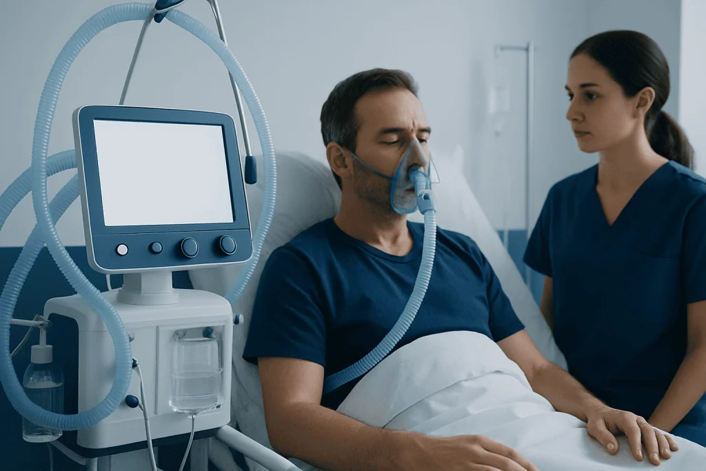
The symptoms of empyema can overlap with pneumonia or other chest illnesses, which is one reason diagnosis may not be immediate. Some people notice that they are not getting better after starting treatment for pneumonia, or that symptoms improve briefly and then worsen again.
Common symptoms may include:
- Fever or chills
- Chest pain, often worse with deep breathing
- Cough
- Shortness of breath
- Fatigue and weakness
- Loss of appetite
- Night sweats
- A general feeling of being unwell
Chest pain from empyema is often described as sharp or aching and may be felt on one side. Shortness of breath can happen because the infected fluid takes up space and limits lung expansion. In older adults or people with weakened immune systems, symptoms may be less typical and can include confusion, low energy, or a slower-than-expected recovery from a recent infection.
Symptoms can vary depending on how much fluid is present, how long the infection has been there, and whether the fluid has become thick and compartmentalized. Because these symptoms can also occur with other lung conditions, medical assessment is important rather than trying to judge the cause at home.
Why empyema develops: causes and risk factors
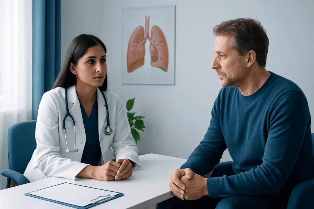
The most common cause of empyema is pneumonia. A bacterial infection in the lung can spread outward into the pleural space, where it triggers inflammation and causes fluid to collect. As germs and inflammatory cells build up, the fluid may become infected and turn into pus. In some patients, this process occurs in stages, beginning with thinner infected fluid and progressing to thicker, loculated collections and scar tissue.
Other possible causes include lung abscess, chest surgery, trauma to the chest, esophageal rupture, and the spread of infection from the bloodstream or nearby organs. Medical procedures involving the chest can rarely introduce infection. People with lung cancer or chronic lung disease may also be at increased risk because their lungs and pleural tissues may already be vulnerable.
Risk factors that can raise the chance of empyema include:
- Recent or severe pneumonia
- Older age
- Diabetes
- Weakened immune system
- Alcohol misuse
- Chronic lung disease
- Recent chest surgery or injury
- Difficulty swallowing or aspiration risk
Empyema itself is not usually a condition that starts without an underlying problem. For that reason, doctors often look not only at the infection around the lung, but also at the reason it developed in the first place. This helps guide treatment and reduce the risk of recurrence.
How doctors diagnose empyema
Diagnosis usually begins with a medical history and physical examination. A doctor will ask about recent pneumonia, fever, chest pain, cough, and breathing difficulties. On examination, breath sounds may be reduced over the affected area, and there may be signs of infection or respiratory distress.
Imaging is central to diagnosis. A chest X-ray may show fluid around the lung, but ultrasound and CT scanning are often more useful because they can show how much fluid is present and whether it is divided into pockets. In many cases, patients may also undergo CT scan imaging to help define the location and extent of the infection and plan drainage.
To confirm empyema, doctors often perform thoracentesis, a procedure that removes some pleural fluid with a needle. The fluid can then be tested for signs of infection, such as bacteria, white blood cells, low glucose, and changes in acidity. These laboratory findings help distinguish empyema from other causes of pleural fluid.
Blood tests may also be done to look for infection and inflammation, and cultures may help identify the responsible organism. In some situations, additional evaluation is needed to search for an underlying cause, especially if there is concern about complicated pneumonia, aspiration, or another lung condition such as pneumonia.
Treatment options and what recovery may involve
Treatment for empyema usually has two goals: clearing the infection and removing the infected fluid. Most patients need antibiotics, often started promptly and then adjusted if culture results identify a specific bacterium. The choice and duration of antibiotics depend on the severity of illness, the suspected source of infection, and how well the person responds.
Because antibiotics may not penetrate thick collections effectively, drainage is often an essential part of treatment. A doctor may remove fluid with a needle, but many people need a chest tube placed into the pleural space so the infection can drain over time. Imaging guidance can help place the tube accurately, especially when the fluid is loculated. In selected cases, procedures used in interventional radiology may support diagnosis or drainage planning.
If the fluid has become thick, divided into multiple pockets, or surrounded by fibrous tissue, more advanced treatment may be needed. This can include medication instilled into the pleural space to help break up loculations or surgery to drain the infection and peel away scar tissue. Some patients are treated with minimally invasive thoracic surgery, while others need a more extensive operation depending on the stage of disease.
Recovery depends on how early treatment begins, the underlying cause, and the person’s overall health. Follow-up imaging is often used to check that the fluid is resolving and the lung is re-expanding. For some patients, care may involve a team that includes pulmonology, infectious disease, radiology, and thoracic surgery specialists, including thoracic surgery when drainage procedures or operative treatment are required.
Prevention and self-care during recovery
The best way to reduce the risk of empyema is to lower the chance of severe chest infection and to seek prompt care for pneumonia symptoms. Not every case can be prevented, but timely treatment of lung infections can reduce the likelihood that infection spreads into the pleural space.
Practical preventive steps may include staying up to date with recommended vaccines, managing chronic conditions such as diabetes or lung disease, avoiding tobacco smoke, and getting medical advice if a cough, fever, or breathing problem is not improving. People at risk of aspiration may also benefit from assessment of swallowing and careful management of feeding and positioning.
During recovery, patients are usually advised to complete prescribed medicines exactly as directed and attend follow-up appointments. Rest, hydration, and gradual return to activity can support healing, but breathing difficulties or persistent fever should not be ignored. Chest discomfort may continue for some time even as the infection improves, so regular review can help confirm that recovery is on track.
In some cases, doctors may recommend breathing exercises or pulmonary rehabilitation support after a significant pleural infection, especially if there has been a long hospital stay. Near the end of treatment planning, patients seeking care across borders may wish to know that Acibadem International’s multidisciplinary specialists and JCI-accredited hospitals diagnose and treat empyema for international patients.
When to seek medical care
Medical care is important if symptoms suggest pneumonia or another chest infection that is not improving. A person should seek prompt assessment for ongoing fever, worsening cough, increasing chest pain, or shortness of breath, especially if these symptoms continue after starting antibiotics for pneumonia.
Urgent care is needed if breathing becomes difficult, lips or fingertips look bluish, confusion develops, or the person appears severely unwell. These symptoms do not always mean empyema, but they do signal that a chest infection or breathing problem may need immediate medical attention.
People who have recently had chest surgery, chest trauma, or a hospital stay should also contact a doctor if they develop fever or new respiratory symptoms. Early evaluation can help identify whether there is fluid around the lung, whether infection is present, and whether drainage or specialist treatment is needed.
Frequently asked questions
Is empyema the same as pneumonia?
No. Pneumonia is an infection in the lung tissue, while empyema is an infection in the pleural space around the lung. Empyema often develops as a complication of pneumonia, but the two conditions are not the same.
Can empyema go away with antibiotics alone?
Sometimes early pleural infection may improve with antibiotics, but many patients with empyema also need drainage of the infected fluid. Thick or loculated pus often does not clear well without a procedure. A doctor decides this based on symptoms, imaging, and fluid testing.
How serious is empyema?
Empyema is a condition that needs prompt medical care because the infection can interfere with breathing and may lead to complications if treatment is delayed. The outlook is often good when it is recognized early and treated appropriately. Recovery can take time, especially after severe infection.
What tests are used to confirm empyema?
Doctors usually use chest imaging such as X-ray, ultrasound, or CT, along with a procedure to sample the pleural fluid. Testing the fluid helps confirm infection and may identify the bacteria involved. Blood tests can also support the diagnosis.
Will surgery always be needed?
No. Many people are treated with antibiotics and chest drainage without major surgery. Surgery is more likely if the infection is advanced, divided into pockets, or if the lung is trapped by scar tissue.
Can empyema come back?
It can recur in some situations, especially if the underlying cause is not fully addressed or if drainage is incomplete. Follow-up care is important to make sure the infection has resolved and the lung has re-expanded. Managing risk factors such as recurrent aspiration or chronic lung disease may also help reduce recurrence.
References
- American Thoracic Society
- National Heart, Lung, and Blood Institute
- Centers for Disease Control and Prevention
- National Health Service
- Merck Manual Consumer Version
This article is for general information only and is not a substitute for professional medical advice. Please consult a qualified doctor about your individual situation.
Explore treatments in Turkey — costs, top hospitals & a free quote
JCI-accredited · board-certified surgeons · reply within 24h
Add us as a Preferred Source to see more of our trusted health content across Google Search, AI Overviews and Discover.