Visual Field Testing for Vision and Eye Health
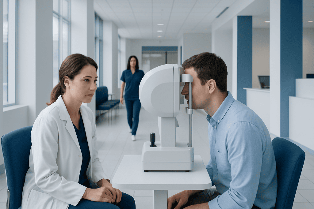
Visual field testing measures sensitivity across the full area a person can see while looking straight ahead. The test is especially important for diagnosing and monitoring glaucoma, which may affect side vision before noticeable symptoms occur.
Key Takeaways
- Visual field testing measures sensitivity across the full area a person can see while looking straight ahead.
- The test is especially important for diagnosing and monitoring glaucoma, which may affect side vision before noticeable symptoms occur.
- Results can be influenced by fatigue, dry eyes, poor concentration, eyelid position, and learning how to perform the test.
- An abnormal visual field result does not confirm a diagnosis by itself; eye examination findings and, when needed, imaging are considered together.
- The test is painless, usually takes only a few minutes per eye, and can be repeated to track change over time.
Visual field testing is a noninvasive eye examination that measures how well a person sees in central and peripheral areas of vision. It is commonly used to detect, assess, and monitor changes caused by glaucoma, retinal conditions, or problems affecting the optic nerve and brain.
What visual field testing measures
Visual field testing, also called perimetry, measures the complete area a person can see while keeping their eyes fixed on one point. This includes central vision, which supports reading and recognizing faces, and peripheral vision, which helps with walking safely, driving, and noticing movement to the side. The examination maps areas where vision is normal, reduced, or absent.
During the most common type of test, a person looks into a bowl-shaped instrument and presses a button whenever they notice small flashes of light. Some lights are brighter or dimmer than others. The pattern of responses helps the eye specialist identify whether any part of the visual field is less sensitive than expected.
Visual field testing does not measure how clearly a person sees letters on an eye chart. Instead, it assesses how well the eye and visual pathways detect light at many points. It is a useful functional test because some eye diseases can produce field changes before a person becomes aware of day-to-day vision loss.
Why an eye specialist may recommend this test
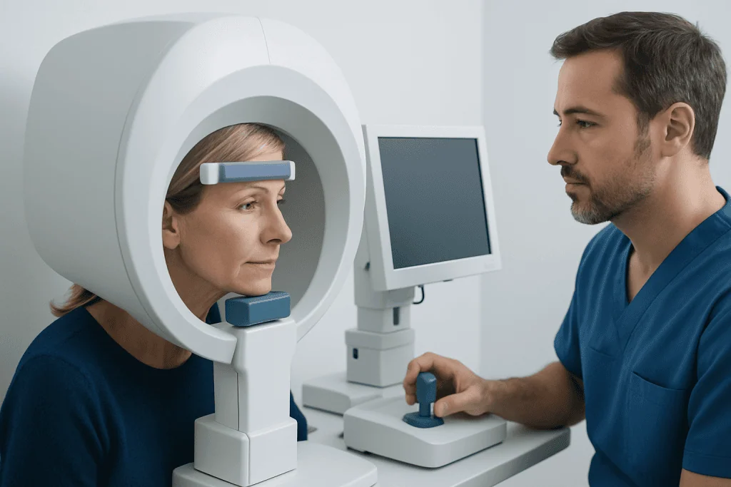
Visual field testing is most often used in the assessment and follow-up of glaucoma. In glaucoma, damage to the optic nerve can gradually create characteristic areas of reduced peripheral vision. A glaucoma assessment commonly combines visual field results with eye pressure measurement, optic nerve examination, and imaging of the retinal nerve fiber layer.
The test may also be recommended when there are signs of optic nerve disease, retinal disorders, or conditions affecting the brain pathways responsible for vision. For example, a stroke, pituitary gland problem, head injury, or other neurologic condition can sometimes cause loss affecting the same side of the visual field in both eyes. The shape and location of a defect can help guide further evaluation.
People may have a visual field test after reporting bumping into objects, missing objects to one side, difficulty navigating unfamiliar spaces, or a new shadow in vision. It may also be ordered as part of routine monitoring when an eye specialist sees changes in the optic nerve or identifies risk factors for glaucoma, even if vision seems normal.
What happens during a visual field test
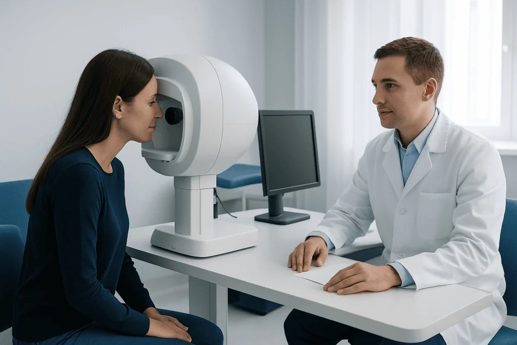
Before testing begins, the person usually sits comfortably with their chin and forehead supported by the machine. One eye is tested at a time while the other is covered. Corrective lenses may be used when appropriate, especially for tests focused on central vision. The technician explains where to look and when to press the response button.
In automated static perimetry, the most widely used format, small lights appear at different locations and brightness levels. The person should continue looking at the central fixation target and press the button whenever a light is seen. It is normal not to see every light, because some are intentionally very dim and a few may be presented to check the consistency of responses.
Other approaches include confrontation visual field testing, in which a clinician compares the patient’s side vision with their own, and kinetic perimetry, in which a moving target enters the field of view. These may be useful in selected situations, such as when a person cannot complete standard automated testing or when a broader field pattern is needed.
The examination is painless and does not involve radiation. It often takes several minutes for each eye, although the exact duration depends on the testing program and whether repeats are needed. Most people can resume usual activities immediately afterward.
How results are interpreted
A visual field report is a map rather than a simple pass-or-fail result. It shows light sensitivity at different points and compares the pattern with results expected for someone of a similar age. The report also includes reliability indicators, such as whether the person maintained fixation and whether responses suggest missed signals or responses given when no light was presented.
Specialists look for both the pattern and the consistency of any reduced areas. Glaucoma may cause defects that follow the arrangement of nerve fibers in the retina, while neurologic causes may produce a different, more symmetrical pattern across the two eyes. A single test rarely tells the whole story, particularly when changes are small.
First-time testing can be challenging because it requires attention and familiarity with the task. Tiredness, anxiety, dry eye symptoms, a drooping eyelid, small pupils, an uncorrected refractive error, or poor positioning can affect accuracy. For this reason, an eye specialist may repeat the test before concluding that a true change has occurred.
When a defect is confirmed, the next steps depend on the clinical context. These may include a detailed dilated eye examination, optic nerve imaging, measurement of eye pressure, retinal testing, or referral for neurologic assessment. Results should be interpreted by a qualified eye care professional rather than viewed in isolation.
Preparing for the appointment and improving test reliability
In most cases, no special preparation is necessary. People should bring their usual glasses, a current medication list, and information about previous eye tests if these were performed elsewhere. Contact lenses can often be worn, but the clinic may advise otherwise for particular examinations or if the eyes are irritated.
Getting adequate rest, eating normally, and allowing enough time for the appointment can make it easier to concentrate. If the eyes feel dry, uncomfortable, or unusually sensitive to light, telling the technician is helpful. The test can sometimes be paused briefly if needed, and the team can check whether the person is positioned comfortably.
During testing, the best approach is to look steadily at the central target without searching for lights. The person should press the button only when a light is genuinely noticed. Trying to anticipate flashes or worrying about missing them can make results less reliable; occasional missed lights are expected.
People who have difficulty maintaining position because of mobility concerns, tremor, hearing loss, language needs, or anxiety should mention this in advance. Clinics can often adapt instructions, pacing, and support to help patients complete the examination as accurately as possible.
What abnormal findings may mean and what treatment involves
An abnormal visual field finding means that an area of vision may be less sensitive than expected, but it does not automatically identify the cause. Some findings reflect test variability or correctable issues such as a refractive error or eyelid obstruction. Others may indicate a condition involving the eye, optic nerve, or visual pathways and need timely clinical review.
If glaucoma is diagnosed, the main goal of treatment is to lower eye pressure and reduce the risk of further optic nerve damage. Depending on the individual situation, management may include prescription eye drops, laser procedures, or glaucoma surgery. Treatment decisions are based on the full eye examination, disease stage, rate of change, general health, and the person’s preferences.
Visual field loss related to retinal, optic nerve, or neurologic disease is managed by addressing the underlying cause. This may require coordination between ophthalmology, neurology, endocrinology, or other specialties. Early assessment is valuable because some causes benefit from prompt treatment or monitoring.
For international patients, Acibadem International’s multidisciplinary specialists and JCI-accredited hospitals can evaluate visual field changes and coordinate appropriate eye and neurologic care. A visual field test may be one component of a broader eye evaluation, including glaucoma treatment when indicated.
When to seek medical care
People should arrange an eye examination if they notice persistent difficulty seeing to one side, frequent collisions with objects, new trouble with night driving, or a gradual narrowing of peripheral vision. These symptoms can have many causes, and an assessment can clarify whether visual field testing or other investigations are appropriate.
Urgent medical care is important for sudden vision loss, a new curtain or dark shadow across vision, sudden loss of vision on one side, new double vision, or vision changes accompanied by severe headache, weakness, numbness, speech difficulty, confusion, or facial drooping. These symptoms may require rapid assessment to protect vision and general health.
People already being monitored for glaucoma or optic nerve disease should attend scheduled follow-up visits, even when there are no noticeable symptoms. Visual field testing is particularly useful for comparing results over time and helping the clinician decide whether the current care plan remains appropriate.
Frequently asked questions
Is visual field testing painful?
No. Visual field testing is noninvasive and painless. It involves looking at a fixed target and responding when small lights are seen, without injections, radiation, or contact with the eye.
How long does a visual field test take?
Testing time varies by the type of examination and the program used. Many standard automated tests take several minutes per eye, with additional time for instructions, positioning, and rest if needed.
Can a visual field test detect glaucoma early?
Visual field testing can detect functional vision changes from glaucoma, including changes a person may not yet notice. However, early glaucoma may not always cause an identifiable field defect, so clinicians also assess eye pressure, the optic nerve, and imaging results.
Why might a visual field test need to be repeated?
Visual field testing depends on attention, comfort, and understanding of the task, so results can vary. Repeating a test helps confirm whether an apparent defect is genuine and allows the specialist to assess whether change is occurring over time.
Can anxiety or tiredness affect visual field testing?
Yes. Fatigue, anxiety, dry eyes, and trouble maintaining focus can make responses less consistent. Patients should tell the technician if they need a short pause or are experiencing discomfort.
Do dilating eye drops affect visual field test results?
In some circumstances, dilating drops or blurred near vision may affect the ability to see the test stimuli clearly. The clinic will determine the best order for examinations and provide instructions if drops are needed.
This article is for general information only and is not a substitute for professional medical advice. Please consult a qualified doctor about your individual situation.
Explore treatments in Turkey — costs, top hospitals & a free quote
JCI-accredited · board-certified surgeons · reply within 24h
Add us as a Preferred Source to see more of our trusted health content across Google Search, AI Overviews and Discover.

More from the Health Library
Anal Bleaching: An Evidence-Based Guide for Patients

Cheese — Explained by Medical Evidence, Not Myths

Motegrity: A Complete Medical Overview

Penises: A Complete Medical Overview

Tarsal: What Patients Need to Know