Bluish Mole — Explained by Medical Evidence, Not Myths

A bluish mole is often a benign blue nevus, not a bruise or trapped blood. Its blue appearance results from how light scatters through pigment located deeper in the skin.
Key Takeaways
- A bluish mole is often a benign blue nevus, not a bruise or trapped blood.
- Its blue appearance results from how light scatters through pigment located deeper in the skin.
- A spot that is new in adulthood, changing, uneven, painful, itchy, bleeding, or rapidly growing should be checked promptly.
- Dermatologists assess suspicious moles with clinical examination and dermoscopy; a biopsy may be needed for a definite diagnosis.
- Do not try to remove, burn, freeze, or cut off a bluish mole at home.
A bluish mole is commonly a blue nevus, a usually harmless mole that looks blue, blue-gray, or blue-black because pigment cells are located deeper in the skin. Most are stable and do not need treatment, but any new, changing, irregular, or symptomatic pigmented spot deserves a medical assessment.
What Is a Bluish Mole?
A bluish mole is a pigmented skin spot that appears blue, blue-gray, slate-colored, or blue-black. In many cases, it is a blue nevus, a benign growth made of melanocytes—the cells that produce melanin, the pigment responsible for skin, hair, and eye color. Blue nevi can be present from childhood or develop later, often as a single small spot.
The color does not usually mean that the mole contains blue pigment. Instead, melanin lies deeper in the skin than it does in many brown moles. Light passing through the skin is scattered in a way that makes the pigment look blue or gray to the eye. This is a normal optical effect and is not, by itself, a sign of cancer.
However, not every blue or dark spot is a blue nevus. Some skin cancers, including melanoma, can be dark blue-black or develop within an existing mole. A clinician can help distinguish a typical benign lesion from one that needs closer evaluation.
How a Blue Nevus May Look and Feel
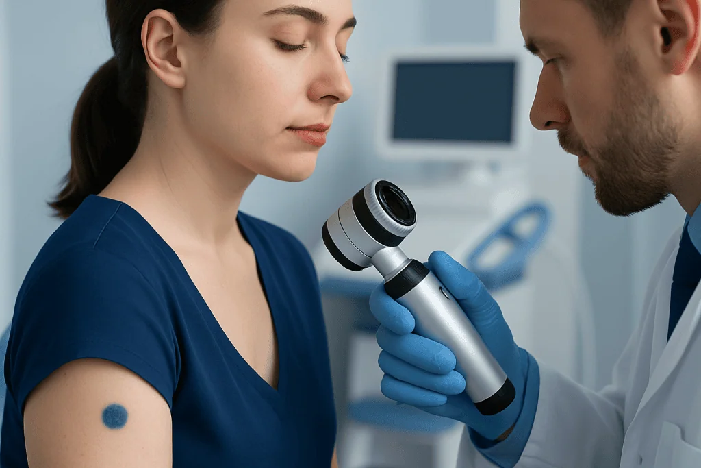
A typical blue nevus is round or oval, well defined, and usually smooth. It often measures only a few millimeters across and may be flat, slightly raised, or dome-shaped. Common locations include the backs of the hands and feet, scalp, face, buttocks, and lower legs, although it can occur almost anywhere on the body.
Many blue nevi have a uniform color, ranging from blue-gray to deep blue-black, and remain unchanged for years. They are usually painless and do not itch, bleed, crust, or form an open sore. Their stable appearance is one reason they are commonly benign.
There are different types of blue nevus. A common blue nevus is typically small and uniform. A cellular blue nevus may be larger, raised, and more often found on the buttocks, lower back, or scalp. Cellular blue nevi are still usually benign, but their appearance can overlap with other pigmented lesions, so professional assessment may be recommended.
Skin appearance alone cannot reliably establish a diagnosis. Lighting, skin tone, the location of the lesion, and whether a person has many moles can all affect how noticeable a bluish mole looks.
Why Bluish Moles Develop

Blue nevi develop when melanocytes are located in the deeper layer of the skin, called the dermis. The reason this happens is usually not known. They are not caused by poor hygiene, touching another person’s mole, or eating particular foods, and they are not contagious.
Some people may have a tendency to develop moles due to inherited factors, but most blue nevi occur sporadically. They can arise during childhood, adolescence, or adulthood. Hormonal changes may make existing skin pigment more noticeable in some people, but a new or changing lesion should not automatically be attributed to hormones.
Ultraviolet exposure from sunlight is an important risk factor for several forms of skin cancer and can contribute to overall mole and pigment changes. It is sensible to protect skin from excessive sun exposure, but sun exposure does not explain every blue nevus. A person should not assume that a dark spot is harmless simply because it appears after time outdoors.
A bruise can also look blue or purple, but it usually follows injury and fades over days to weeks. A bluish mole generally remains in the same place and does not fade like a bruise. If there is uncertainty, a medical review is safer than relying on appearance alone.
How Doctors Evaluate a Bluish Mole
A dermatologist or another qualified clinician begins by asking when the spot appeared, whether it has changed, and whether there are symptoms such as itching, tenderness, bleeding, or crusting. They may also ask about personal or family history of melanoma, previous skin cancers, severe sunburns, immune suppression, and the number of moles a person has.
The skin examination includes looking at the mole’s size, shape, borders, color pattern, and surrounding skin. A handheld device called a dermatoscope can magnify the lesion and reveal structures below the skin surface. Dermoscopy often helps clinicians recognize features of a typical blue nevus and identify lesions that require further testing.
If the diagnosis remains uncertain, or if the mole has concerning features, the clinician may recommend a biopsy or complete removal. The tissue is examined under a microscope by a pathologist. This is the most reliable way to determine whether a lesion is benign or whether further care is needed.
Photographs and mole mapping may be useful for people with numerous moles or lesions that need monitoring. A photo taken at home can also help document change, but it should support—not replace—a clinical examination. Evaluation of suspicious lesions is part of skin cancer assessment and early detection care.
Treatment and Monitoring Options
A confirmed, typical blue nevus often needs no treatment. If it is stable, symptom-free, and has reassuring clinical features, a clinician may advise simple observation. People can become familiar with the appearance of their skin and report meaningful changes rather than checking a mole repeatedly or anxiously.
Removal may be considered if a lesion is difficult to diagnose, is changing, repeatedly irritated by clothing or shaving, or causes cosmetic concern. The method depends on the lesion’s size, depth, and location. Excision allows the removed tissue to be examined, which is particularly important when melanoma must be ruled out.
It is not safe to remove a mole with over-the-counter acids, home freezing products, cutting tools, or other home remedies. These methods can cause burns, scarring, infection, or delay diagnosis of a potentially serious lesion. Professional dermatology evaluation provides a safer route when removal or testing is appropriate.
If a biopsy identifies melanoma or another skin cancer, care is individualized and may involve surgery and, in some circumstances, additional treatments and follow-up. The diagnosis of melanoma cannot be made from a photograph or description alone, which is why timely examination of suspicious spots matters.
Skin Awareness and Everyday Prevention
There is no proven way to prevent every mole, but regular sun protection supports long-term skin health. This includes seeking shade when practical, wearing protective clothing and a wide-brimmed hat, and using broad-spectrum sunscreen on exposed skin as directed. Avoiding tanning beds is also important because they expose skin to ultraviolet radiation.
A practical approach is to check the skin periodically in good light, including the scalp, soles of the feet, nails, and areas not easily seen. A partner or clinician can help inspect the back and scalp. The goal is not to diagnose a mole at home, but to notice a spot that is new or different from the rest.
The ABCDE guide can help identify changes worth discussing with a clinician:
- Asymmetry: one half does not resemble the other.
- Border irregularity: edges are uneven, blurred, or notched.
- Color variation: more than one color or a changing color.
- Diameter: a lesion that is enlarging, especially beyond about 6 mm.
- Evolving: any ongoing change in size, shape, color, or symptoms.
These signs are guides, not diagnostic rules. Some harmless moles may meet one criterion, and some melanomas may be smaller or look different. An “ugly duckling”—a spot that clearly looks unlike a person’s other moles—also warrants attention.
When to Seek Medical Care
A person should arrange a medical appointment for a bluish mole that is new in adulthood, noticeably enlarging, becoming raised, changing color, developing uneven borders, or looking different from other moles. Prompt assessment is also appropriate if it bleeds without clear injury, forms a crust or sore, becomes painful, or has persistent itching.
It is especially important to seek advice sooner for anyone with a personal history of melanoma or other skin cancer, a strong family history of melanoma, many atypical moles, or a weakened immune system. These factors do not mean a bluish mole is cancerous, but they can lower the threshold for professional review.
Most bluish moles will prove benign, and seeking assessment is a sensible precaution rather than a reason to expect the worst. Acibadem International’s multidisciplinary specialists and JCI-accredited hospitals assess and treat skin conditions for international patients when expert evaluation is needed.
Frequently asked questions
Is a bluish mole usually cancerous?
No. A bluish mole is often a benign blue nevus, especially when it is small, uniform, and unchanged over time. Still, color alone cannot rule out melanoma, so a new or changing blue-black spot should be assessed by a qualified clinician.
Why does a mole look blue instead of brown?
A blue nevus looks blue because melanin is located deeper in the skin. The way light scatters through the skin changes the apparent color, even though the pigment itself is not truly blue.
Can a blue nevus turn into melanoma?
Most blue nevi remain benign and stable. Melanoma arising in association with a blue nevus is rare, but changes in size, shape, color, surface, or symptoms should be examined rather than assumed to be harmless.
Should a stable bluish mole be removed?
A stable mole with typical benign features often does not need removal. A clinician may recommend removal if diagnosis is uncertain, the lesion changes or is irritated, or the person wants it removed for cosmetic reasons.
How can someone tell a bluish mole from a bruise?
A bruise usually appears after pressure or injury and gradually changes color and fades within days or weeks. A bluish mole typically remains in the same place with a more consistent shape and color; uncertainty should be discussed with a clinician.
Can a dermatologist diagnose a blue mole without a biopsy?
A dermatologist can often make a likely diagnosis using a skin examination and dermoscopy. If the lesion has unusual features or cannot be confidently identified, a biopsy may be recommended to obtain a definite diagnosis.
This article is for general information only and is not a substitute for professional medical advice. Please consult a qualified doctor about your individual situation.
Explore treatments in Turkey — costs, top hospitals & a free quote
JCI-accredited · board-certified surgeons · reply within 24h
Add us as a Preferred Source to see more of our trusted health content across Google Search, AI Overviews and Discover.

More from the Health Library

BMI Chart Women: A Complete Medical Overview

Iliac: An Evidence-Based Guide for Patients

Ataxia Telangiectasia: A Complete Medical Overview

Coffee Ground Emesis — Explained by Medical Evidence, Not Myths

HPV in Men: What Patients Need to Know