Malignant Melanoma Images: Early Signs, Risk Factors, and How It Is Treated

Melanoma may develop in an existing mole or appear as a new spot on otherwise normal-looking skin. Images are educational tools, but a skin examination and biopsy are needed to diagnose melanoma.
Key Takeaways
- Melanoma may develop in an existing mole or appear as a new spot on otherwise normal-looking skin.
- Images are educational tools, but a skin examination and biopsy are needed to diagnose melanoma.
- The ABCDE rule and the “ugly duckling” sign can help people notice concerning changes.
- Ultraviolet exposure, a history of sunburn, fair skin, many moles, and family history can increase risk.
- Early assessment matters because melanoma is often more manageable when found before it has spread.
Malignant melanoma images can be useful for learning common warning patterns, but photographs cannot confirm whether a mole or skin mark is cancerous. Any new, changing, unusual, or bleeding spot should be assessed promptly by a qualified clinician, ideally a dermatologist.
What malignant melanoma images can—and cannot—show
Malignant melanoma images commonly show a dark or multicolored skin mark with an uneven shape, irregular edges, or a pattern that looks different from a person’s other moles. They may help people recognize changes worth checking, including a new spot that grows or a long-standing mole that begins to change. However, melanoma can look very different from one person to another, including on different skin tones.
A photograph cannot diagnose melanoma. Benign moles, harmless pigmented spots, inflamed skin lesions, and other forms of skin cancer can sometimes resemble melanoma. Conversely, some melanomas are subtle and may not look like the examples commonly shown online. A clinician examines the lesion directly, often with a dermatoscope, and confirms the diagnosis through a biopsy when appropriate.
Melanoma is a cancer that begins in melanocytes, the cells that make pigment in the skin. It can occur anywhere on the body, including areas with limited sun exposure such as the soles of the feet, palms, under the nails, scalp, eyes, and genital area. For a broader overview, readers can visit melanoma information.
How to read melanoma warning patterns
Clinicians often use the ABCDE guide to help patients remember changes that deserve attention. “A” stands for asymmetry, meaning one half of a spot does not match the other. “B” refers to a border that is uneven, notched, blurred, or poorly defined. “C” means color variation, such as shades of brown, black, tan, red, pink, blue, gray, or white within one lesion.
“D” is diameter: a spot larger than about 6 millimeters can warrant review, although melanomas can be smaller. “E” is evolving, which is especially important. Evolution may include a change in size, shape, color, surface texture, sensation, or bleeding. A spot that persists and changes over weeks or months should not be ignored.
Another useful concept is the “ugly duckling” sign. Most people’s normal moles tend to share a general pattern. A lesion that looks noticeably different from the others—for example, much darker, more raised, pinker, or more irregular—may need examination. Taking well-lit, dated photographs for personal comparison can be helpful, but this should complement rather than replace professional skin checks.
- New pigmented mark in adulthood
- Mole that is enlarging or changing appearance
- Spot that itches, hurts, crusts, or bleeds without clear injury
- Dark streak or discoloration under a nail, particularly if widening
- Non-healing patch or nodule that differs from nearby skin
Melanoma appearances on different skin tones and body areas

Melanoma can occur in every skin tone. In people with darker skin, it may be diagnosed later when changes are less easily noticed or when the lesion develops in a less visible location. It is important to check the entire body rather than focusing only on sun-exposed areas.
Acral lentiginous melanoma is a subtype that can develop on the palms, soles, or beneath fingernails and toenails. It may appear as a slowly enlarging dark patch, an irregular brown-to-black area, or a longitudinal dark streak in a nail. A new or changing nail streak should be assessed, especially if pigment extends onto the surrounding skin or the nail becomes distorted.
Some melanomas contain little or no dark pigment. These are called amelanotic melanomas and can appear pink, red, skin-colored, or as a persistent scaly or raised area. Because their appearance may resemble eczema, a pimple, a scar, or another benign condition, any lesion that continues to grow, bleed, or fail to heal deserves medical attention.
Risk factors and what causes melanoma
Melanoma develops when genetic changes allow melanocytes to grow abnormally. Ultraviolet radiation from sunlight and artificial tanning devices is an important avoidable risk factor. Intense intermittent sun exposure and blistering sunburns, particularly earlier in life, are associated with increased risk, although melanoma can develop without a known history of heavy sun exposure.
Other factors that may increase risk include having fair skin that burns easily, light eyes or hair, freckles, many common moles, atypical moles, a personal history of melanoma or other skin cancers, and a close family history of melanoma. A weakened immune system can also increase the risk of several skin cancers. These factors do not mean a person will develop melanoma, and people without them can still be affected.
Regular awareness is useful for everyone. People with multiple risk factors may benefit from discussing an individualized skin-surveillance plan with a dermatologist. This may include professional full-skin examinations and guidance on monitoring specific moles over time.
How melanoma is diagnosed and staged
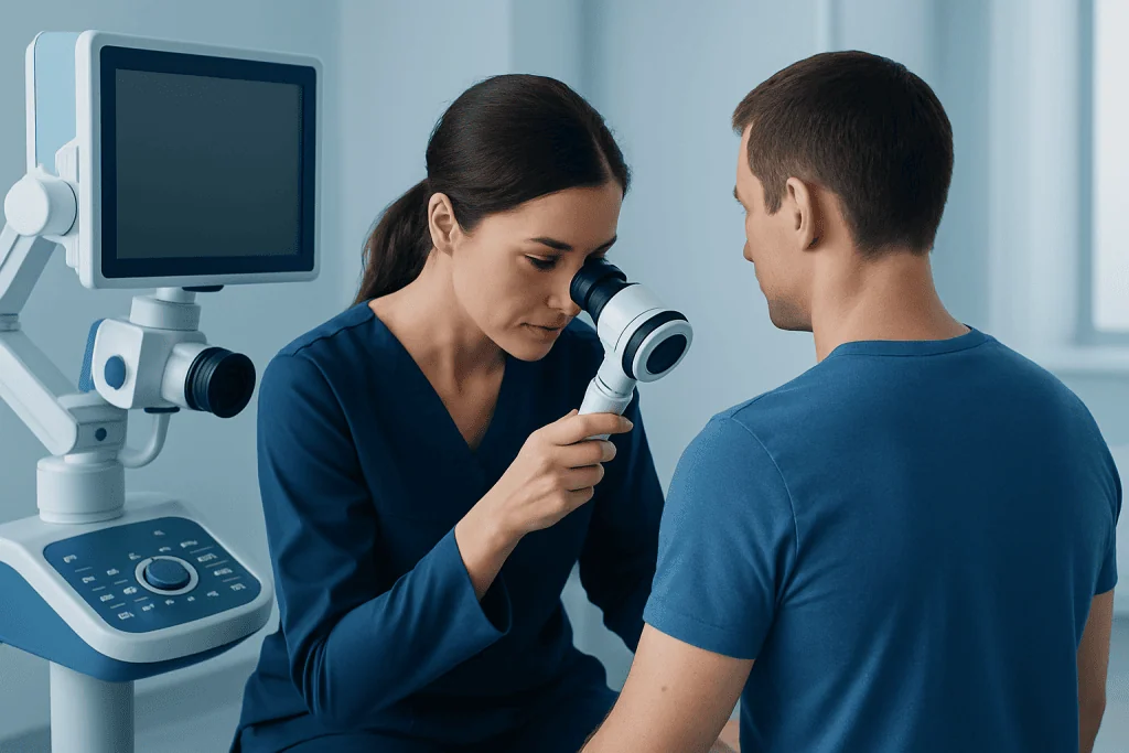
Diagnosis begins with a medical history and a careful skin examination. A dermatologist may use dermoscopy, a handheld device that magnifies skin structures and helps evaluate pigment patterns that are not visible to the naked eye. Dermoscopy supports clinical judgment but does not replace tissue testing when a lesion is concerning.
If melanoma is suspected, the clinician usually removes all or part of the lesion in a biopsy. A pathologist examines the sample under a microscope to determine whether melanoma is present and, if so, to describe features that guide treatment. These features may include tumor thickness, ulceration, and how actively cells are dividing.
After diagnosis, staging assesses whether the cancer appears limited to the original skin site or has spread to lymph nodes or other organs. Depending on the melanoma’s features, this may involve a physical examination, lymph node assessment, sentinel lymph node biopsy, and selected imaging tests. Staging helps the care team recommend the most suitable approach and follow-up schedule.
Treatment options for malignant melanoma
For melanoma that is confined to the skin, surgery is generally the main treatment. The surgeon removes the melanoma along with a margin of surrounding normal-looking skin. The size of that margin is based on the depth and location of the melanoma. In many early cases, surgery may be the only treatment needed.
For selected melanomas with a higher likelihood of reaching nearby lymph nodes, a sentinel lymph node biopsy may be discussed at the time of surgery. If melanoma has spread or carries a higher risk of recurrence, treatment may include immunotherapy, targeted therapy for tumors with particular gene changes, radiation therapy in selected circumstances, or participation in clinical trials. The treatment plan is individualized according to the stage, tumor biology, general health, and patient preferences.
Care may involve dermatology, surgical oncology, medical oncology, pathology, radiology, and other specialists. Information about skin cancer treatment can help patients understand the multidisciplinary approaches used for different types and stages of skin cancer. Acibadem International’s multidisciplinary specialists and JCI-accredited hospitals diagnose and treat melanoma for international patients.
Prevention, skin awareness, and follow-up care
Sun protection can reduce exposure to ultraviolet radiation. Practical measures include seeking shade when the sun is strongest, wearing protective clothing and a broad-brimmed hat, using sunglasses, and applying broad-spectrum sunscreen with an SPF of 30 or higher to exposed skin. Sunscreen should be reapplied as directed, especially after swimming, sweating, or towel drying. Indoor tanning devices should be avoided.
Monthly self-checks can help people become familiar with their normal skin. A full check may include the face, ears, scalp, neck, trunk, arms, hands, legs, feet, soles, and the skin between the toes. Mirrors or help from a trusted person can make it easier to inspect the back, scalp, and other difficult-to-see areas.
After melanoma treatment, follow-up appointments are important because recurrence and new skin cancers can occur. The frequency and content of follow-up depend on the melanoma stage and individual circumstances. A care team can explain which symptoms to report and how often professional skin examinations are recommended.
When to seek medical care
A person should arrange a prompt medical assessment for a new or changing mole, an “ugly duckling” spot, or a lesion that bleeds, crusts, itches persistently, becomes painful, or does not heal. It is also sensible to seek care for a new dark mark on the palm, sole, or under a nail, particularly if it is enlarging or irregular.
Most skin changes are not melanoma, and seeking an assessment does not mean cancer is likely. However, early evaluation can provide reassurance when a lesion is benign and allows timely treatment when it is needed. People with a previous melanoma, many atypical moles, or a strong family history should ask a dermatologist about appropriate regular surveillance.
Frequently asked questions
Can malignant melanoma images diagnose a suspicious mole?
No. Images can illustrate warning features, but many harmless lesions can look concerning and some melanomas may appear subtle. A clinician needs to examine the skin lesion and may recommend a biopsy to confirm or exclude melanoma.
What does early melanoma usually look like?
Early melanoma may look like a new or changing mole with asymmetry, an irregular border, more than one color, or ongoing evolution. It may also appear as a pink, red, or skin-colored lesion rather than a dark spot. A lesion that looks different from a person’s other moles should be checked.
Can melanoma be flat?
Yes. Many early melanomas are flat patches or macules rather than raised lumps. A flat spot that enlarges, develops irregular edges, or changes in color can still require evaluation.
Is every dark line under a nail melanoma?
No. Nail streaks may result from harmless pigment changes, injury, medications, or other causes. Still, a new, widening, irregular, or changing dark streak, especially one extending onto surrounding skin, should be examined by a clinician.
How quickly can melanoma change?
The rate of change varies. Some melanomas evolve gradually over months or years, while others may become noticeable more quickly. Any persistent change in a mole or new skin lesion should be reviewed rather than monitored indefinitely at home.
What happens if a doctor suspects melanoma?
The doctor will examine the lesion, often using dermoscopy, and usually recommends a biopsy if it has concerning features. A pathologist examines the tissue sample to establish the diagnosis. If melanoma is found, further evaluation helps determine the stage and appropriate treatment.
References
- American Academy of Dermatology
- American Cancer Society
- National Cancer Institute
- World Health Organization
- National Comprehensive Cancer Network
This article is for general information only and is not a substitute for professional medical advice. Please consult a qualified doctor about your individual situation.
Melanoma in Turkey — costs, top hospitals & a free quote
JCI-accredited · board-certified surgeons · reply within 24h
Add us as a Preferred Source to see more of our trusted health content across Google Search, AI Overviews and Discover.

Medical Oncology Department
Medical treatment of cancer with chemotherapy, immunotherapy and targeted therapies under a multidisciplinary tumor board.
60 specialists in this unit