How Hernia Is Diagnosed: Tests, Imaging & What to Expect

Many hernias can be diagnosed during a focused physical examination. Ultrasound is commonly used for a suspected groin or abdominal-wall hernia when the finding is not clear on examination.
Key Takeaways
- Many hernias can be diagnosed during a focused physical examination.
- Ultrasound is commonly used for a suspected groin or abdominal-wall hernia when the finding is not clear on examination.
- CT and MRI provide more detailed images and may help identify small, recurrent or complex hernias.
- A painful bulge that cannot be pushed back, especially with vomiting or abdominal swelling, needs urgent medical assessment.
- Diagnosis helps the surgical team determine whether monitoring, planned repair or urgent treatment is appropriate.
Hernia diagnosis is often made through a medical history and physical examination, particularly when a bulge becomes more noticeable with standing, coughing or straining. Imaging tests such as ultrasound, CT or MRI are used when the examination is unclear, symptoms suggest a complication, or the hernia may be located deeper in the abdomen or groin.
Overview: How Hernia Diagnosis Works
Hernia diagnosis usually begins with a clinician listening to the person’s symptoms and examining the area of concern. A hernia occurs when tissue, often fat or part of the intestine, pushes through a weak point in muscle or connective tissue. This may produce a visible or felt lump in the groin, abdomen, around a previous surgical scar, or less commonly in other locations.
In many cases, a diagnosis can be made without scans. The clinician may ask the person to stand, cough, strain gently or change position because these actions increase pressure inside the abdomen and can make a hernia easier to detect. Imaging is helpful when symptoms are present but no lump is clear, when a hernia is deep or recurrent, or when another condition may be causing similar discomfort.
The purpose of assessment is not only to confirm whether a hernia is present. It also helps identify the likely type, location and size of the hernia, assess whether its contents can move back into the abdomen, and look for signs that prompt treatment is needed. For an overview of hernia types and management, readers can review hernia information.
Symptoms and History the Clinician Will Ask About
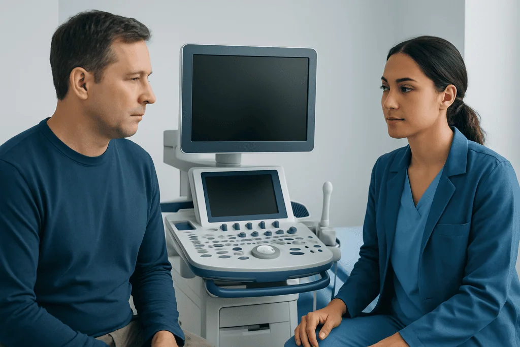
A hernia may cause a bulge, heaviness, pressure, pulling sensation or localized discomfort. Symptoms often become more apparent during lifting, coughing, exercise, prolonged standing or bowel movements. Some people notice a lump that disappears when lying down, while others have discomfort without an obvious bulge.
During the consultation, the clinician will ask when symptoms began, whether the lump changes in size, and what activities bring it on or relieve it. They may also ask about nausea, vomiting, constipation, fever, reduced appetite, difficulty passing stool or gas, and whether the bulge can be gently reduced. These details can help distinguish a straightforward hernia from a possible complication.
Medical history matters as well. Previous abdominal or pelvic surgery, an earlier hernia repair, pregnancy, chronic cough, constipation, urinary straining, heavy physical work, obesity and conditions affecting connective tissue can all be relevant. The clinician will also consider medicines, smoking status and any health conditions that could influence future treatment planning.
Physical Examination for a Suspected Hernia

The physical examination is the central part of hernia diagnosis. It is usually performed in a private setting and involves inspection and gentle palpation of the abdomen, groin or scar area. The person may be examined while standing and lying down, as some hernias become less visible when abdominal pressure falls.
The clinician may ask the person to cough, take a deep breath or perform a gentle bearing-down movement. This can reveal an impulse or bulge at a weak point in the abdominal wall. In suspected inguinal hernia, the groin and nearby structures are assessed carefully; examination techniques are adapted to the person’s anatomy, symptoms and comfort.
The clinician may also check whether a bulge is reducible, meaning it returns into the abdomen with gentle pressure or when lying down. A non-reducible bulge is not always an emergency, but it requires careful assessment. Severe tenderness, skin color change, marked abdominal distension or symptoms of bowel obstruction can indicate that urgent evaluation is needed.
Imaging Tests: Ultrasound, CT and MRI
Imaging is not necessary for every hernia. It is most useful when the examination does not provide a clear answer, when symptoms persist despite a normal examination, or when a deeper, small, recurrent or complicated hernia is suspected. The most suitable scan depends on the body area involved, the clinical question and the person’s overall health.
Ultrasound uses sound waves to create real-time images and does not involve radiation. It is often used to assess a suspected groin or abdominal-wall hernia, especially when a lump appears only with movement or straining. The sonographer may ask the person to stand, cough or tighten the abdominal muscles so the area can be assessed dynamically.
CT scanning uses X-rays and computer processing to show detailed cross-sectional images of the abdomen and pelvis. It may be recommended when pain is significant, a complex abdominal-wall hernia is suspected, prior surgery has changed the anatomy, or there is concern about bowel obstruction or another abdominal condition. Contrast dye may be used in some cases, depending on the reason for the scan.
MRI provides detailed soft-tissue images without ionizing radiation. It can be valuable for certain occult groin hernias, athletic pubalgia-like symptoms, pelvic conditions or situations where ultrasound and CT have not clarified the cause of pain. Access to timely MRI or other imaging can support a more efficient diagnostic pathway when clinically appropriate.
Other Tests and Conditions That Can Resemble a Hernia
There is no routine blood test that confirms a hernia. However, blood tests may be ordered if a person is unwell, has severe pain, fever, vomiting, or possible infection or bowel obstruction. These tests can help the medical team understand the person’s general condition and plan urgent care safely when needed.
Several conditions can cause groin or abdominal lumps or pain. Depending on the location and symptoms, the clinician may consider enlarged lymph nodes, lipoma, muscle strain, fluid collections, testicular or scrotal conditions, ovarian or pelvic conditions, varicose veins, or bowel and urinary problems. Imaging can be especially useful when the diagnosis is uncertain.
Not all groin pain is caused by a hernia, and not every abdominal bulge needs surgery. A careful evaluation avoids unnecessary treatment while ensuring that a true hernia is appropriately monitored or managed. If a diagnosis remains unclear, a referral to a general surgeon or another relevant specialist may be recommended.
What Happens After Diagnosis: Treatment Planning
Once a hernia is confirmed, the next step is to decide whether observation, planned repair or urgent treatment is best. This decision takes account of the hernia type, symptoms, size, ability to reduce the bulge, impact on daily life, work and exercise, previous repairs, and the person’s general health. Some minimally symptomatic hernias may be monitored with medical guidance, while symptomatic or enlarging hernias are often assessed for repair.
Hernia repair aims to return protruding tissue to its usual position and strengthen the weak area. Depending on the hernia and the individual, surgery may be performed through an open incision or with minimally invasive laparoscopic or robotic techniques. Mesh is commonly used in many repairs to reinforce tissue, although the approach is individualized.
For patients considering a procedure, hernia surgery information can explain treatment approaches and preparation in more detail. Candidacy is determined after review of symptoms, examination findings, imaging where needed, medical history and anesthesia considerations. The surgical team will discuss expected benefits, alternatives and potential risks before a decision is made.
On the day of surgery, the team confirms the plan, provides anesthesia as appropriate, performs the repair and monitors recovery before discharge. Many uncomplicated repairs are performed as day procedures, although the timeline varies by hernia type, technique, health status and whether surgery is urgent or planned.
Recovery, Benefits and Possible Risks of Hernia Repair
Recovery after hernia repair varies. People are usually encouraged to walk gently soon after surgery, while gradually returning to normal activities according to their surgeon’s advice. Mild pain, bruising, swelling and tiredness can occur in the early recovery period. Desk-based work may be possible sooner than strenuous work, but individual guidance is important.
The main potential benefit of repair is relief of hernia-related symptoms and prevention of further enlargement or complications. Surgery may also improve confidence with movement and everyday activity for people who are limited by discomfort. However, no procedure is right for every person at every time, which is why diagnosis and shared decision-making are important.
Potential risks include bleeding, infection, fluid collection, temporary urinary difficulty, injury to nearby structures, persistent discomfort, recurrence and anesthesia-related complications. The likelihood and relevance of these risks differ by procedure and patient factors. A surgeon can explain how they apply to an individual situation and how recovery instructions can reduce avoidable problems.
Acibadem International’s multidisciplinary specialists and JCI-accredited hospitals assess and treat hernias for international patients, including diagnostic imaging and surgical planning when indicated.
When to Seek Medical Care
A person should arrange a medical appointment for a new or persistent groin, abdominal or scar-area bulge, particularly if it grows, causes discomfort, affects activity or is associated with digestive symptoms. Early assessment can clarify the cause and help determine whether monitoring or treatment is appropriate.
Urgent medical care is important if a known or suspected hernia becomes suddenly very painful, firm, tender or unable to be pushed back when it was previously reducible. Vomiting, fever, worsening abdominal swelling, inability to pass stool or gas, or red, purple or dark skin over the bulge also require prompt evaluation because they may indicate trapped or compromised tissue.
People should avoid trying to force a painful bulge back into place. They should seek professional advice instead, especially if symptoms are new, severe or worsening. A qualified clinician can assess the urgency and arrange appropriate examination and imaging.
Frequently asked questions
Can a doctor diagnose a hernia without a scan?
Yes. Many hernias are diagnosed through symptoms and a physical examination, particularly when a bulge becomes noticeable with coughing, standing or straining. A scan is usually considered when the examination is uncertain, the hernia may be deeper, or another cause of symptoms needs to be excluded.
What is the best imaging test for a hernia?
There is no single best test for every situation. Ultrasound is often useful for suspected groin and abdominal-wall hernias, while CT can provide detailed information in complex or urgent abdominal cases. MRI may help detect certain small or occult hernias and evaluate soft tissues in greater detail.
Does a hernia ultrasound hurt?
Ultrasound is generally painless and does not use radiation. The probe may apply gentle pressure over a tender area, and the sonographer may ask the person to cough or strain briefly to make a hernia more visible.
Can a CT scan show a hernia that cannot be felt?
Yes, CT can identify many hernias that are not obvious on examination, particularly deeper abdominal-wall or internal hernias. It can also show surrounding structures and help assess for bowel obstruction or other causes of abdominal pain.
What should someone do before a hernia imaging test?
Preparation depends on the scan. Ultrasound often requires little or no preparation, while CT or MRI instructions can vary depending on the body area and whether contrast is planned. The imaging department will provide specific guidance about eating, drinking, medicines and metal items before the appointment.
Is a painful hernia always an emergency?
Not always, but new or worsening pain should be assessed by a clinician. Sudden severe pain, a hard or non-reducible bulge, vomiting, fever, abdominal swelling, or inability to pass stool or gas may indicate an urgent problem and require immediate medical care.
References
- National Institute of Diabetes and Digestive and Kidney Diseases
- National Health Service
- American College of Surgeons
- European Hernia Society
- Radiological Society of North America
This article is for general information only and is not a substitute for professional medical advice. Please consult a qualified doctor about your individual situation.
Persistent digestive symptoms? Get evaluated in Turkey
JCI-accredited · board-certified surgeons · reply within 24h
Add us as a Preferred Source to see more of our trusted health content across Google Search, AI Overviews and Discover.