Upj Stone: What Patients Need to Know

A UPJ stone sits at the junction between the kidney and ureter, where even a small stone can interfere with urine flow. Typical symptoms include sudden flank pain, nausea, blood in the urine, and urinary urgency, although some stones cause few symptoms.
Key Takeaways
- A UPJ stone sits at the junction between the kidney and ureter, where even a small stone can interfere with urine flow.
- Typical symptoms include sudden flank pain, nausea, blood in the urine, and urinary urgency, although some stones cause few symptoms.
- Urine and blood tests plus imaging help confirm the stone, assess obstruction, and identify infection or reduced kidney function.
- Treatment depends on stone size, location, symptoms, infection risk, kidney drainage, and the person’s overall health.
- Fever or chills with suspected kidney stone symptoms requires urgent medical assessment because it may indicate an infected obstruction.
A UPJ stone is a kidney stone located at the ureteropelvic junction (UPJ), the narrow area where urine leaves the kidney and enters the ureter. Because this location can obstruct urine drainage, evaluation is important, especially when pain, fever, vomiting, or reduced urine output occurs.
What Is a UPJ Stone?
A UPJ stone is a urinary stone lodged at the ureteropelvic junction, often shortened to UPJ. This is the point where the renal pelvis, the urine-collecting area inside the kidney, narrows to become the ureter, the tube that carries urine to the bladder. A stone at this narrow passage may slow or block the flow of urine from the kidney.
Most UPJ stones are kidney stones that formed from minerals and salts in concentrated urine, then moved into this outlet area. The term describes the stone’s location rather than its chemical type. Stones may contain calcium compounds, uric acid, struvite, cystine, or a combination of materials.
When urine cannot drain normally, the kidney may become stretched, a finding called hydronephrosis. A temporary blockage can be painful but is often treatable. However, persistent obstruction or obstruction combined with infection can threaten kidney health, so a suspected UPJ stone should be assessed by a qualified clinician.
How a UPJ Stone May Feel
The most familiar symptom is renal colic: sudden, intense pain in the side or back below the ribs. Pain may come in waves as the ureter contracts around the stone. It can travel toward the lower abdomen, groin, or genital area, although the exact pain pattern varies from person to person.
Other possible symptoms include nausea, vomiting, sweating, restlessness, burning during urination, frequent urination, or visible or microscopic blood in the urine. Blood may make urine look pink, red, or brown, but normal-looking urine does not rule out a stone.
Some UPJ stones are found incidentally on imaging performed for another reason. A stone may cause little pain when it is not obstructing drainage. Symptoms also do not reliably show a stone’s size, so imaging is important when a clinician suspects obstruction or another cause of pain.
- Side, back, or upper abdominal pain
- Nausea or vomiting
- Blood in the urine
- Urinary frequency, urgency, or discomfort
- Cloudy or foul-smelling urine, particularly if infection is present
Why Stones Form at the UPJ

A stone can lodge at the UPJ because it is a naturally narrow transition point. Kidney stones develop when urine contains more crystal-forming substances than the fluid can dilute, allowing crystals to join and grow. Low fluid intake is a common contributor, especially in hot environments or during illnesses that cause fluid loss.
Risk factors differ according to stone type. They may include a personal or family history of stones, high dietary salt intake, diets high in animal protein, obesity, certain bowel disorders or surgeries, recurrent urinary infections, gout, and some medications. Metabolic conditions that alter calcium, uric acid, oxalate, or citrate in the urine can also increase risk.
An anatomical narrowing at the UPJ, known as ureteropelvic junction obstruction, may sometimes affect urine drainage and be identified during evaluation. This is not the same as a UPJ stone, although both can contribute to hydronephrosis. Clinicians consider anatomy, imaging findings, and the stone’s position before deciding whether the stone alone explains the obstruction.
How Doctors Diagnose a UPJ Stone
Diagnosis begins with a discussion of symptoms, medical history, medications, previous stones, infections, and family history. A clinician will also perform an examination to look for flank tenderness, dehydration, fever, or signs that another condition may be causing the pain.
A urine test can identify blood, signs of infection, crystals, and urine acidity. Blood tests may assess kidney function, inflammation, and factors relevant to stone formation. If infection is suspected, a urine culture may be needed to identify the responsible bacteria and guide antibiotic treatment.
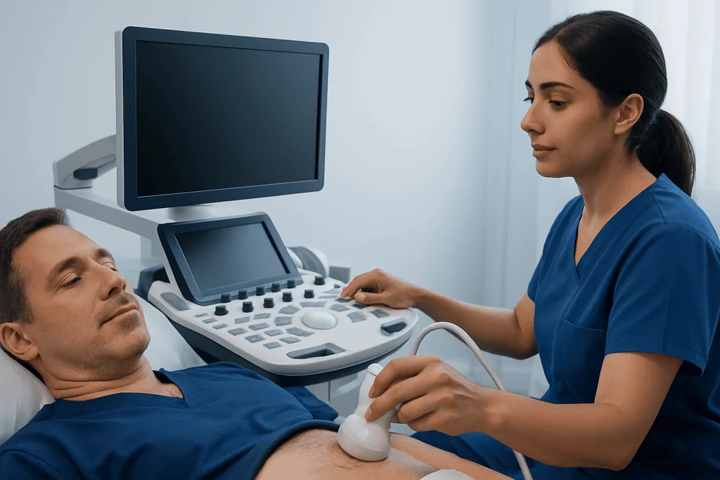
Imaging shows the stone’s location and size and helps evaluate kidney swelling. Non-contrast computed tomography is commonly used for acute suspected stones because it is highly accurate. Ultrasound may be preferred in some situations, including pregnancy or when reducing radiation exposure is especially important. X-rays and specialized imaging may also be useful in selected cases.
After a stone episode, especially if stones recur, are numerous, or occur at a young age, clinicians may recommend stone analysis and urine testing over 24 hours. This evaluation can help identify modifiable causes and support a prevention plan. Broader information about kidney stones may also help patients understand stone formation and recurrence.
Treatment Options for a UPJ Stone
Treatment is individualized. The key questions are whether urine is draining, whether there is infection, how severe symptoms are, how large the stone is, and whether kidney function is affected. Small stones may pass without a procedure, while larger stones or stones causing ongoing obstruction often need active treatment.
For a stable person without infection or significant obstruction, a clinician may recommend observation, pain and nausea management, and follow-up imaging. In selected patients, medication may be used to relax the ureter and support stone passage. Patients should follow individualized guidance about fluids; forcing excessive amounts of water during severe pain or a complete obstruction is not a substitute for assessment.
If the kidney needs urgent drainage, a urologist may place a ureteric stent or a nephrostomy tube. These measures relieve obstruction but do not necessarily remove the stone. Urgent drainage is particularly important when obstruction occurs with infection, fever, sepsis concerns, declining kidney function, or a single functioning kidney.
Definitive treatment may include ureteroscopy, in which a small instrument is passed through the urinary tract to remove or break up a stone, or shock wave lithotripsy, which uses focused sound waves to fragment suitable stones. Larger or more complex stones may require other procedures. The urology team discusses benefits, limitations, recovery, and follow-up based on the person’s anatomy and stone characteristics.
Preventing Future Stones and Supporting Recovery
Prevention starts with understanding the stone type and individual risk factors. For many people, regular fluid intake that keeps urine relatively pale is helpful, unless a clinician has advised fluid restriction for another medical condition. The most suitable daily amount depends on climate, activity, kidney and heart health, and other personal factors.
General prevention measures may include moderating salt intake, maintaining a balanced diet, and avoiding unnecessary high-dose supplements. People should not routinely eliminate dietary calcium without medical advice; normal dietary calcium can be important for bone health and may reduce absorption of oxalate from food. Dietary recommendations should be tailored for recurrent stone formers and for people with specific stone compositions.
If a stone is passed, a clinician may ask the patient to strain urine and save the stone for laboratory analysis. Follow-up is important after an obstructing UPJ stone to confirm that the kidney has drained and that no stone fragments remain. Further testing can guide targeted measures, such as dietary changes or preventive medication when appropriate.
When to Seek Medical Care
Anyone with new severe flank pain, blood in the urine, or symptoms suggestive of a stone should contact a healthcare professional for advice. Similar symptoms can occur with urinary infection, appendicitis, gallbladder disease, ovarian conditions, and other problems that need different treatment. Imaging and laboratory testing help clarify the cause safely.
Urgent medical care is needed for fever, chills, confusion, severe weakness, persistent vomiting, inability to pass urine, worsening pain that is not controlled, or symptoms in pregnancy. These features can indicate infection, dehydration, or significant obstruction. A blocked urinary system with infection is an emergency and should not be managed at home.
People with one kidney, known chronic kidney disease, a transplanted kidney, diabetes, immune suppression, or a history of urinary tract obstruction should seek prompt assessment for possible stone symptoms. Acibadem International’s multidisciplinary specialists and JCI-accredited hospitals evaluate and treat urinary stone conditions for international patients.
Frequently asked questions
Is a UPJ stone dangerous?
A UPJ stone can become serious if it blocks urine drainage for a prolonged period or if infection develops behind the blockage. Many stones are treated successfully, but prompt assessment is important when symptoms are severe or accompanied by fever, vomiting, or reduced urine output.
Can a UPJ stone pass on its own?
Some small UPJ stones may move into the ureter and pass naturally, but passage depends on size, shape, location, and individual anatomy. A clinician may monitor selected stones with symptom management and follow-up imaging. Stones causing persistent obstruction or uncontrolled symptoms may need a procedure.
What size UPJ stone needs surgery?
There is no single size that automatically requires surgery. The decision also depends on pain, obstruction, infection, kidney function, stone density, anatomy, and whether the stone is moving. A urologist uses imaging findings and the person’s clinical situation to recommend observation or treatment.
Can a UPJ stone cause hydronephrosis?
Yes. A stone at the ureteropelvic junction can restrict the flow of urine from the kidney, causing urine to collect and stretch the kidney’s drainage system. This is called hydronephrosis and may improve once the obstruction resolves or is treated.
Should a person drink a lot of water during UPJ stone pain?
Maintaining normal hydration is generally sensible, but drinking very large quantities of water does not force a stone through and may worsen discomfort if urine flow is blocked. People with severe pain, vomiting, fever, or difficulty urinating should seek medical advice rather than attempting to manage the problem with fluids alone.
How long does recovery take after UPJ stone treatment?
Recovery depends on the procedure, stone burden, whether a stent is used, and the person’s overall health. Many people resume light activities relatively soon after minimally invasive treatment, while symptoms such as urinary discomfort or mild blood in the urine may occur temporarily. The treating team provides procedure-specific recovery and follow-up instructions.
References
- National Institute of Diabetes and Digestive and Kidney Diseases
- European Association of Urology
- American Urological Association
- National Health Service
- Mayo Clinic
This article is for general information only and is not a substitute for professional medical advice. Please consult a qualified doctor about your individual situation.
Explore treatments in Turkey — costs, top hospitals & a free quote
JCI-accredited · board-certified surgeons · reply within 24h
Add us as a Preferred Source to see more of our trusted health content across Google Search, AI Overviews and Discover.

More from the Health Library



Related Specialists

Prof. Dr. Altan Göktaş
Ophthalmology
Assoc. Prof. Dr. Keramettin Yanık
Clinical Laboratory
Prof. Dr. Tarık Zafer Nursal
General Surgery
Prof. Dr. Hacer Kuzu Okur
Pulmonary Medicine