PET Scan Cerebral: Preparation, Procedure and Results
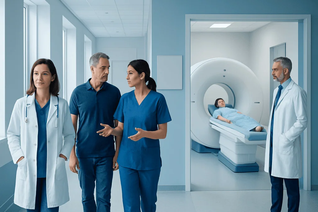
A cerebral PET scan evaluates brain function and metabolism rather than anatomy alone. Preparation often includes fasting and avoiding strenuous exercise, but individual instructions may differ.
Key Takeaways
- A cerebral PET scan evaluates brain function and metabolism rather than anatomy alone.
- Preparation often includes fasting and avoiding strenuous exercise, but individual instructions may differ.
- The scan itself is painless; total appointment time commonly includes tracer uptake and imaging time.
- PET results are interpreted alongside symptoms, examination findings, and other brain tests.
- Most people return to normal activities shortly after the appointment, with simple radiation-safety advice.
A pet scan cerebral, also called a brain PET scan, is an imaging test that shows how active different areas of the brain are by tracking a small amount of radioactive tracer. It may help clinicians investigate neurological symptoms, plan treatment, or clarify findings from MRI, CT, EEG, or other tests.
Overview: What Is a PET Scan Cerebral?
A pet scan cerebral is a positron emission tomography (PET) examination of the brain. It uses a very small amount of radioactive tracer, usually injected into a vein, to produce images showing metabolic activity in brain tissue. Areas that use more or less of the tracer can provide information about how the brain is functioning.
Unlike MRI or CT, which primarily show brain structure, PET is often used to assess function. Depending on the tracer and clinical question, it may support the assessment of memory disorders, epilepsy, brain tumors, inflammation, infection, or movement-related neurological conditions. A PET scan is not usually interpreted in isolation; it is considered together with a person’s symptoms, medical history, neurological examination, and other imaging or laboratory results.
The most common brain PET tracer is fluorodeoxyglucose (FDG), which behaves similarly to glucose. Because brain cells need glucose for energy, FDG-PET can show patterns of reduced or increased metabolism. Other specialized tracers may be selected for particular questions, such as evaluating certain types of dementia or tumor activity.
How Does a Brain PET Scan Work and Who May Need One?
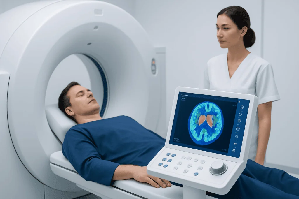
After the tracer enters the bloodstream, it travels to the brain and is taken up by tissues according to their activity. A PET scanner detects signals released by the tracer and a computer converts them into detailed images. PET is often combined with CT or MRI so functional information can be matched precisely to brain anatomy.
A doctor may recommend a brain PET scan when symptoms or earlier tests leave an important question unanswered. For example, it can contribute to the evaluation of progressive memory or thinking changes, help locate a seizure focus when epilepsy surgery is being considered, assess a known or suspected brain tumor, or distinguish active tumor tissue from treatment-related changes in selected situations.
Not everyone with headaches, memory lapses, dizziness, or seizures needs PET imaging. The most appropriate test depends on the symptoms, examination, and previous investigations. MRI, CT, EEG, blood tests, cognitive assessment, or other studies may be more suitable first steps. A neurologist, radiologist, oncologist, or referring physician can explain whether cerebral PET is likely to add useful information.
What Should I Do to Prepare for a PET Scan of the Brain?

Preparation instructions should always come from the imaging center because they can vary according to the tracer, the reason for the scan, and a person’s health conditions. For many FDG brain PET scans, patients are asked to fast for several hours beforehand. Plain water is usually allowed and can help maintain hydration, but the care team will give specific directions about food, drinks, and medications.
It is important to tell the team about diabetes, pregnancy, breastfeeding, kidney disease, allergies, recent illness, and all medicines or supplements. Blood glucose may be checked before an FDG-PET scan because high or unstable glucose can affect tracer distribution and image quality. People with diabetes should not change insulin, diabetes tablets, or meal schedules without individualized medical advice.
Patients should bring previous scans, reports, a medication list, and referral information if requested. Comfortable clothing without metal is helpful. Jewelry, hair accessories, removable dental items, hearing aids, and other metal objects may need to be removed before imaging, particularly if PET is performed with MRI or CT.
- Follow fasting and medication instructions exactly.
- Drink water unless advised otherwise.
- Tell staff about diabetes, pregnancy, breastfeeding, and recent treatments.
- Arrive on time so there is adequate time for preparation and tracer uptake.
What to Avoid 24 Hours Before a PET Scan?
Many imaging departments advise avoiding strenuous exercise for about 24 hours before an FDG-PET scan. Intense activity can increase glucose use in muscles and may affect tracer distribution. Gentle everyday movement is generally fine unless the healthcare team gives different instructions.
Patients may also be asked to avoid alcohol and to limit caffeine or nicotine before the study, especially when the scan is focused on brain function or when sedation is planned. These substances can affect alertness, blood flow, or brain activity. The exact restrictions vary, so a person should follow the instructions provided for their specific appointment rather than applying a general rule.
Do not stop prescribed medicines independently. Some medicines, including certain drugs for seizures, anxiety, diabetes, or neurological conditions, may influence the examination or require timing adjustments. The referring clinician and PET department can advise whether a medicine should be taken as usual, delayed, or documented for interpretation.
Why Do You Have to Sit in a Dark Room Before a PET Scan?
For many cerebral PET examinations, patients rest quietly in a dim, calm room after the tracer injection. This period allows the tracer to distribute while reducing unnecessary stimulation of the brain. Bright light, reading, conversation, music, phone use, anxiety, and active thinking can change activity in particular brain regions and may make images harder to interpret.
The goal is not to test a person’s ability to remain still or awake. It is to capture a consistent resting pattern of brain metabolism. Staff may ask the patient to relax with their eyes open or closed, depending on the scan protocol, and to avoid talking or using electronic devices during the uptake period.
People who have anxiety, claustrophobia, pain, difficulty lying still, or a condition that causes restlessness should mention this before the appointment. The imaging team can discuss practical support and, when appropriate, coordinate a plan with the referring doctor. Sedation is not routinely required and may alter brain activity, so it is considered carefully.
How Long Does a PET Scan of the Brain Typically Take?
The scanning portion of a brain PET examination often takes around 20 to 45 minutes, although this varies with the scanner, tracer, and whether PET is combined with CT or MRI. The full visit is longer because it includes registration, preparation, tracer injection, and a quiet uptake period before imaging.
After injection, the uptake period commonly lasts about 30 to 60 minutes. During the scan, the patient lies on a padded table with the head positioned carefully to minimize movement. The machine is generally open around the body, and the patient can communicate with staff through an intercom.
Patients should plan for several hours at the imaging center unless their appointment team gives a more precise time. Delays can sometimes occur because tracer production and scheduling are closely coordinated. Staff will explain what to expect on the day and can answer questions about transport, companions, or special needs.
Procedure, Recovery Timeline, Benefits and Possible Risks
Before imaging, a healthcare professional places a small intravenous line and injects the tracer. The injection may feel similar to a routine blood test. After resting in the designated low-stimulation area, the patient lies on the scanning table. Keeping the head still is important, and supports may be used for comfort. If CT is included, it is usually completed during the same visit; contrast material is only used when clinically needed.
Most people can leave shortly after the scan and return to ordinary activities, including eating and drinking, unless they received sedation or have been given different instructions. Drinking extra water over the rest of the day can help the tracer leave the body through urine. The amount of radiation is limited and the tracer naturally decays, but patients may be advised to follow brief precautions around pregnant people or very young children, depending on the tracer and local protocol.
Brain PET can offer valuable functional information that is not always visible on structural scans. Risks are generally low and include minor discomfort or bruising at the injection site, rare allergic reactions, and exposure to a small amount of radiation. PET should be avoided or carefully considered during pregnancy unless the expected benefit is important; breastfeeding advice depends on the tracer used. The care team can discuss individual benefits and limitations before the test.
In complex neurological or cancer-related cases, results may be reviewed by specialists from radiology, neurology, neurosurgery, oncology, and nuclear medicine. Acibadem International’s multidisciplinary specialists and JCI-accredited hospitals diagnose and treat neurological conditions for international patients.
Results and When to Seek Medical Care
A nuclear medicine physician or radiologist analyzes the images and prepares a report for the referring doctor. Results may describe areas of reduced metabolism, increased uptake, or no concerning abnormality, but these findings do not automatically confirm a specific diagnosis. A pattern can have more than one explanation, and its meaning depends on the full clinical picture.
The referring clinician will discuss what the findings mean, whether more testing is needed, and whether they change the care plan. For example, results may help guide follow-up for cognitive symptoms, seizure evaluation, tumor assessment, or treatment planning. Patients should ask when and how they will receive their report, and should keep a copy for future care where possible.
When to seek medical care: urgent assessment is appropriate for sudden weakness or numbness on one side, new trouble speaking, severe sudden headache, loss of consciousness, a first seizure, prolonged seizure activity, or sudden major confusion. These symptoms may have causes that need prompt evaluation and should not wait for an outpatient PET scan. New or worsening neurological symptoms should also be discussed with a qualified doctor, even if a scan has already been scheduled.
Frequently asked questions
Is a pet scan cerebral painful?
The scan itself is painless. Most people only feel a brief needle prick when the intravenous line is placed and the tracer is injected. Remaining still on the scanning table can be mildly uncomfortable for some people, but staff can help with positioning.
Can a brain PET scan diagnose dementia?
A brain PET scan can show metabolic or tracer-uptake patterns that may support the evaluation of dementia. However, it does not diagnose every form of dementia on its own. Doctors interpret it alongside symptoms, cognitive testing, examination findings, blood tests, and structural brain imaging.
Can I take my regular medicines before a brain PET scan?
Many medicines can be taken as usual, but the answer depends on the medicine and reason for the scan. Diabetes medicines and some neurological medicines may need individualized timing instructions. Patients should ask the PET department or referring clinician before making any changes.
Do I need someone to drive me home after a brain PET scan?
Most people do not need a driver if they have not received sedation and feel well after the scan. A companion or arranged transport may be appropriate for people who are unwell, have cognitive symptoms, or receive medicine for anxiety or claustrophobia. The imaging center can provide specific advice.
How soon are brain PET scan results available?
Timing varies by imaging center and by the complexity of the study. Images need careful review, often with comparison to MRI, CT, EEG, or previous scans. The referring doctor usually discusses the report once it has been finalized.
Is the radiation from a brain PET scan safe?
A brain PET scan uses a small amount of radiation from the tracer, and sometimes additional radiation if it is combined with CT. Clinicians recommend the examination when the expected diagnostic value outweighs this limited exposure. Patients who are pregnant, may be pregnant, or are breastfeeding should tell the team before the scan.
References
- Radiological Society of North America
- Society of Nuclear Medicine and Molecular Imaging
- National Institute of Neurological Disorders and Stroke
- American College of Radiology
This article is for general information only and is not a substitute for professional medical advice. Please consult a qualified doctor about your individual situation.
Explore treatments in Turkey — costs, top hospitals & a free quote
JCI-accredited · board-certified surgeons · reply within 24h
Add us as a Preferred Source to see more of our trusted health content across Google Search, AI Overviews and Discover.