Glioblastoma Surgery: Procedure, Recovery and Results
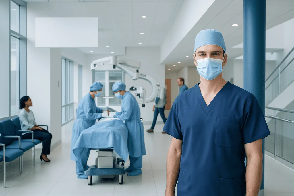
Glioblastoma surgery is commonly performed to confirm the diagnosis and reduce the amount of tumor present. The safest possible extent of tumor removal is generally the goal, but complete removal is not always possible.
Key Takeaways
- Glioblastoma surgery is commonly performed to confirm the diagnosis and reduce the amount of tumor present.
- The safest possible extent of tumor removal is generally the goal, but complete removal is not always possible.
- Surgery is usually followed by radiotherapy and chemotherapy because glioblastoma cells can extend beyond what is visible on scans.
- Recovery varies with the tumor location, the operation performed, pre-existing symptoms and the person's overall health.
- Glioblastoma can recur after surgery, so regular imaging and follow-up with a neuro-oncology team are essential.
Glioblastoma surgery is usually the first major treatment step when the tumor can be reached safely. Its purpose is to obtain tissue for diagnosis and remove as much visible tumor as possible while protecting important brain functions, followed by personalized ongoing cancer care.
Overview: what glioblastoma surgery involves
Glioblastoma surgery is an operation to remove as much of a glioblastoma brain tumor as can be done safely. It also provides a tissue sample that allows specialists to confirm the diagnosis and perform molecular testing, which helps guide treatment planning. In many cases, surgery is followed by radiotherapy and chemotherapy because microscopic tumor cells may remain in brain tissue even after a successful operation.
The operation is most often a craniotomy, in which a neurosurgeon temporarily removes a small section of skull to reach the tumor. The aim is called maximal safe resection: reducing the tumor burden while avoiding unacceptable harm to functions such as movement, speech, vision, memory or sensation. When removal would carry too much risk, a smaller operation or a biopsy may be recommended instead.
Glioblastoma is a fast-growing primary brain tumor in adults. Treatment decisions are best made by a multidisciplinary team that may include neurosurgeons, neuro-oncologists, radiation oncologists, neuroradiologists, neuropathologists, rehabilitation specialists and supportive-care professionals. More information about the disease is available in this overview of glioblastoma.
Who may be a candidate and how the procedure is planned
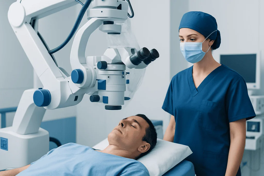
Whether surgery is appropriate depends on the tumor’s size, location and growth pattern, as well as the person’s symptoms, general health and goals of care. Surgery may be considered when imaging suggests that a tumor can be approached with an acceptable level of risk. It can relieve pressure inside the skull and may improve symptoms caused by mass effect, such as headache, weakness, seizures or changes in speech.
Before surgery, patients usually have detailed brain imaging, most often magnetic resonance imaging (MRI). The care team reviews how close the tumor is to areas controlling speech, movement, vision and other important abilities. In selected cases, advanced imaging, functional mapping, neuropsychological assessment or tractography may help plan the safest route to the tumor.
Some tumors are located deep in the brain or within highly functional areas, making extensive removal unsafe. In these situations, a stereotactic biopsy may be used to collect a small tissue sample, allowing diagnosis and molecular testing without attempting broad removal. The team should explain the expected benefits, possible limitations and alternatives before a decision is made.
Patients may be asked to review medicines before surgery, especially blood thinners, diabetes treatments and supplements. Steroid medicine may be prescribed to reduce brain swelling, and anti-seizure medicine may be used when clinically appropriate. Individual medication instructions should always come directly from the treating team.
How glioblastoma surgery is performed, step by step
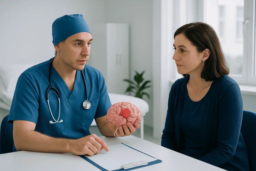
Glioblastoma surgery is performed in a hospital operating room under general anesthesia in most cases. The surgical team positions the patient carefully, prepares the scalp and makes an incision. A small bone flap is temporarily removed to create access to the brain, and the neurosurgeon uses operating microscopes, navigation systems and other tools to locate the tumor precisely.
During tumor removal, surgeons work to separate abnormal tissue from nearby healthy brain structures as safely as possible. Image guidance, ultrasound, intraoperative MRI or fluorescence-guided techniques may be used in some centers to help identify tumor tissue. These tools can support decision-making, but they do not eliminate the need to stop removal when functionally important tissue is at risk.
For tumors near language or movement centers, an awake craniotomy may be considered for selected patients. During part of this approach, the patient can perform simple speaking or movement tasks while the surgical team maps critical brain areas. This can help the surgeon preserve important functions while removing tumor tissue where safe.
At the end of the procedure, the bone flap is replaced and secured, and the scalp is closed. Removed tissue is examined by a neuropathologist. Results commonly include the tumor type and grade as well as molecular markers, such as IDH status and MGMT promoter methylation, which can inform prognosis and treatment choices.
Benefits, limits and risks of glioblastoma surgery
The principal benefits of glioblastoma surgery are establishing an accurate diagnosis, reducing the volume of tumor and helping relieve symptoms caused by pressure or swelling. Removing more tumor when it can be done safely is generally associated with better disease control than limited removal alone. However, surgery is one part of treatment rather than a stand-alone cure.
Glioblastoma cells can infiltrate surrounding brain tissue beyond the edges that can be seen on MRI or identified during surgery. For this reason, even when postoperative imaging shows that all visible enhancing tumor has been removed, further treatment is usually advised. This commonly includes radiotherapy combined with chemotherapy, followed by additional chemotherapy when appropriate.
Potential risks vary according to the tumor location and the person’s health. They can include bleeding, infection, blood clots, seizures, fluid buildup, wound problems, stroke-like injury and reactions to anesthesia. Neurological changes may include weakness, numbness, language difficulty, vision changes, balance problems, cognitive changes or personality changes. Some symptoms are temporary and improve as swelling settles, while others may persist.
The surgical team balances these risks against the potential value of removing tumor. An open discussion about likely functional outcomes, rehabilitation needs and the person’s priorities is an important part of informed decision-making. Specialists can also explain options for brain tumor surgery in the context of an individual treatment plan.
Recovery timeline after glioblastoma surgery
After surgery, patients are monitored closely in a recovery area or intensive care setting, often for the first night. The team checks alertness, strength, speech, pupils and other neurological functions. A CT scan or MRI may be performed soon after surgery to assess the surgical area and provide a baseline for future comparisons.
Hospital stay often lasts several days, but the timeline varies. Some people need additional care for weakness, balance problems, speech changes or fatigue. Physical therapy, occupational therapy, speech and language therapy and neuropsychology support can help patients regain independence and adapt to changes in function.
In the first weeks at home, fatigue, headaches, scalp tenderness and emotional ups and downs can occur. Recovery is not always linear, and activity should increase gradually according to the surgeon’s advice. Driving, work, travel, exercise and bathing restrictions differ between individuals, particularly for people who have had seizures or neurological symptoms.
Radiotherapy and chemotherapy are commonly planned after the surgical wound has had time to heal, often within several weeks. Follow-up appointments review pathology findings, assess recovery and coordinate next steps. A structured neurorehabilitation program may be valuable for people with ongoing physical, cognitive or communication difficulties.
How successful is surgery for glioblastoma?
Success in glioblastoma surgery is measured in more than one way. A successful operation may obtain enough tissue for a clear diagnosis, remove a large amount of visible tumor without causing major new neurological deficits, relieve pressure-related symptoms and allow timely treatment with radiotherapy and chemotherapy. The appropriate goal differs from person to person.
When maximal safe removal is possible, it is generally linked with improved outcomes compared with biopsy alone or more limited resection. However, the tumor’s location may make complete visible removal unsafe. In that situation, protecting speech, movement, independence and quality of life can be more important than pursuing a larger resection.
It is important to recognize that glioblastoma surgery does not guarantee that the cancer has been eliminated. Outcomes are influenced by age, functional status, tumor location, the amount removed, molecular features and response to subsequent treatment. The treating team can provide the most meaningful interpretation after reviewing surgical findings, pathology and follow-up imaging.
Can you survive glioblastoma after surgery?
Yes. People can live after glioblastoma surgery, and surgery is often an important part of treatment. Survival varies widely, and no single operation result can predict an individual’s course. Care generally continues after surgery with radiotherapy, chemotherapy, regular MRI scans and management of symptoms or treatment side effects.
Glioblastoma remains a serious condition because its cells tend to grow into nearby brain tissue and may return despite treatment. Still, many factors affect outlook, including the patient’s overall health, age, tumor characteristics, the safety and extent of surgery and how the tumor responds to further therapy. Molecular testing of the tissue sample is especially useful in personalizing discussions about treatment and prognosis.
Supportive care is also part of active treatment. Seizure management, rehabilitation, nutrition support, counseling and practical help for families can improve day-to-day well-being. Patients are encouraged to ask their team about clinical trials when relevant, as these may provide access to carefully monitored emerging approaches.
How fast does glioblastoma come back after surgery and when to seek medical care
Glioblastoma can recur because microscopic cells may remain after surgery and additional therapy. The timing of recurrence is highly variable: it may be detected within months for some people, while others have a longer period of disease control. Regular MRI monitoring is needed because recurrence can sometimes appear on scans before it causes clear symptoms.
New or worsening symptoms do not always mean tumor recurrence. They can also result from postoperative healing, brain swelling, seizures, medication effects, infection or treatment-related changes. The care team may use repeat MRI, specialized imaging, clinical examination and sometimes further testing to clarify what is happening.
Medical advice should be sought promptly for a new seizure, severe or rapidly worsening headache, repeated vomiting, sudden weakness or numbness, new difficulty speaking, confusion, major vision changes, fainting, fever with wound redness or drainage, or a marked change in alertness. Emergency care is appropriate for sudden severe neurological symptoms or loss of consciousness.
Acibadem International’s multidisciplinary specialists and JCI-accredited hospitals support international patients requiring coordinated diagnosis and treatment for glioblastoma. Ongoing care may include radiotherapy and other neuro-oncology treatments based on the individual’s pathology and clinical needs.
Frequently asked questions
How successful is surgery for glioblastoma?
Glioblastoma surgery can be successful when it confirms the diagnosis, safely removes a substantial amount of visible tumor and helps reduce symptoms. It is usually followed by radiotherapy and chemotherapy because surgery cannot reliably remove microscopic cells that may have spread into surrounding brain tissue. The result depends greatly on tumor location, molecular features and the person’s overall health.
Can you survive glioblastoma after surgery?
Yes, people can survive glioblastoma surgery and continue treatment afterward. However, glioblastoma is a serious tumor with a variable outlook, and surgery alone is not considered curative. The care team uses pathology, molecular testing, imaging and treatment response to discuss an individual prognosis.
How long does it take to recover from glioblastoma surgery?
Initial hospital recovery commonly takes several days, while healing and adjustment at home often continue for weeks. Some people recover more quickly, while others need rehabilitation for weakness, speech changes, balance difficulties, fatigue or cognitive symptoms. Further treatment is often organized within weeks once recovery and wound healing allow.
How fast does glioblastoma come back after surgery?
Glioblastoma recurrence can occur at different times, often within months but sometimes after a longer period of disease control. The timing depends on tumor biology, the extent of safe tumor removal and response to radiotherapy and chemotherapy. Scheduled MRI scans are important because recurrence may be found before symptoms become noticeable.
Is glioblastoma surgery done while the patient is awake?
Most glioblastoma operations are performed under general anesthesia. An awake craniotomy may be used for selected tumors near areas responsible for speech, movement or other essential functions. During awake mapping, the surgical team checks these functions to help preserve them as much as possible.
What happens after glioblastoma surgery?
The tumor tissue is examined to confirm the diagnosis and identify molecular markers that guide treatment. Patients usually have early postoperative imaging and follow-up with a neuro-oncology team. Radiotherapy and chemotherapy are commonly recommended after surgery, along with rehabilitation or supportive care when needed.
References
- National Cancer Institute
- National Institute of Neurological Disorders and Stroke
- American Brain Tumor Association
- European Association of Neuro-Oncology
- National Comprehensive Cancer Network
This article is for general information only and is not a substitute for professional medical advice. Please consult a qualified doctor about your individual situation.
Oncology care in Turkey — second opinion and treatment plan
JCI-accredited · board-certified surgeons · reply within 24h
Add us as a Preferred Source to see more of our trusted health content across Google Search, AI Overviews and Discover.