Testicular Rupture Ultrasound: Preparation, Procedure and Results
Testicular rupture is an emergency that may occur after a direct blow or other significant scrotal injury. Ultrasound with Doppler imaging assesses testicular shape, internal tissue and blood flow without radiation.
Key Takeaways
- Testicular rupture is an emergency that may occur after a direct blow or other significant scrotal injury.
- Ultrasound with Doppler imaging assesses testicular shape, internal tissue and blood flow without radiation.
- Irregular testicular contours, disrupted tissue and surrounding blood collection can suggest rupture.
- A concerning ultrasound result or strong clinical suspicion may lead to prompt surgical exploration.
- Severe pain, swelling, bruising, nausea or a high-riding testicle after injury requires urgent medical assessment.
A testicular rupture ultrasound is usually the first imaging test used after significant scrotal trauma. It can quickly identify changes that suggest a tear in the testicle, internal bleeding or impaired blood flow, helping clinicians decide whether urgent surgery is needed.
Testicular Rupture Ultrasound: An Overview
A testicular rupture ultrasound is a focused scrotal scan performed after an injury to assess whether the protective outer covering of a testicle, called the tunica albuginea, may have torn. It uses sound waves rather than radiation and commonly includes Doppler imaging, which shows blood flow within the testicle and nearby tissues.
Testicular rupture is uncommon but time-sensitive because early assessment and, when necessary, surgical repair may help preserve testicular tissue and function. Ultrasound is highly useful in this setting, but it is interpreted alongside the physical examination, the type of injury and the person’s symptoms.
A painful, swollen scrotum can also result from bruising, a collection of blood called a hematoma, infection, or twisting of the spermatic cord. For example, testicular torsion can also cause sudden severe pain and requires urgent evaluation. A clinician should not rely on symptoms alone to distinguish these conditions.
How Is Testicular Rupture Diagnosed?
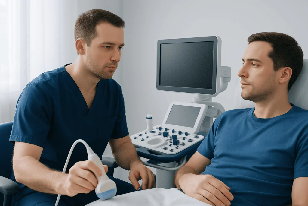
Testicular rupture is diagnosed through urgent clinical assessment supported by scrotal ultrasound. A clinician will ask how the injury happened, when symptoms began and whether there is severe pain, nausea, bruising, swelling, blood in the urine or difficulty urinating. Direct impacts during sports, falls, accidents, assaults and straddle injuries are possible causes.
During the examination, the clinician checks the scrotum for swelling, discoloration, tenderness, open wounds and an abnormal testicular position or shape. Examination can be difficult when swelling and pain are substantial, which is one reason ultrasound is valuable.
Ultrasound can provide important information quickly. However, if the scan is unclear and the examination strongly suggests rupture, urologists may recommend surgical exploration rather than delaying care. Surgery is both diagnostic and therapeutic because it allows the injured testicle to be examined and repaired when possible.
Depending on the injury, urine testing, blood tests or imaging of the pelvis and abdomen may also be needed to check for associated injuries. A suspected rupture should be assessed in an emergency department or by an urgent urology service.
What Are the Ultrasound Findings of Testicular Rupture?
The most important ultrasound finding suggesting testicular rupture is an irregular or disrupted outer contour of the testicle. This may indicate a tear in the tunica albuginea, allowing internal testicular tissue to protrude or become distorted.
Other possible findings include a heterogeneous, or uneven, appearance of the testicular tissue due to bruising and bleeding; a focal defect in the tissue; and a hematocele, which is blood collecting around the testicle. The scan may also show an intratesticular hematoma, meaning bleeding within the testicular tissue.
Color Doppler ultrasound evaluates blood flow. Reduced, patchy or absent flow can suggest damage to the blood supply and may increase concern for tissue injury. Normal flow does not always completely exclude rupture, particularly when other imaging or examination findings are concerning.
Ultrasound can also identify injuries that may be managed differently, such as an isolated hematoma, fluid collection or inflammation of the epididymis. The radiologist or urologist considers the complete pattern of findings rather than relying on one feature alone.
How Should I Prepare for a Testicular Ultrasound?
Most people need very little preparation for a testicular ultrasound. Eating and drinking are usually allowed, and there is no need for fasting unless a healthcare professional gives different instructions because of other tests or a possible procedure.
Wear comfortable, loose-fitting clothing if possible. The patient will generally change into a gown or remove clothing from the waist down in a private examination area. A sheet or towel is used to preserve dignity, and the penis is positioned away from the scanning area.
It is helpful to tell the care team about the injury, including when it occurred, whether symptoms are worsening and any medications that may affect bleeding, such as anticoagulants. The patient should also mention prior scrotal surgery, implanted devices, known testicular conditions or medication allergies, although these rarely prevent ultrasound.
Severe pain should not be endured in order to prepare for the scan. Clinicians can provide appropriate pain relief while arranging urgent assessment. If there is an open wound, heavy bleeding, fainting, vomiting or other major trauma, emergency care should take priority over routine imaging arrangements.
What to Expect During a Testicular Ultrasound?
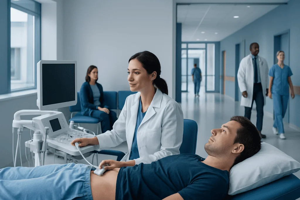
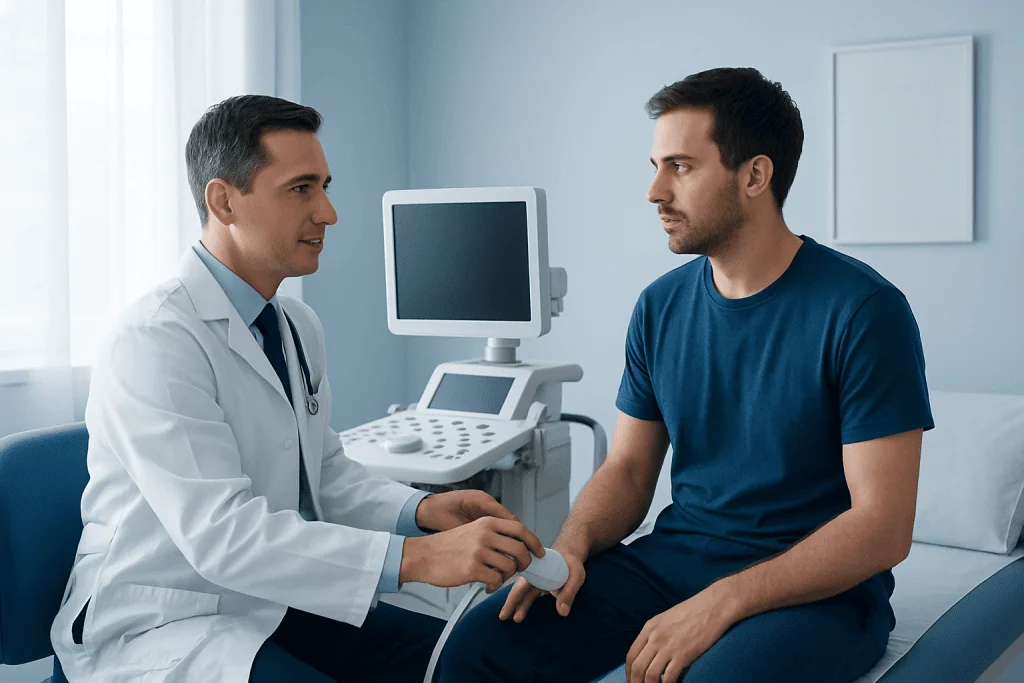
A scrotal ultrasound is performed by a sonographer, radiologist or other trained imaging professional. The patient lies on their back, and the scrotum is gently supported with a towel. Warm, clear gel is applied to the skin to help transmit sound waves.
A small handheld device called a transducer is moved over the scrotum. Images are taken of both testicles for comparison, as well as the epididymis and surrounding tissues. Doppler imaging may be used to display and measure blood flow. The test itself often takes about 15 to 30 minutes, although timing can vary with the extent of injury and the need for additional assessment.
Ultrasound does not use needles, injections or radiation. The main discomfort is usually the gentle pressure of the transducer on an already tender area. The sonographer may adjust pressure and positioning as much as possible for comfort, but should be told immediately if pain is difficult to tolerate.
In emergency settings, results may be reviewed quickly by the treating team. If findings suggest rupture, reduced blood supply or another urgent condition, a urology specialist may recommend prompt next steps, including testicular rupture surgery.
Benefits, Limits and Recovery After the Scan
The main benefit of testicular rupture ultrasound is that it is fast, noninvasive and free of ionizing radiation. It helps clinicians assess the structure of the testicle, detect bleeding and evaluate blood flow, supporting decisions about observation, repeat imaging or urgent surgery.
The scan itself does not require a recovery period. The patient can generally return to normal activities immediately after the examination unless the injury, pain medication, hospital evaluation or planned treatment requires otherwise. The care team will explain activity limits, scrotal support and follow-up based on the diagnosis.
There are no major medical risks from ultrasound. In an acute injury, pressure from the transducer may temporarily worsen discomfort. A limitation is that extensive swelling, large blood collections or severe tenderness can make images harder to interpret; therefore, an apparently reassuring scan may not override a strong clinical concern.
If surgery is needed, recovery varies according to the severity of injury and the procedure performed. In general, clinicians may advise rest, supportive underwear, ice packs wrapped in cloth for short intervals, and avoiding strenuous activity until cleared. Individualized follow-up is important to monitor healing and testicular function.
When to Seek Medical Care
Medical care should be sought urgently after a significant blow, crush injury or penetrating injury to the scrotum, especially when pain is severe or symptoms do not settle quickly. It is safest to seek emergency assessment rather than waiting to see whether substantial swelling or bruising improves at home.
Warning signs include persistent or increasing pain, rapid swelling, marked bruising, nausea or vomiting, fever, dizziness, an abnormal testicular position, a cut or puncture wound, or blood in the urine. Sudden pain without a clear injury also needs urgent evaluation because torsion and other conditions can threaten the testicle.
Until assessed, the person may support the scrotum with close-fitting underwear and avoid strenuous movement. Applying a cold pack wrapped in a cloth for short periods may help swelling, but it should not delay emergency care. Do not attempt to push a testicle back into position or drain swelling.
Acibadem International’s multidisciplinary specialists and JCI-accredited hospitals evaluate scrotal trauma and coordinate urgent imaging, urology care and surgery for international patients when appropriate.
Frequently asked questions
Is testicular rupture visible on ultrasound?
Ultrasound can often show findings that strongly suggest testicular rupture, including an irregular testicular outline, disrupted tissue and bleeding around the testicle. Doppler imaging also assesses blood flow. However, the scan is interpreted together with the examination, and surgery may still be advised if concern remains high.
Does a testicular ultrasound hurt after an injury?
Ultrasound itself is noninvasive, but the transducer may place mild pressure on a tender area. The care team can position the scrotum carefully and adjust pressure to reduce discomfort. Patients should tell the sonographer if pain is severe.
How quickly should ultrasound be done for suspected testicular rupture?
Suspected testicular rupture should be assessed urgently, usually through an emergency department or urgent urology service. Ultrasound is commonly arranged promptly when it can help guide management. It should not delay surgery when clinical findings indicate an immediate need for exploration.
Can a testicle heal without surgery after trauma?
Some minor testicular injuries, such as small bruises or selected hematomas, may be treated without surgery with close medical follow-up. A true rupture often requires urgent surgical repair to remove damaged tissue, control bleeding and preserve viable testicular tissue when possible. Only a clinician can determine the appropriate approach after examination and imaging.
What happens if a testicular rupture is not treated?
Without timely assessment and treatment, a rupture may lead to ongoing bleeding, tissue loss, infection, chronic pain or reduced testicular function. Fertility and hormone production can be affected in some cases, particularly if both testicles are injured. Prompt care offers the best opportunity to address the injury appropriately.
Will I need follow-up after a scrotal injury?
Follow-up is often recommended, particularly after a hematoma, surgery or an ultrasound finding that needs monitoring. A clinician may repeat examination or ultrasound to confirm healing and evaluate blood flow. The follow-up plan depends on the injury severity, symptoms and treatment provided.
References
- American Urological Association
- European Association of Urology
- American College of Radiology
- Merck Manual Professional Edition
This article is for general information only and is not a substitute for professional medical advice. Please consult a qualified doctor about your individual situation.
Ultrasound in Turkey — costs, top hospitals & a free quote
JCI-accredited · board-certified surgeons · reply within 24h
Add us as a Preferred Source to see more of our trusted health content across Google Search, AI Overviews and Discover.

Check-up & Preventive Medicine
Comprehensive health screenings designed for early detection and prevention.
1 specialists in this unit