Pediatric Ultrasound: Preparation, Procedure and Results

Pediatric ultrasound does not use ionizing radiation and is widely used for infants, children, and teenagers. Preparation varies by scan type; fasting may be needed for abdominal or gallbladder examinations, while a full bladder may be needed for pelvic or urinary scans.
Key Takeaways
- Pediatric ultrasound does not use ionizing radiation and is widely used for infants, children, and teenagers.
- Preparation varies by scan type; fasting may be needed for abdominal or gallbladder examinations, while a full bladder may be needed for pelvic or urinary scans.
- The test is usually painless, takes about 15 to 45 minutes, and does not require recovery time.
- A radiologist interprets the images and sends a report to the doctor who requested the examination.
- Ultrasound results may identify a condition, guide further testing, or provide reassurance when findings are normal.
Pediatric ultrasound is a painless imaging examination that uses sound waves to create pictures inside a child’s body. Preparation depends on the body area being examined, and most children can return to normal activities immediately afterward.
Overview: What Is a Pediatric Ultrasound?
Pediatric ultrasound is an imaging test that helps doctors view structures inside a child’s body without surgery or radiation. It uses a handheld device called a transducer to send high-frequency sound waves through the skin. These waves return as echoes, which a computer converts into real-time images.
It is commonly used to examine the abdomen, kidneys, bladder, pelvis, thyroid, brain in young infants, hips, testicles, joints, muscles, and soft tissues. Doppler ultrasound, a specialized form of the test, can also assess blood flow through blood vessels and organs.
The examination is tailored to the child’s age, symptoms, and clinical question. Pediatric ultrasound protocols may differ from adult protocols because children have smaller body structures, different normal measurements, and particular comfort needs during imaging.
What Does a Pediatric Ultrasound Do?
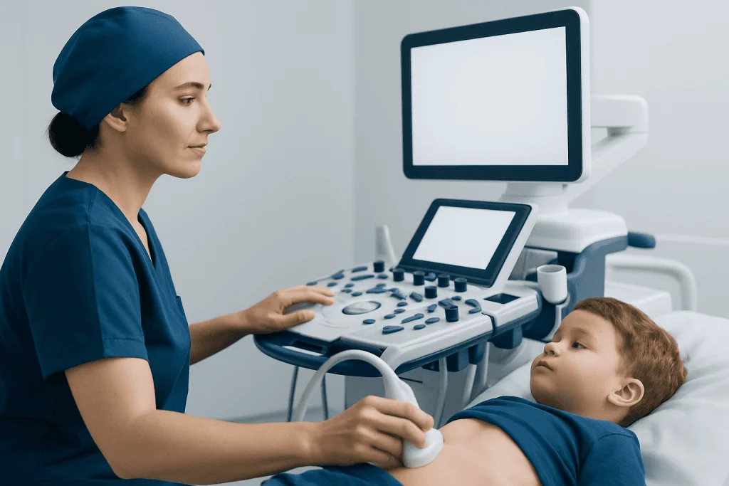
A pediatric ultrasound creates moving images that can help doctors investigate symptoms, monitor a known condition, or evaluate an abnormality found during a physical examination or another test. It may help assess pain, swelling, vomiting, urinary symptoms, jaundice, a lump, an injury, or concerns about organ development.
For example, an abdominal ultrasound may examine the liver, gallbladder, spleen, pancreas, kidneys, bladder, and bowel-related findings. Doctors may request it when evaluating recurrent abdominal pain, suspected kidney stones, urinary tract concerns, or possible appendicitis. It can also support assessment of conditions such as kidney stones when clinically appropriate.
Ultrasound can show the size, shape, and texture of organs and may detect fluid collections, cysts, inflammation, enlarged structures, or changes in blood flow. However, it does not answer every medical question. Gas in the bowel, body position, and the area being examined can sometimes limit the images, so other tests may occasionally be needed.
Is Ultrasound Safe for a 4 Year Old?

Yes. Ultrasound is considered safe for a 4-year-old and for children of other ages when it is medically indicated and performed by trained professionals. Unlike X-rays and CT scans, ultrasound does not use ionizing radiation. It works with sound waves that are not felt by the child.
Healthcare teams follow established pediatric ultrasound guidelines and use the lowest appropriate ultrasound settings for the shortest necessary time. This approach is sometimes described as the ALARA principle: keeping exposure “as low as reasonably achievable” while still obtaining useful diagnostic images.
The gel placed on the skin is generally well tolerated. Some children may feel mild pressure from the transducer, especially if the area is tender, but the scan itself should not be painful. Sedation is not usually needed; caregivers can often remain nearby to help a young child feel secure.
Do You Need to Do Anything to Prep for an Ultrasound?
Preparation depends on the type of pediatric ultrasound. The imaging department should provide specific instructions in advance, and these instructions take priority because requirements can differ by the child’s age, symptoms, and the organs being assessed.
For pediatric abdominal ultrasound preparation, a child may be asked not to eat for several hours before the appointment. Fasting can reduce bowel gas and allow clearer assessment of structures such as the gallbladder, liver, pancreas, and upper abdomen. Infants and young children may receive modified fasting instructions to avoid unnecessary discomfort or dehydration.
For an ultrasound of the bladder, kidneys, or pelvis, the child may instead be asked to drink water beforehand and avoid urinating until after the scan. A comfortably full bladder can improve visibility. Children should generally wear loose, comfortable clothing, and caregivers may wish to bring a familiar toy, book, or soothing item.
- Confirm whether food, drinks, or regular medicines should be adjusted before the test.
- Bring relevant referral forms, previous imaging reports, and a list of medicines if requested.
- Explain in simple terms that a camera-like device will move over the skin with warm gel.
- Tell the imaging team about pain, developmental needs, allergies, or difficulties staying still.
Why Can't You Drink Milk Before an Ultrasound?
Milk may be restricted before some abdominal ultrasounds because it is considered a meal rather than a clear liquid. Eating or drinking milk can cause the gallbladder to contract and may increase digestive activity, making certain abdominal structures harder to evaluate clearly.
This instruction does not apply to every pediatric ultrasound. For some scans, a child may be allowed clear fluids or may need to drink water to fill the bladder. Very young children and infants need individualized instructions, since prolonged fasting may not be appropriate.
Caregivers should not assume that all foods and drinks are restricted. If instructions are unclear, they should contact the imaging center before the appointment. Following the requested preparation helps reduce the chance that the scan needs to be postponed or repeated.
What Happens During the Procedure and How Are Results Given?
At the appointment, a sonographer or radiology professional confirms the child’s details and explains the examination. The child is positioned on an examination bed, and gel is placed on the skin over the body area being scanned. The gel helps sound waves travel effectively between the transducer and the skin.
The professional moves the transducer gently over the area and may ask the child to change position, take a breath, or remain still briefly. Caregivers can often stay in the room, depending on local policies. The scan commonly lasts 15 to 45 minutes, though timing varies with the body area and the complexity of the examination.
There is no recovery period after a standard ultrasound. The child can usually eat, drink, go to school, and resume normal activities right away unless the doctor gives other instructions. A radiologist reviews the images and prepares a report for the referring clinician, who explains what the findings mean in the context of the child’s symptoms and medical history.
A normal result can be reassuring, while an abnormal or unclear result does not automatically mean a serious problem. It may indicate a condition that can be treated, a harmless variation, or a need for follow-up imaging, blood tests, or specialist review.
Benefits, Limitations, and When to Seek Medical Care
The main benefits of pediatric ultrasound are that it is noninvasive, radiation-free, usually painless, and able to show real-time movement and blood flow. It is especially valuable for evaluating many soft tissues and abdominal or urinary concerns. Ultrasound may also be used to guide certain procedures when a doctor considers this appropriate.
Its limitations are important to understand. Ultrasound does not show every part of the body equally well, and bones, air-filled bowel, and deep structures can make images harder to interpret. The quality of the examination also depends on the child’s ability to remain still and the clinical question being investigated.
Parents or caregivers should seek prompt medical assessment rather than waiting for an outpatient scan if a child has severe or worsening abdominal pain, difficulty breathing, persistent vomiting with signs of dehydration, unusual sleepiness, fainting, a significant injury, a swollen or painful testicle, or blood in urine or stool. Emergency care is appropriate for severe symptoms or whenever a caregiver believes the child may be seriously unwell.
Acibadem International’s multidisciplinary specialists and JCI-accredited hospitals provide diagnostic imaging and coordinated care for international patients when ultrasound findings require further evaluation or treatment.
Frequently asked questions
How long does a pediatric ultrasound take?
Most pediatric ultrasound examinations take about 15 to 45 minutes. The exact time depends on the body part being examined, whether blood-flow imaging is needed, and how easily the child can stay in a suitable position. Additional time may be needed for check-in and preparation.
Does a pediatric ultrasound hurt?
Ultrasound is usually painless. The child may feel the cool or warm gel and mild pressure from the transducer. If the scanned area is already sore, the pressure may be briefly uncomfortable, and the sonographer can adjust technique where possible.
Can a parent stay with a child during an ultrasound?
In many imaging departments, a parent or caregiver can remain with the child to offer reassurance. Local safety and infection-control policies may vary, so it is helpful to ask when booking. Bringing a familiar comfort item may also help younger children cooperate.
When will pediatric ultrasound results be available?
A radiologist reviews the images and sends a report to the doctor who ordered the test. Timing varies by facility and by how urgently the scan was requested. The referring clinician is best placed to explain the result and whether any follow-up is needed.
Can a child eat before an abdominal ultrasound?
Some abdominal ultrasounds require fasting, while others do not. Food and milk can affect the appearance of the gallbladder and digestive system, so the imaging center may provide age-specific instructions. Caregivers should follow the instructions given for that particular appointment.
What should I bring to a pediatric ultrasound appointment?
Bring the referral or appointment details, identification or insurance documents if applicable, and previous imaging reports or relevant medical records when requested. A list of medicines and a comfort item for the child can also be useful. The child should usually wear loose clothing that allows easy access to the area being scanned.
References
- American College of Radiology
- Radiological Society of North America
- Society for Pediatric Radiology
- U.S. Food and Drug Administration
- National Institute of Biomedical Imaging and Bioengineering
This article is for general information only and is not a substitute for professional medical advice. Please consult a qualified doctor about your individual situation.
Ultrasound in Turkey — costs, top hospitals & a free quote
JCI-accredited · board-certified surgeons · reply within 24h
Add us as a Preferred Source to see more of our trusted health content across Google Search, AI Overviews and Discover.

Check-up & Preventive Medicine
Comprehensive health screenings designed for early detection and prevention.
1 specialists in this unit