Normal Pressure Hydrocephalus MRI: Preparation, Procedure and Results
MRI is an important part of assessing suspected normal pressure hydrocephalus, but it cannot confirm the diagnosis alone. NPH commonly involves walking difficulty, thinking changes, and urinary urgency or incontinence, although symptoms vary.
Key Takeaways
- MRI is an important part of assessing suspected normal pressure hydrocephalus, but it cannot confirm the diagnosis alone.
- NPH commonly involves walking difficulty, thinking changes, and urinary urgency or incontinence, although symptoms vary.
- The scan is noninvasive and does not use ionizing radiation; contrast is not always needed.
- MRI can help doctors rule out stroke, tumors, bleeding, brain shrinkage, and other explanations for symptoms.
- People with new or rapidly worsening neurological symptoms should seek medical assessment promptly.
A <a href="https://acibademinternational.com/diseases/normal-pressure-hydrocephalus/”>normal pressure hydrocephalus MRI is a painless brain scan that can show patterns of enlarged fluid spaces associated with NPH and help exclude other conditions. MRI findings are interpreted alongside symptoms, examination results, and sometimes cerebrospinal fluid testing before treatment is considered.
Normal Pressure Hydrocephalus MRI: an answer-first overview
A normal pressure hydrocephalus MRI is a detailed brain scan used to look for enlarged ventricles, the fluid-filled spaces within the brain, and for imaging patterns that may support a diagnosis of normal pressure hydrocephalus (NPH). It also helps clinicians look for other conditions that can cause similar changes in walking, memory, and bladder control.
NPH is a potentially treatable cause of neurological symptoms, most often seen in older adults. The term “normal pressure” refers to the fact that pressure measured during a lumbar puncture may be within a typical range, even though the movement and absorption of cerebrospinal fluid are disturbed.
MRI is usually one part of a broader assessment. A neurologist, geriatrician, neurosurgeon, radiologist, and rehabilitation specialists may consider the person’s symptom history, physical and cognitive assessment, MRI appearance, and response to cerebrospinal fluid drainage testing when deciding whether NPH is likely and whether treatment may help.
How MRI works and who may need it

MRI uses a powerful magnetic field and radiofrequency waves to produce detailed images of the brain. Unlike CT scanning and standard X-rays, MRI does not use ionizing radiation. Different MRI sequences show brain tissue, cerebrospinal fluid, blood vessels, and signs of prior injury in different ways, allowing a detailed review of the ventricles and surrounding structures.
A doctor may recommend MRI when a person develops a gradual, unexplained combination of slowed or unsteady walking, difficulties with attention or planning, and urinary urgency or leakage. Not everyone has all three symptoms, and these concerns are common in several neurological, urinary, and mobility conditions. MRI helps clarify whether NPH should remain under consideration.
Most people can undergo MRI safely. Before the appointment, the imaging team will check for implanted devices, metal fragments, some clips or stimulators, and other factors that may require special safety planning. Many modern implants are MRI-conditional, meaning scanning may still be possible under specified conditions. Pregnancy, kidney disease, and prior contrast reactions should also be discussed if contrast material is being considered.
Preparing for and having a normal pressure hydrocephalus MRI

For a routine brain MRI, people can usually eat, drink, and take prescribed medicines as normal unless their care team gives different instructions. Comfortable clothing without metal fasteners may be useful, although many centers provide a gown. It is important to remove jewelry, watches, hearing aids, removable dental appliances, credit cards, and other metal or magnetic items before entering the scan room.
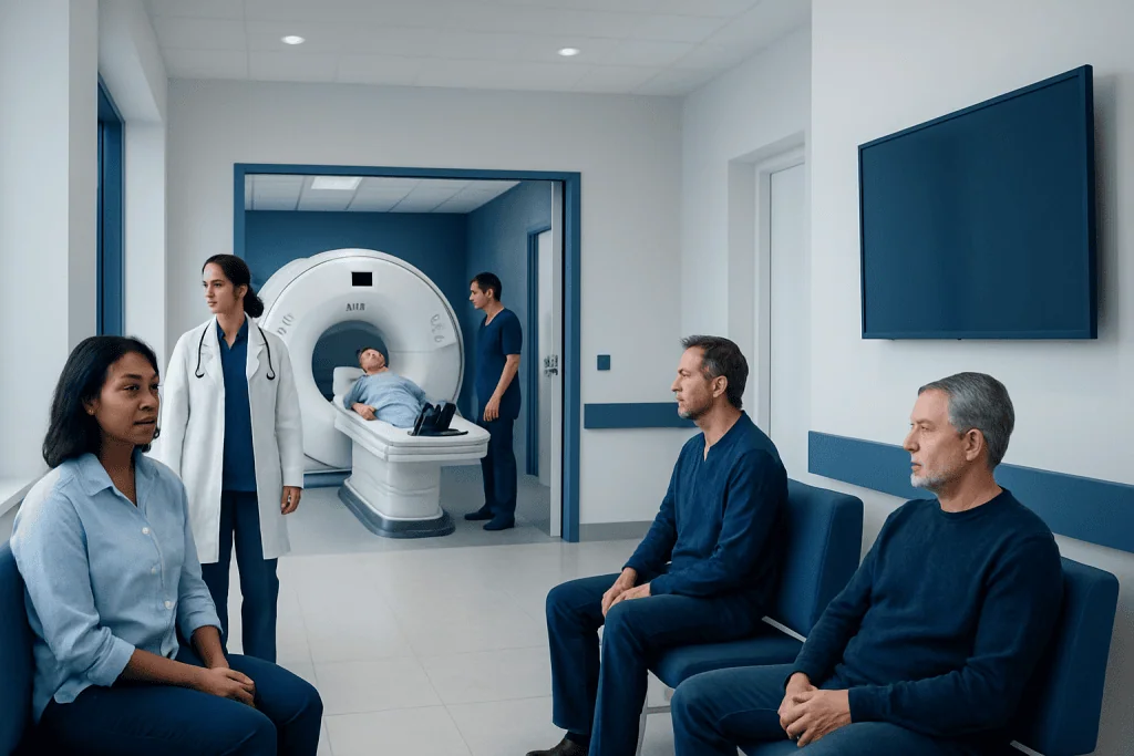
At the appointment, a radiographer will review the safety questionnaire and help the person lie on a padded scanning table. A lightweight head coil is placed around the head to improve image quality. The table then moves into the MRI scanner. The machine makes loud tapping and knocking sounds, so earplugs or headphones are provided.
During the scan, remaining as still as possible helps prevent blurred images. A brain MRI often takes around 30 to 60 minutes, depending on the sequences requested and whether additional imaging is needed. The radiographer can communicate throughout the examination, and an alarm device is available if the person needs assistance.
Some people find the enclosed scanner uncomfortable. They should tell the referring clinician or imaging team in advance if they have claustrophobia, severe pain when lying flat, dementia-related distress, or movement difficulties. Practical measures, a support person where permitted, or carefully planned medication may make the examination more manageable.
What are the typical findings on an MRI for normal pressure hydrocephalus?
Typical MRI findings in suspected NPH include ventriculomegaly, meaning enlargement of the brain’s ventricles that seems greater than expected from normal age-related brain volume loss alone. Radiologists may assess ventricular size using measurements such as the Evans index, but no single measurement establishes the diagnosis.
Another supportive pattern is disproportionate enlargement of the subarachnoid spaces, sometimes called DESH. This may include widened cerebrospinal fluid spaces near the Sylvian fissures with comparatively narrow grooves over the upper part of the brain. A reduced callosal angle and changes in tissue around the ventricles may also be described.
These patterns are helpful but not definitive. Enlarged ventricles can occur for other reasons, including brain atrophy, previous stroke, obstruction of cerebrospinal fluid flow, infection, trauma, or other forms of hydrocephalus. MRI therefore helps clinicians distinguish NPH from possible alternatives, while the final assessment remains clinical rather than based on an image alone.
A report may also identify findings that influence treatment planning, such as extensive small-vessel disease, old strokes, bleeding, masses, or signs of another neurodegenerative disorder. These findings do not automatically exclude NPH, but they can affect expectations and the choice of further tests.
Would hydrocephalus show up on MRI?
Yes. Hydrocephalus generally shows up on MRI as enlargement of one or more ventricles, although the appearance and cause can vary. MRI can show whether the pattern suggests impaired cerebrospinal fluid circulation, an obstruction, changes after bleeding or infection, or ventricular enlargement related to brain tissue loss.
For suspected NPH, MRI is particularly useful because it provides a detailed view of both the ventricles and the spaces around the brain. It can also assess brain tissue and blood vessels for other explanations of symptoms. In urgent situations, CT may be used first because it is faster and more widely available, with MRI often performed later for further detail.
Imaging must always be interpreted in context. A person can have enlarged ventricles without having symptomatic hydrocephalus, and a person with symptoms suggestive of NPH needs a full evaluation even if the MRI findings are not straightforward. Depending on the case, clinicians may recommend formal gait testing, cognitive testing, a lumbar puncture or extended drainage trial, and specialist review.
Benefits, limitations, and possible next steps
The main benefit of normal pressure hydrocephalus MRI is that it is noninvasive and offers high-detail information that supports diagnosis and treatment planning. It can identify features associated with NPH, evaluate possible coexisting brain conditions, and establish a baseline that clinicians may compare with future imaging if needed.
MRI does have limitations. It cannot reliably predict on its own who will improve after treatment, and it does not replace a symptom assessment or cerebrospinal fluid testing. Findings may overlap with age-related changes or other neurological disorders. A normal or uncertain scan does not mean a person’s symptoms should be dismissed.
If the overall evaluation supports NPH, a clinician may discuss a spinal tap test, also called a large-volume lumbar puncture, or temporary lumbar drainage. These tests assess whether removing cerebrospinal fluid leads to measurable short-term improvement in walking or other symptoms. When appropriate, a hydrocephalus treatment plan may include surgical placement of a shunt to divert excess cerebrospinal fluid.
The MRI itself has few physical risks. The most common challenges are noise, remaining still, and anxiety in enclosed spaces. Contrast agents are not routinely required for every NPH MRI; when used, they are generally well tolerated but require individualized screening, especially for people with significant kidney disease or a history of contrast reactions.
What are the three W's of hydrocephalus?
The “three W’s” are a simple memory aid sometimes used for the classic symptom pattern of normal pressure hydrocephalus: walking difficulty, worsening cognition, and wetting, meaning urinary urgency or incontinence. The order is often important: gait changes may begin before noticeable memory or bladder symptoms.
Walking changes can include short, shuffling steps, trouble starting to walk, a broad-based gait, difficulty turning, or a feeling that the feet are “stuck” to the floor. Cognitive changes may involve slowed thinking, reduced attention, apathy, or trouble with planning rather than memory loss alone. Urinary symptoms may start as increased urgency and frequency.
These symptoms are not unique to NPH. Arthritis, Parkinsonian disorders, medication effects, prostate or pelvic-floor conditions, urinary infections, vascular disease, and dementia can all contribute to similar difficulties. A careful medical assessment is therefore important, especially because more than one condition may be present.
How urgent is normal pressure hydrocephalus? When to seek medical care
Normal pressure hydrocephalus is usually not an emergency in the same way as sudden-onset hydrocephalus caused by acute bleeding or blockage. However, it should not be ignored. Gradually worsening gait, thinking, or bladder symptoms deserve timely medical assessment because NPH may be treatable and earlier evaluation can help clarify options.
Urgent medical care is needed for sudden severe headache, repeated vomiting, new confusion, fainting, seizures, fever with severe neck stiffness, a sudden inability to walk, new weakness or numbness on one side, facial drooping, speech difficulty, or rapid loss of consciousness. These symptoms can indicate conditions requiring emergency evaluation, such as stroke, infection, bleeding, or acute pressure changes in the brain.
A person should arrange a non-emergency medical appointment if they or a family member notices progressive balance problems, unexplained falls, changes in daily functioning, new urinary urgency alongside gait changes, or slower thinking. Bringing a family member or caregiver to the appointment can be helpful because symptom changes may develop gradually.
Acibadem International’s multidisciplinary specialists and JCI-accredited hospitals can assess suspected NPH for international patients, coordinating neurology, neuroradiology, neurosurgery, and rehabilitation input where appropriate.
Frequently asked questions
Is an MRI enough to diagnose normal pressure hydrocephalus?
No. MRI provides important supportive evidence, but NPH is diagnosed by considering imaging together with symptoms, neurological examination, and often cerebrospinal fluid testing. Doctors also use MRI to look for other conditions that may explain or contribute to symptoms.
Does a normal pressure hydrocephalus MRI require contrast?
Not always. Many MRI examinations for suspected NPH can be performed without contrast because the key assessment involves ventricular size and brain fluid-space patterns. Contrast may be added if the clinician needs to investigate a tumor, inflammation, infection, or another possible cause.
How long does an MRI for suspected NPH take?
A brain MRI commonly takes about 30 to 60 minutes, although timing varies by scanner and the imaging protocol. Additional sequences, contrast administration, or movement-related repeat images can extend the appointment.
Can MRI distinguish NPH from Alzheimer’s disease?
MRI can show patterns that support NPH and can identify changes that may be associated with other brain disorders. However, it cannot always distinguish NPH from Alzheimer’s disease with certainty, and both conditions can occur in the same person. Clinical assessment and, when appropriate, further testing are needed.
What happens after an MRI suggests normal pressure hydrocephalus?
The clinician reviews the scan together with the person’s symptoms and examination findings. Further steps may include gait and cognitive testing, a lumbar puncture test, temporary cerebrospinal fluid drainage, and consultation with a neurosurgeon about whether shunt treatment is appropriate.
Can normal pressure hydrocephalus improve with treatment?
Some people experience improvement after appropriate treatment, particularly in walking, but the degree of benefit varies. Treatment decisions are individualized because symptoms may have more than one cause and procedures have potential risks. A specialist team can discuss expected benefits and limitations for the individual situation.
References
- National Institute of Neurological Disorders and Stroke
- Hydrocephalus Association
- Radiological Society of North America
- National Institute on Aging
- American College of Radiology
This article is for general information only and is not a substitute for professional medical advice. Please consult a qualified doctor about your individual situation.
Explore treatments in Turkey — costs, top hospitals & a free quote
JCI-accredited · board-certified surgeons · reply within 24h
Add us as a Preferred Source to see more of our trusted health content across Google Search, AI Overviews and Discover.

More from the Health Library



Related Specialists

Dr. Özge Kaymaz Yılmaz
Gynecology & Obstetrics
Prof. Dr. Öner Gülcan
Cardiovascular Surgery
Prof. Dr. Sertaç Arslan
Pulmonary Medicine
Dr. Bülent Bolat
Cardiovascular Surgery