Heart Valve Anatomy: An Evidence-Based Patient Guide

The heart has four valves that act as one-way doors for blood flow. The mitral and tricuspid valves sit between heart chambers, while the aortic and pulmonary valves control blood leaving the heart.
Key Takeaways
- The heart has four valves that act as one-way doors for blood flow.
- The mitral and tricuspid valves sit between heart chambers, while the aortic and pulmonary valves control blood leaving the heart.
- Valve disease may involve narrowing, leaking, infection, or structural changes present from birth.
- Echocardiography is the main test used to assess valve structure and function.
- Some valve conditions need monitoring only, while severe disease may require repair or replacement.
Heart valve anatomy consists of four valves—the tricuspid, pulmonary, mitral, and aortic valves—that open and close in sequence to direct blood through the heart and lungs. Understanding their locations and roles can help patients make sense of symptoms, test results, and treatment discussions.
Heart Valve Anatomy: The Essential Answer
Heart valve anatomy refers to the four one-way valves inside the heart that guide blood through its chambers and into the lungs and body. These valves open when pressure pushes blood forward and close tightly afterward to prevent blood from moving backward.
The four valves are the tricuspid valve, pulmonary valve, mitral valve, and aortic valve. Together, they support an efficient circulation cycle: blood travels from the body to the right side of the heart, moves to the lungs for oxygen, returns to the left side of the heart, and is then pumped to the rest of the body.
A heart valve anatomy diagram commonly shows the right side of the heart on the left side of the page. This can be confusing at first, but it reflects the view of a person facing the reader. A heart valve anatomy labeled illustration can help show how each valve relates to the atria, ventricles, major arteries, and veins.
The Four Heart Valves and What They Do
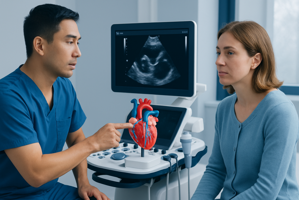
The tricuspid valve lies between the right atrium and right ventricle. It has three leaflets, or flaps. It opens to allow oxygen-poor blood returning from the body to enter the right ventricle, then closes when the ventricle pumps blood toward the lungs.
The pulmonary valve sits between the right ventricle and the pulmonary artery. It opens when the right ventricle contracts, allowing blood to travel to the lungs for oxygen. It then closes to keep blood from returning to the ventricle.
The mitral valve is located between the left atrium and left ventricle. It normally has two leaflets and allows oxygen-rich blood from the lungs to move into the heart’s main pumping chamber. The aortic valve lies between the left ventricle and aorta; it opens as the left ventricle pumps blood to the body and closes between heartbeats.
- Atrioventricular valves: the tricuspid and mitral valves, located between the atria and ventricles.
- Semilunar valves: the pulmonary and aortic valves, located at the exits of the ventricles.
How Heart Valves Work During Each Heartbeat

Heart valves do not actively open and close like muscles. Instead, they respond to changing pressure inside the heart. When pressure behind a valve becomes greater than pressure ahead of it, the valve opens. When pressure reverses, the valve closes.
During the filling phase of a heartbeat, called diastole, the mitral and tricuspid valves open so blood can move from the atria into the ventricles. During the pumping phase, called systole, these valves close while the aortic and pulmonary valves open to send blood out of the heart.
The mitral and tricuspid valves are supported by thin, cord-like structures called chordae tendineae, which attach to papillary muscles in the ventricles. These supporting structures help prevent the valve leaflets from turning backward during ventricular contraction. The aortic and pulmonary valves have three pocket-shaped cusps and do not have chordae tendineae.
What Can Affect Heart Valve Function?
Valve disease develops when a valve does not open fully, does not close completely, or has an abnormal shape. Stenosis means narrowing, which makes it harder for blood to pass through. Regurgitation, also called insufficiency or leakage, means a valve allows blood to flow backward.
Some valve conditions are present at birth. Others develop later due to age-related calcification, rheumatic heart disease, infection of the heart lining or valves, previous chest radiation, connective tissue disorders, or changes in heart size. A bicuspid aortic valve, for example, has two cusps instead of the usual three and may be present from birth.
Valve disorders can affect one valve or more than one. Examples include heart valve disease, aortic stenosis, mitral regurgitation, and infective endocarditis. The impact depends on the valve involved, how severe the problem is, how quickly it developed, and how well the heart muscle is functioning.
Heart valves guidelines from major cardiology organizations generally emphasize regular follow-up, imaging tailored to disease severity, and shared decision-making when intervention is being considered. A cardiologist can explain which recommendations apply to the individual patient.
Symptoms, Diagnosis, and Monitoring
Mild valve disease may not cause symptoms and may be discovered during a routine examination when a clinician hears a heart murmur. A murmur is a sound caused by turbulent blood flow; it does not always mean that treatment is needed, but it may lead to further assessment.
When valve disease becomes more significant, possible symptoms include shortness of breath, reduced exercise tolerance, unusual tiredness, chest discomfort, palpitations, dizziness, fainting, ankle swelling, or a sensation of a fast or irregular heartbeat. These symptoms can also have causes unrelated to the valves, so medical assessment is important.
Echocardiography is the main test for evaluating heart valves. It uses sound waves to show valve shape, movement, blood flow, and the effects of valve disease on heart chambers. Depending on the situation, clinicians may also use electrocardiography, chest imaging, exercise testing, transesophageal echocardiography, cardiac CT, cardiac MRI, or cardiac catheterization.
Monitoring is individualized. A person with a mild, stable valve problem may need periodic clinical reviews and echocardiograms, whereas moderate or severe disease usually requires closer follow-up. Keeping scheduled appointments helps identify changes before they place unnecessary strain on the heart.
Heart Valve Repair and Replacement: Patient Education
Not every valve condition requires a procedure. Treatment may include monitoring, managing blood pressure or heart rhythm problems, treating fluid buildup, preventing complications in selected situations, and addressing the underlying cause where possible. Medicines can ease symptoms or support heart function, but they usually cannot reverse a severely narrowed valve.
When a valve becomes severely narrowed or leaky, a specialist team may consider valve repair or replacement. Repair preserves the person’s own valve and is often preferred when it can provide a durable result, particularly for some mitral and tricuspid valve problems. Replacement may be recommended when repair is not suitable or when the valve is too damaged.
For heart valve replacement patient education, it is helpful to know that replacement valves may be mechanical or biological. Mechanical valves are highly durable but commonly require long-term anticoagulant treatment. Biological valves are made from animal tissue or donated human tissue and may not require lifelong anticoagulation solely because of the valve, although they can wear out over time. The best choice depends on age, health, anatomy, future pregnancy plans, bleeding risk, preferences, and other clinical factors.
Options may include open-heart surgery or, for carefully selected patients, less invasive catheter-based procedures. Heart valve replacement planning typically involves a heart team that may include cardiologists, cardiac surgeons, imaging specialists, anesthesiologists, and other professionals. Candidacy is based on symptoms, imaging findings, valve anatomy, overall health, and expected benefit.
What Happens During Treatment and Recovery?
The exact steps depend on whether the procedure is surgical or catheter-based. In surgical repair or replacement, the patient receives anesthesia, the heart is accessed through an incision, and the surgeon repairs or replaces the affected valve. Some operations can be performed through smaller incisions in appropriate patients, but this is not suitable for every type of valve problem.
In a catheter-based procedure, a specialist guides a thin tube through a blood vessel, often from the groin, to reach the heart. A replacement valve or repair device may then be positioned using imaging guidance. These procedures are used for selected conditions and are not interchangeable with surgery in every case.
Recovery also varies. Hospital stays after catheter-based treatment are often shorter than after open surgery, while recovery following surgical procedures commonly takes several weeks and continues gradually at home. Follow-up appointments, imaging, wound care when relevant, activity guidance, and cardiac rehabilitation may all be part of recovery.
Potential benefits include improved blood flow, relief of symptoms, better ability to be active, and protection of heart function. Risks depend on the procedure and individual health but can include bleeding, infection, abnormal heart rhythms, stroke, blood clots, kidney problems, valve leakage, or a need for another procedure. The care team discusses expected benefits and risks before treatment.
When to Seek Medical Care
A person should arrange medical evaluation for new or worsening breathlessness, reduced ability to exercise, repeated dizziness, fainting, swelling in the legs or abdomen, persistent palpitations, or chest discomfort. These symptoms do not always indicate valve disease, but they deserve timely assessment.
Urgent medical care is needed for severe chest pain, sudden severe shortness of breath, fainting with ongoing symptoms, blue or gray lips, confusion, or symptoms that may suggest a stroke, such as facial drooping, arm weakness, or difficulty speaking. Local emergency services should be contacted rather than driving oneself to care.
People with known valve disease should follow their cardiologist’s monitoring plan and ask before starting strenuous exercise, becoming pregnant, or having non-cardiac surgery. Good oral hygiene and regular dental care are also sensible because they support overall health; antibiotic prevention before dental work is recommended only for certain high-risk heart conditions.
Acibadem International’s multidisciplinary specialists and JCI-accredited hospitals assess and treat heart valve conditions for international patients, with care plans based on individual anatomy, symptoms, and diagnostic findings.
Frequently asked questions
How many valves are in the heart?
The heart has four valves: tricuspid, pulmonary, mitral, and aortic. They work in sequence to keep blood moving in one direction through the heart, lungs, and body.
Which heart valve is most commonly affected by disease?
Any valve can be affected, but aortic and mitral valve conditions are commonly diagnosed in adults. Aortic stenosis may develop with age-related valve calcification, while mitral regurgitation can have several causes involving the valve, heart muscle, or supporting structures.
Can a damaged heart valve heal on its own?
A structurally damaged or severely calcified valve usually does not return to normal on its own. However, some mild abnormalities remain stable for years, and treatment of related conditions can reduce symptoms or slow strain on the heart.
Does a heart murmur always mean valve disease?
No. Some murmurs are harmless and occur when blood flows more quickly through a normal heart. A clinician may recommend an echocardiogram if the murmur sounds abnormal or if symptoms or other findings suggest a valve problem.
Can heart valve disease be prevented?
Not all valve disease can be prevented, especially conditions present from birth or age-related changes. Managing blood pressure, avoiding smoking, treating infections appropriately, maintaining dental health, and attending recommended medical follow-up can support heart health.
When is heart valve replacement needed?
Replacement may be considered when valve narrowing or leakage is severe, causes symptoms, affects heart function, or is likely to lead to complications. The timing and approach are decided after detailed imaging and discussion with a specialist heart team.
References
- American Heart Association
- American College of Cardiology
- European Society of Cardiology
- National Heart, Lung, and Blood Institute
- Mayo Clinic
This article is for general information only and is not a substitute for professional medical advice. Please consult a qualified doctor about your individual situation.
Explore treatments in Turkey — costs, top hospitals & a free quote
JCI-accredited · board-certified surgeons · reply within 24h
Add us as a Preferred Source to see more of our trusted health content across Google Search, AI Overviews and Discover.