Bone Tumor CT Scan: Preparation, Procedure and Results
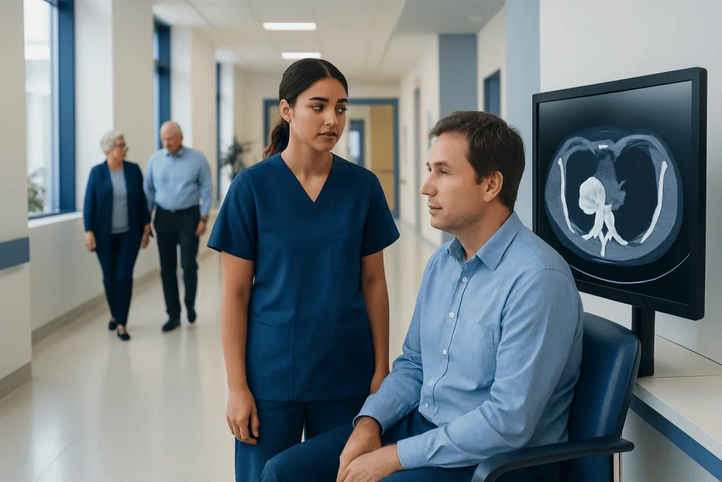
A CT scan provides detailed images of bone structure and can show features of a bone lesion, including its size, location and effect on surrounding tissue. CT findings may suggest whether a lesion is more likely benign or malignant, but biopsy is often needed for a definite diagnosis.
Key Takeaways
- A CT scan provides detailed images of bone structure and can show features of a bone lesion, including its size, location and effect on surrounding tissue.
- CT findings may suggest whether a lesion is more likely benign or malignant, but biopsy is often needed for a definite diagnosis.
- Preparation depends on whether contrast material is planned; patients should follow the imaging centre's instructions about eating, drinking and medicines.
- The scan itself is usually quick, painless and non-invasive, with most people returning to normal activities immediately afterward.
- Results are interpreted alongside symptoms, examination findings and other tests such as X-rays, MRI, blood tests or nuclear medicine scans.
A bone tumor CT scan is an imaging test that uses X-rays and computer processing to produce detailed pictures of bone and nearby tissues. It can help clinicians assess a suspicious bone lesion, evaluate its extent and plan further tests, but it usually cannot confirm cancer without a biopsy.
Bone Tumor CT Scan: What It Shows
A bone tumor CT scan is a computed tomography examination used to create detailed, cross-sectional images of a bone and the tissues around it. It may be requested after an X-ray identifies an unusual area, when a person has persistent bone pain or swelling, or when a clinician needs a clearer view of a known bone lesion.
CT is particularly useful for showing the hard outer layer of bone, small areas of mineralisation, subtle bone destruction and the relationship between a lesion and nearby joints or structures. If cancer is suspected or already diagnosed, it may help assess the local extent of disease, identify suitable biopsy routes and assist surgical planning.
A CT scan is only one part of assessment. Radiologists and treating clinicians interpret it together with medical history, physical examination and, where needed, other imaging such as MRI or a bone cancer assessment. A tissue biopsy is commonly required to establish the exact diagnosis.
How a CT Scan for a Bone Tumor Works
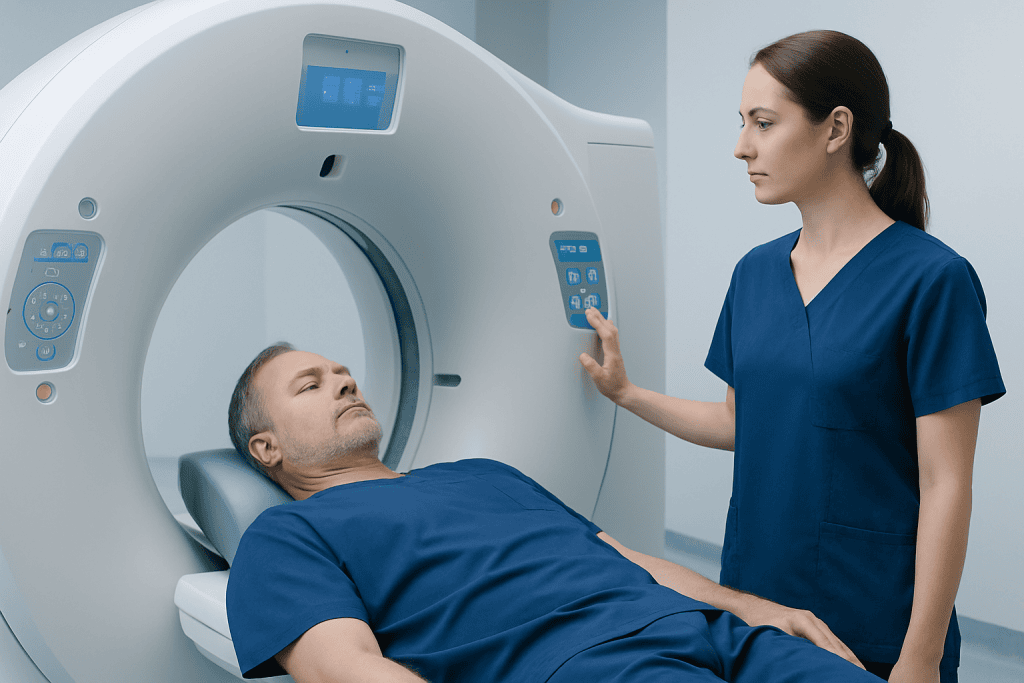
CT scanners use a rotating X-ray source and detectors to collect images from many angles around the body. A computer then reconstructs these images into thin slices, which can be viewed individually or combined into detailed three-dimensional images.
For bone lesions, CT can clarify the lesion’s margins, internal pattern and density. It can also show whether the surrounding bone has reacted by forming new bone, whether the cortex is thinned or broken, and whether there is involvement of adjacent soft tissue. These details can help narrow the range of possible diagnoses.
Some examinations use an iodine-based contrast material, usually injected into a vein. Contrast can make blood vessels and certain soft-tissue changes more visible. It is not needed for every bone CT, so the radiology team will decide based on the clinical question.
CT exposes patients to ionising radiation. The team uses the lowest practical dose needed to obtain useful images, and the expected benefit of answering an important clinical question generally outweighs this small risk.
Who May Need a Bone Tumor CT Scan
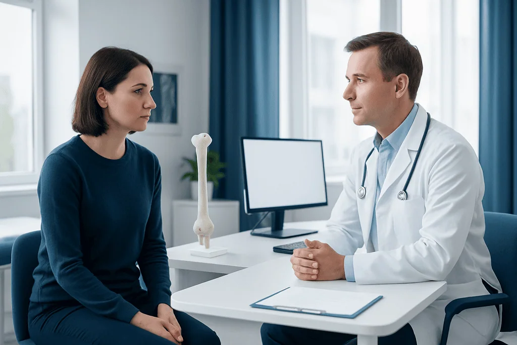
A clinician may recommend a bone tumor CT scan for a person with an unexplained bone lesion on an X-ray, persistent localised bone pain, swelling, a lump near a bone, or a fracture that occurred with little force. It may also be used to investigate symptoms in someone with a history of cancer, although symptoms do not necessarily mean a lesion is cancerous.
CT is often selected when precise detail of the bone itself is needed. MRI may be preferred to assess bone marrow, muscles, nerves and other soft tissues, while nuclear medicine imaging may be used to look for changes in multiple bones. These tests answer different questions and may be complementary rather than interchangeable.
People who are pregnant or may be pregnant should tell their doctor and radiology team before the scan. The examination can often be postponed or adjusted, depending on the urgency. Patients should also report kidney disease, previous reactions to contrast material, thyroid conditions and all medicines they take if contrast is being considered.
- Known allergy or previous reaction to iodine-based contrast
- Reduced kidney function or dialysis treatment
- Diabetes medicines or anticoagulants that may require individual advice
- Difficulty lying flat or remaining still because of pain, anxiety or limited mobility
Preparation and Step-by-Step Procedure
The imaging centre provides preparation instructions based on the body area being examined and whether contrast is planned. For a CT scan without contrast, many people can eat and drink normally. If contrast is planned, patients may be asked to avoid food for a few hours beforehand, while clear fluids may still be allowed. Individual instructions from the radiology department should always take priority.
Before the examination, the patient may change into a gown and remove jewellery, belts, removable dental items or other metal near the area being scanned. The radiographer reviews safety questions and, if contrast is used, may place a small intravenous cannula in the arm or hand.
During the scan, the patient lies on a padded table that moves slowly through the circular opening of the CT scanner. The machine is open at both ends and does not usually cause the enclosed feeling associated with some MRI scans. The radiographer can see and speak with the patient throughout the procedure.
The actual image acquisition often takes only a few minutes, although the full appointment may take longer for preparation, positioning and contrast administration. Remaining still is important because movement can blur the images. Most patients do not feel anything during the scan; contrast may cause a brief warm sensation or a metallic taste.
What Should You Not Do Before a Bone Scan?
The term “bone scan” often refers to a nuclear medicine test rather than a CT scan. A nuclear bone scan uses a small amount of radioactive tracer to show areas of increased bone activity, whereas a CT scan shows detailed anatomy. Preparation instructions are therefore different, and patients should confirm which test has been scheduled.
Before a nuclear bone scan, patients should not stop prescribed medicines unless their clinician specifically advises it. They should tell the department about pregnancy, breastfeeding, recent imaging tests and any supplements or medicines they use. Some centres may give specific instructions about products containing bismuth or recent contrast studies, as these can occasionally affect certain imaging tests.
Before a bone tumor CT scan, people should not eat or drink contrary to the department’s instructions, particularly if intravenous contrast is planned. They should also avoid arriving without information about allergies, kidney conditions or prior contrast reactions. Comfortable clothing and early arrival can make the visit easier.
Can a CT Scan Tell if a Bone Lesion Is Cancerous?
A CT scan can show features that help radiologists judge whether a bone lesion appears more likely to be benign or concerning. For example, its border, pattern of bone change, growth into nearby tissue and associated fracture can provide important clues. However, imaging appearance alone cannot reliably determine the nature of every lesion.
Some benign conditions can look active or unusual on imaging, and some cancers can initially appear less distinctive. For this reason, clinicians consider the person’s age, symptoms, medical history and findings from X-ray, MRI or other tests. Follow-up imaging may be appropriate for selected lesions that have reassuring features.
When a lesion remains suspicious or its exact type will change treatment, a biopsy may be recommended. Image-guided biopsy is planned carefully so that the sample is accurate and does not interfere with possible future surgery. Discussion by radiology, orthopaedic oncology, pathology and medical oncology teams is often valuable for complex cases.
Benefits, Risks and Recovery After CT
The main benefit of a bone tumor CT scan is the detailed information it can provide quickly. It can help define the anatomy of a lesion, identify complications such as a weakened bone or fracture, guide a safe biopsy approach and support surgical or other treatment planning. It may also provide a useful baseline for future comparison.
The scan is generally painless and does not require recovery time. Once the examination is complete, most people can return to usual activities immediately. If contrast was used, the team may advise drinking fluids afterward unless the patient has been told to limit fluid intake for another medical reason.
Risks are uncommon but include exposure to a small amount of radiation and, when contrast is used, nausea, itching or a rash. Serious allergic-type reactions are rare, and radiology teams are prepared to manage them. Contrast can occasionally affect kidney function, especially in people with significant pre-existing kidney disease; screening questions and, when appropriate, blood tests help reduce this risk.
If there is persistent rash, breathing difficulty, facial swelling or other concerning symptoms after leaving the department, urgent medical help is needed. Patients should also contact their clinician if they develop redness, pain or swelling around the injection site.
What Happens if You Eat 2 Hours Before a CT Scan?
Eating two hours before a CT scan is often not a problem for an examination performed without contrast, but requirements vary by scan type and local protocol. For CT scans involving intravenous contrast, some departments ask patients to avoid solid food for a specified period to reduce the chance of nausea or vomiting.
If a patient has eaten, they should tell the radiology team rather than cancelling the appointment themselves. The team can decide whether the scan can proceed, whether a short delay is needed or whether no change is necessary. Water is commonly allowed, but patients should follow their own appointment instructions.
Fasting instructions may also differ if sedation, anaesthesia or another procedure is planned on the same day. People with diabetes, children and those with medical conditions that make fasting difficult may need an individual plan from their clinical team.
How Long Does It Take to Get Results From a Bone Scan for Cancer?
For a CT scan, the images are usually available immediately after the examination, but they must be reviewed by a radiologist. A formal report may be sent to the referring clinician within a few days, although timing varies by hospital, urgency and whether comparison with earlier studies is needed.
A nuclear medicine bone scan may involve two stages: receiving the tracer and returning for images after an uptake period. The images are then interpreted by a nuclear medicine physician or radiologist. Results are commonly discussed at a follow-up appointment rather than at the scanning visit.
If a test was requested urgently, the referring clinician may receive preliminary information sooner. Patients should ask when and how they will receive results, and should avoid drawing conclusions from image portals without a clinician’s interpretation. An abnormal scan does not by itself diagnose cancer.
When to Seek Medical Care
Medical assessment is advisable for bone pain that persists, becomes worse, wakes a person from sleep, or is associated with a swelling, lump or reduced movement of a nearby joint. A doctor should also assess an unexplained fracture, especially after a minor injury, and new or worsening symptoms in someone with a current or previous cancer diagnosis.
Urgent medical attention is appropriate for severe pain after an injury, inability to bear weight, new weakness or numbness, loss of bladder or bowel control, or signs of a serious allergic reaction after contrast. These symptoms can have several causes, but prompt assessment is important.
Acibadem International’s multidisciplinary specialists and JCI-accredited hospitals support diagnosis and treatment planning for international patients with suspected or confirmed bone tumors. Care may include coordinated imaging, pathology review and bone tumor treatment planning when clinically appropriate.
Frequently asked questions
Is a bone tumor CT scan painful?
The CT scan itself is painless. The patient needs to lie still on a table while it moves through the scanner. If contrast is injected, there may be a brief warm feeling or metallic taste, which usually passes quickly.
How long does a bone tumor CT scan take?
The image-taking portion is often completed within minutes. The total visit may take around 30 minutes or longer because of registration, safety checks, positioning and possible contrast administration. The exact time depends on the body area and scan protocol.
Do I need contrast for a CT scan of a bone lesion?
Not always. CT without contrast can provide excellent information about the bone itself. Contrast may be added when the clinician needs a clearer view of nearby blood vessels, soft tissues or possible tumour extension.
Can CT distinguish a benign bone tumor from a malignant one?
CT can identify patterns that make a lesion appear more or less concerning, but it cannot definitively classify every lesion. Other imaging, follow-up scans or a biopsy may be needed. A specialist team interprets the findings in the context of the person's symptoms and history.
Can I drive home after a CT scan?
Most people can drive and return to normal activities after a routine CT scan. If sedation was used, which is uncommon for CT, the patient will need someone else to take them home. People who feel unwell after contrast should follow the advice of the imaging team.
What should I bring to my CT appointment?
Patients should bring their referral information, identification, relevant previous imaging reports or discs if requested, and a list of medicines and allergies. It is also helpful to mention kidney disease, pregnancy or a past reaction to contrast material. The radiology department may provide additional instructions before the appointment.
References
- American College of Radiology
- Radiological Society of North America
- National Cancer Institute
- National Institute of Biomedical Imaging and Bioengineering
- World Health Organization
This article is for general information only and is not a substitute for professional medical advice. Please consult a qualified doctor about your individual situation.
Oncology care in Turkey — second opinion and treatment plan
JCI-accredited · board-certified surgeons · reply within 24h
Add us as a Preferred Source to see more of our trusted health content across Google Search, AI Overviews and Discover.

More from the Health Library


Related Specialists

Prof. Dr. Coşkun Çeltik
Pediatrics
Embriyolog Gülsüm Tüysüz
Vitro Fertilization and Reproductive Medicine Center
Dr. Şahiner Sezgin
Internal Medicine
Dr. Olcayto İncedere
Cardiology