

Lung Cancer Xray Pictures: Preparation, Procedure and Results
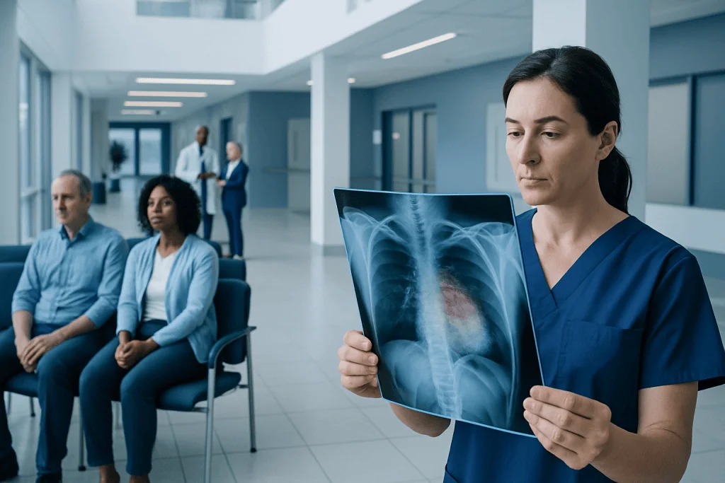
A chest X-ray is often an early test for respiratory symptoms, but it is not a screening test that can reliably exclude lung cancer. Lung cancer may appear as a white shadow, round nodule, enlarged area near the center of the chest, fluid around the lung, or partial lung collapse.
Key Takeaways
- A chest X-ray is often an early test for respiratory symptoms, but it is not a screening test that can reliably exclude lung cancer.
- Lung cancer may appear as a white shadow, round nodule, enlarged area near the center of the chest, fluid around the lung, or partial lung collapse.
- Small, early cancers may not be visible on an X-ray, particularly when hidden by the heart, ribs, or other chest structures.
- CT scanning provides more detailed images and is commonly the next step after an abnormal chest X-ray.
- Only examination of a tissue sample, called a biopsy, can confirm a diagnosis of lung cancer.
Lung cancer xray pictures can sometimes reveal an abnormal area in the lung, such as a mass, collapse, fluid buildup, or a change in lung tissue. However, a chest X-ray cannot reliably diagnose or rule out lung cancer; further imaging, often a CT scan, and a tissue biopsy are usually needed.
Overview: What Lung Cancer Xray Pictures Can Show
Lung cancer xray pictures are chest X-ray images that may be taken when a person has symptoms such as a persistent cough, coughing up blood, chest discomfort, unexplained weight loss, repeated chest infections, or shortness of breath. The image may show a change that needs further assessment, but it cannot by itself confirm that cancer is present.
On an X-ray, lungs usually look dark because they contain air. Areas that are denser than air, including infection, fluid, scarring, enlarged lymph nodes, or a tumor, can appear lighter or whiter. A radiologist interprets the image alongside the person’s symptoms, medical history, prior scans, and risk factors. An unexpected shadow should be evaluated carefully, but it does not automatically mean cancer.
Chest X-rays are quick and widely available, making them useful as an initial investigation. They are less sensitive than CT scans for small nodules and early-stage disease. When an X-ray is abnormal or symptoms remain concerning despite a normal result, clinicians may recommend further lung cancer assessment and treatment planning through a specialist team.
How a Chest X-Ray Works and Who May Need One
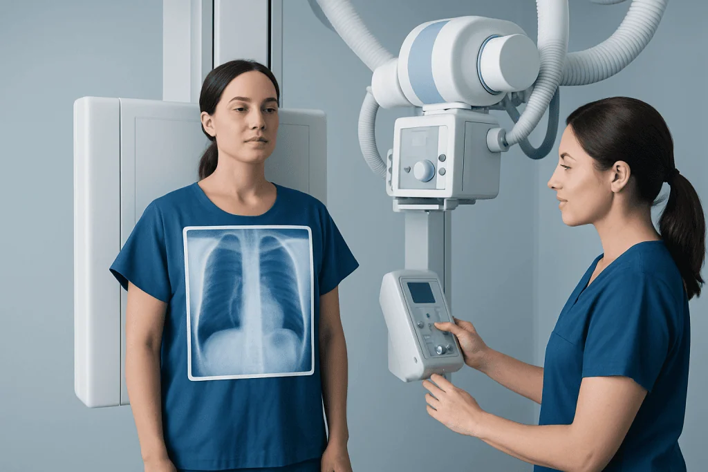
A chest X-ray uses a small amount of ionizing radiation to create a two-dimensional image of the lungs, heart, ribs, and nearby structures. Different tissues absorb different amounts of radiation. Air-filled lungs allow more radiation to pass through and look darker, while bone and denser tissue look lighter.
A clinician may request a chest X-ray for new or ongoing chest symptoms, after an abnormal examination finding, or to investigate possible infection and other lung conditions. It may also be used to monitor a known lung problem. People with a history of smoking, substantial secondhand smoke exposure, occupational exposure to substances such as asbestos, or a personal history of cancer may need particularly prompt review of persistent symptoms.
For people at high risk for lung cancer, low-dose CT screening may be more appropriate than chest X-ray screening. Eligibility depends on factors such as age and smoking history and should be discussed with a qualified clinician. A chest X-ray is not considered an effective stand-alone screening test for people without symptoms.
- Chest X-ray: often used as a first assessment for symptoms.
- CT scan: provides detailed cross-sectional images and detects smaller abnormalities.
- PET-CT: may help assess whether a confirmed or suspected cancer has spread.
- Biopsy: obtains cells or tissue to establish the diagnosis.
How Do I Prepare for a Lung Scan?
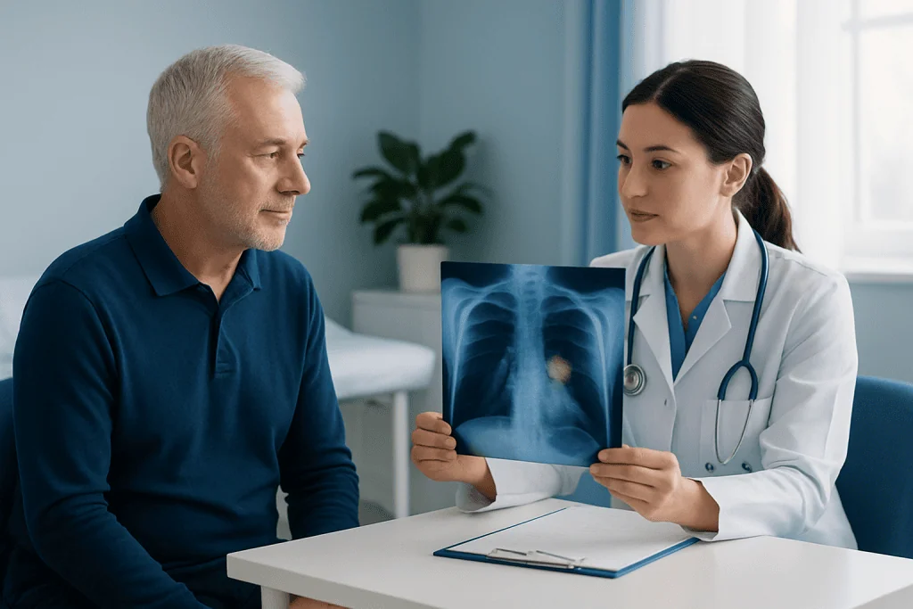
Preparation depends on the type of lung scan being performed. For a standard chest X-ray, most people can eat, drink, and take regular medicines as usual unless their clinician gives different instructions. The examination is generally painless and does not require needles, contrast dye, sedation, or recovery time.
Before the image is taken, the person may be asked to remove jewelry, clothing with metal fasteners, and other items from the chest and neck area, then change into a gown. It is important to tell the radiology team about pregnancy or the possibility of pregnancy, even though the radiation dose from a chest X-ray is low and protective measures can be considered.
A CT scan may involve additional preparation, especially if contrast dye is planned. The imaging department may ask about kidney disease, allergies, diabetes medicines, and previous reactions to contrast material. Some CT examinations require a short period without food beforehand, so patients should follow the instructions provided for their specific appointment.
Step by Step: What Happens During and After the Procedure
For a chest X-ray, a radiographer positions the person standing or sitting against an image detector. Most examinations include a front view and a side view. The person is asked to hold still and briefly hold their breath while each image is captured. The actual exposure lasts only a fraction of a second.
The appointment commonly takes about 10 to 15 minutes, including positioning. A person who is too unwell to stand may have a portable X-ray performed at the bedside. Afterward, normal activities can usually be resumed immediately because there is no anesthesia, contrast injection, or physical recovery period.
The images are reviewed by a radiologist, who sends a report to the requesting clinician. If a possible abnormality is found, the next step may be repeat imaging, CT scanning, referral to a respiratory physician, or review by a multidisciplinary cancer team. The pathway is individualized because many non-cancerous conditions can produce changes on a chest X-ray.
The benefits of chest X-ray include speed, convenience, and the ability to identify many important chest conditions. Its main limitation is that overlapping structures can conceal small lesions. The radiation exposure is low, but imaging is still used when the expected clinical benefit outweighs the small risk.
What Does the First Stage of Lung Cancer Look Like?
Early-stage lung cancer may not cause any symptoms and may not have a distinctive appearance on a chest X-ray. If visible, it can appear as a small rounded spot, called a pulmonary nodule, or as a subtle area of increased density. However, many nodules are benign and may be caused by old infections, inflammation, or harmless scars.
Stage I lung cancer generally means that the cancer is limited to the lung and has not spread to nearby lymph nodes or distant organs. Staging cannot be determined accurately from an X-ray alone. CT imaging, PET-CT in selected cases, biopsy results, and sometimes surgical evaluation are used to define the cancer’s size, location, lymph node involvement, and spread.
Small cancers can be difficult to see on an X-ray because they may be hidden behind the heart, diaphragm, ribs, or central airways. This is one reason clinicians may order CT imaging when symptoms or risk factors remain concerning. Information about lung cancer can help patients understand the diagnostic pathway, possible stages, and treatment discussions that may follow.
How Long Does It Take to Get Results From a Lung Cancer Scan?
The time needed to receive results varies by hospital, the urgency of the request, and the type of scan. A chest X-ray image is available immediately, but the formal radiology report may be sent to the referring clinician on the same day or within several days. Urgent findings are generally communicated more quickly through clinical pathways.
CT and PET-CT results may take longer because they involve more images and detailed interpretation. If an abnormality requires specialist review, the clinician may discuss the results after consultation with respiratory, radiology, pathology, thoracic surgery, and oncology professionals.
A person should ask when and how results will be provided before leaving the appointment. If symptoms worsen while waiting, or if the person has been told the scan was urgent and has not received follow-up within the expected timeframe, contacting the requesting clinician is appropriate.
What Confirms Lung Cancer?
Lung cancer is confirmed by examining cells or tissue under a microscope. This is called a biopsy. A pathologist assesses the sample to determine whether cancer is present and, if it is, identifies the type of lung cancer. Molecular and genetic tests may also be performed on the tumor sample to help guide treatment decisions.
The way a biopsy is obtained depends on where the abnormality is located and a person’s overall health. Options can include bronchoscopy, an ultrasound-guided needle sample from lymph nodes, a CT-guided needle biopsy through the chest wall, or a surgical biopsy. The care team explains the expected benefits, limitations, and possible risks of the recommended approach.
Imaging remains essential before and after diagnosis. CT, PET-CT, brain imaging in selected situations, and lung function tests may help establish stage and support treatment planning. Management can include surgery, radiation therapy, systemic treatment, or a combination, depending on the cancer type, stage, molecular features, and the individual’s preferences and health.
At Acibadem International, multidisciplinary specialists in JCI-accredited hospitals support international patients through diagnostic evaluation and individualized treatment planning for lung cancer and other thoracic conditions.
When to Seek Medical Care
A person should arrange a medical assessment for a cough that lasts more than a few weeks, a cough that changes or worsens, unexplained breathlessness, chest or shoulder pain, recurring chest infections, persistent hoarseness, unexplained fatigue, or unintentional weight loss. These symptoms are common and often have causes other than cancer, but they deserve professional assessment when persistent or unexplained.
Urgent medical care is appropriate for coughing up blood, severe or rapidly worsening shortness of breath, new severe chest pain, blue or gray lips, fainting, or confusion. These symptoms can occur with a range of serious conditions and should not be managed by waiting for a routine imaging appointment.
People should not rely on online image comparisons to interpret their own scans. Lung cancer Xray pictures can look different from one person to another, and accurate interpretation requires trained review of the complete image, clinical context, and often additional tests.
Frequently asked questions
Can a chest X-ray detect lung cancer?
A chest X-ray can detect some changes that may be caused by lung cancer, including a mass, nodule, fluid around the lung, or collapse of part of the lung. However, it can miss small or early cancers and cannot confirm the diagnosis. CT imaging and a biopsy are often needed.
Can lung cancer be missed on an X-ray?
Yes. Small tumors or tumors located behind the heart, ribs, diaphragm, or central chest structures may not be visible on a chest X-ray. A normal X-ray does not always exclude lung cancer when symptoms or risk factors remain concerning.
What does lung cancer look like on an X-ray picture?
It may look like a white or gray shadow, a rounded nodule, an area of collapse, or fluid around a lung. These findings are not specific to cancer and can also occur with infection, inflammation, scarring, or other conditions. A radiologist and treating clinician interpret the finding in context.
Is a chest X-ray painful?
No. A chest X-ray is painless and usually takes only a few minutes of positioning and image capture. The person may need to take a deep breath and hold it briefly while the pictures are taken.
Do I need to fast before a chest X-ray?
Fasting is usually not needed for a standard chest X-ray. People can generally eat, drink, and take their regular medicines unless they have been given separate instructions. CT scans with contrast may have different preparation requirements.
What happens if a chest X-ray shows a spot on the lung?
A spot does not necessarily mean lung cancer. The clinician may compare older images, arrange a CT scan, recommend follow-up imaging, or refer the person to a respiratory specialist. The appropriate next step depends on the size and appearance of the spot, symptoms, and individual risk factors.
References
- World Health Organization
- American Cancer Society
- National Cancer Institute
- American College of Radiology
- National Health Service
This article is for general information only and is not a substitute for professional medical advice. Please consult a qualified doctor about your individual situation.
Lung Cancer in Turkey — costs, top hospitals & a free quote
JCI-accredited · board-certified surgeons · reply within 24h
Add us as a Preferred Source to see more of our trusted health content across Google Search, AI Overviews and Discover.

Medical Oncology Department
Medical treatment of cancer with chemotherapy, immunotherapy and targeted therapies under a multidisciplinary tumor board.
60 specialists in this unitMore from the Health Library



Related Specialists

Dr. Nevzat Demirhan
Radiology
Fzt. Sena Durmaz
Physical Medicine and Rehabilitation
Dt. Müge Tiralı Türkkan
Oral & Dental Health
Dr. Caner Ünlüer
Neurosurgery