Carotid Body Tumor on Ultrasound: Preparation, Procedure and Results
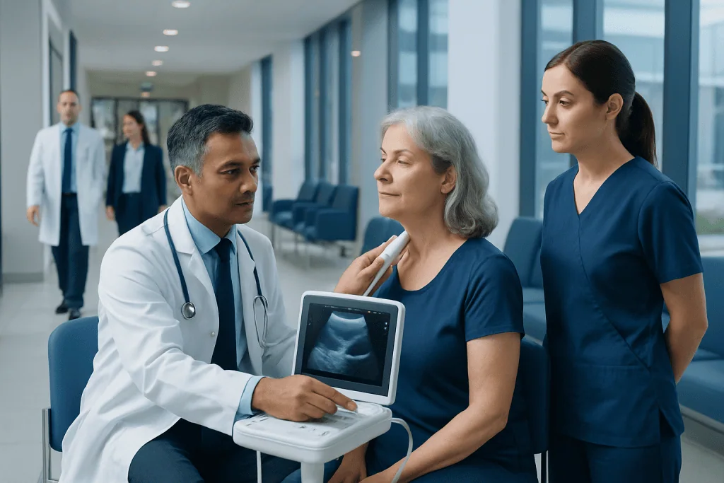
Carotid body tumors are usually slow-growing and most are noncancerous, but they require specialist assessment because of their location beside major blood vessels and nerves. Ultrasound uses sound waves and Doppler blood-flow imaging to assess a neck mass without radiation or injections.
Key Takeaways
- Carotid body tumors are usually slow-growing and most are noncancerous, but they require specialist assessment because of their location beside major blood vessels and nerves.
- Ultrasound uses sound waves and Doppler blood-flow imaging to assess a neck mass without radiation or injections.
- A typical ultrasound finding is a well-defined, blood-flow-rich mass that separates the internal and external carotid arteries.
- Most carotid ultrasound examinations take about 30 to 45 minutes, and preliminary findings may be available the same day.
- CT, MRI and sometimes functional imaging or laboratory tests may be used after ultrasound to clarify the diagnosis and plan care.
- Management may include monitoring, surgery, radiotherapy or a combination, depending on tumor size, growth, symptoms, health status and inherited risk.
A carotid body tumor on ultrasound may be identified as a highly vascular lump near the point where the main carotid artery divides in the neck. Ultrasound is a painless first-line imaging test that can help clinicians assess the mass and plan further evaluation, although additional scans are often needed to confirm its extent and guide treatment.
Overview: What a Carotid Body Tumor on Ultrasound Can Show
A carotid body tumor on ultrasound is often seen as a small-to-large mass at the carotid bifurcation, the area in the upper neck where the common carotid artery divides into the internal and external carotid arteries. The carotid body is a normal cluster of specialized cells that helps the body sense changes in oxygen, carbon dioxide and blood acidity. Tumors arising from this tissue are also called carotid body paragangliomas.
Ultrasound is commonly used when a clinician feels a painless neck lump, hears a blood-flow sound over the neck, or wants to investigate symptoms such as pressure or difficulty swallowing. It can show the location of a mass, its relationship to nearby arteries and the amount of blood flow within it. It does not use ionizing radiation and is generally comfortable and safe.
Although ultrasound can strongly suggest a carotid body tumor, it does not always provide a final diagnosis on its own. Contrast-enhanced MRI or CT angiography may be recommended to define the tumor’s size, blood supply and relationship to nerves and blood vessels. Because these tumors can be very vascular, a needle biopsy is usually avoided unless a specialist considers it necessary and safe.
How Carotid Ultrasound Works and Who May Need It
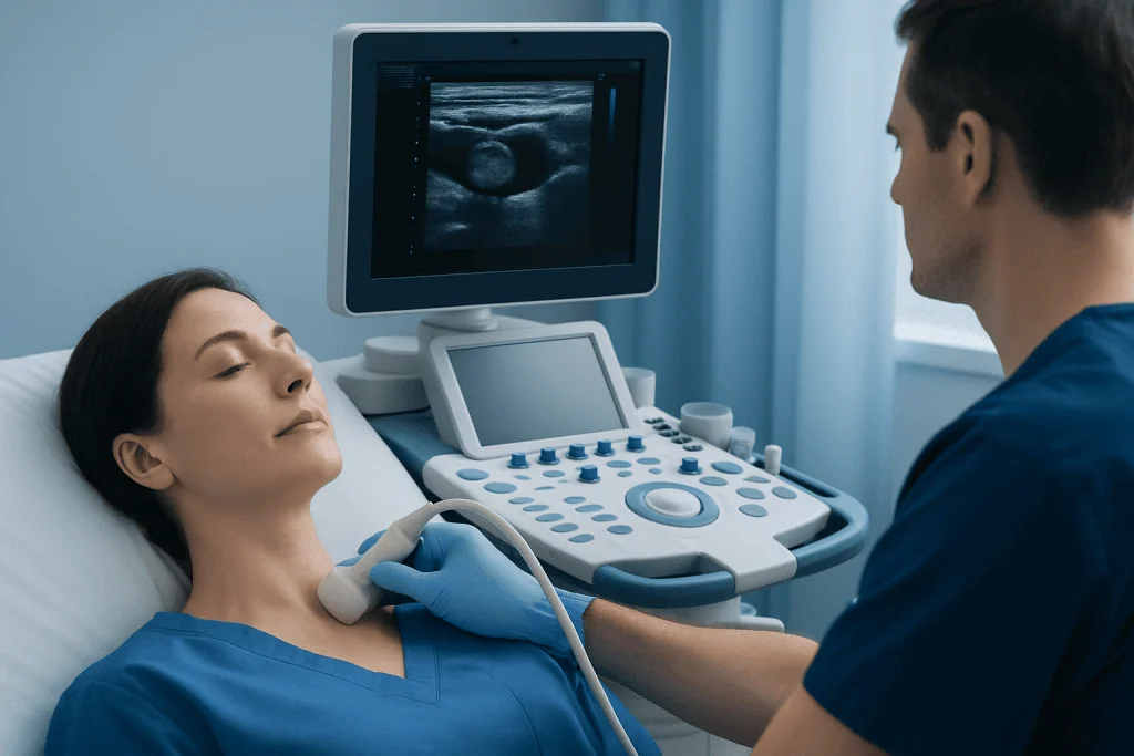
Carotid ultrasound uses a handheld device called a transducer. The transducer sends high-frequency sound waves into the neck and receives returning echoes, which a computer converts into images. Doppler ultrasound adds information about the direction and speed of blood flow, making it particularly useful for identifying the prominent blood supply often present in carotid body tumors.
A clinician may request this test for a persistent lump near the angle of the jaw, a pulsating neck swelling, unexplained neck discomfort, or changes in swallowing or voice. It may also be performed when imaging done for another reason shows a possible mass near the carotid arteries. People with a family history of paraganglioma or certain inherited gene changes may need periodic surveillance imaging as advised by their specialist.
Ultrasound is suitable for most people, including those who cannot receive certain contrast agents or who wish to avoid radiation exposure. However, very deep tumors, masses extending toward the skull base, or complex anatomy may be assessed more completely with MRI or CT. The choice of imaging is individualized and depends on the clinical question.
Preparation and Step-by-Step Procedure
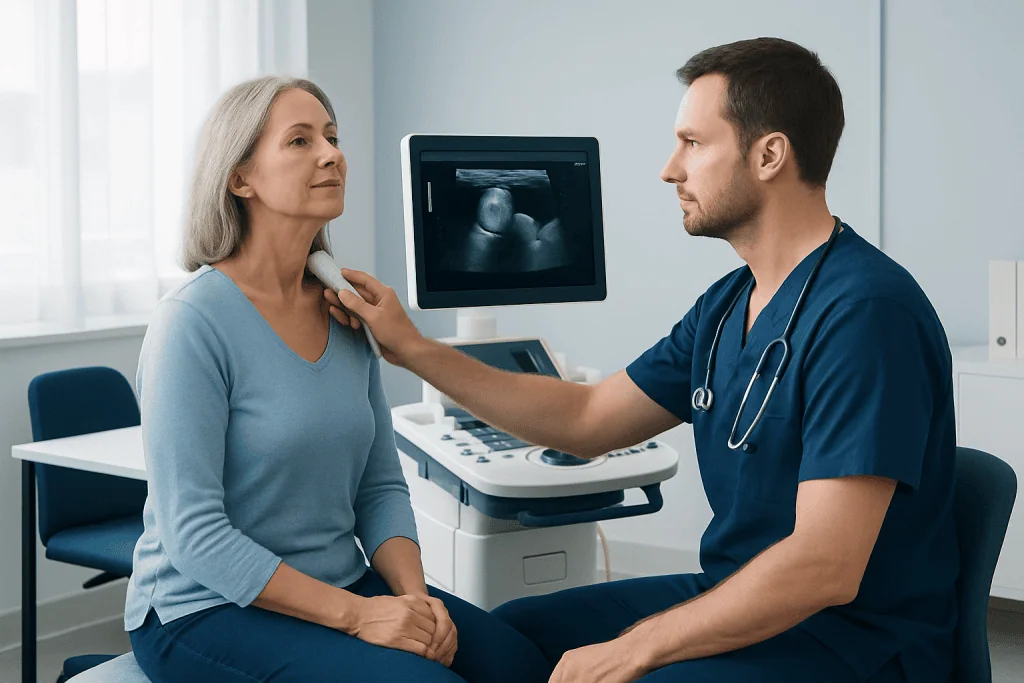
In most cases, no special preparation is needed for a carotid artery ultrasound. A person can usually eat, drink and take regular medicines as normal unless the healthcare team gives different instructions. Wearing an open-collar shirt or a top that allows easy access to the neck can make the appointment more comfortable. Necklaces and other jewelry should be removed before scanning.
During the examination, the person lies on their back with the head turned slightly away from the side being examined. A sonographer applies clear gel to the skin of the neck. The gel helps sound waves travel efficiently and does not stain clothing. The transducer is moved gently along the neck to produce images of the arteries and nearby tissues.
Images are captured in different positions, and Doppler settings are used to map blood flow. Gentle pressure may be needed, but the examination should not be painful. The scan generally takes around 30 to 45 minutes, though timing can vary when both sides of the neck or a detailed mass assessment is required. Normal activities can usually be resumed immediately afterward.
- Before the scan: remove neck jewelry and share relevant medical history.
- During the scan: lie still and follow simple instructions about head position or breathing.
- After the scan: wipe away the gel and wait for the report process explained by the imaging team.
What Are the Typical Characteristics of a Carotid Body Tumor on Ultrasound?
On ultrasound, a carotid body tumor often appears as a solid, well-defined mass near the carotid bifurcation. A key feature is splaying, or separation, of the internal and external carotid arteries as the mass grows between them. This pattern is sometimes described as the Lyre sign on imaging because of the shape made by the separated vessels.
Color and spectral Doppler ultrasound commonly show marked internal blood flow. This reflects the tumor’s vascular nature and helps distinguish it from some other neck lumps. The lesion may look darker than surrounding soft tissue on grayscale ultrasound, although appearance can vary with tumor size, equipment and individual anatomy.
Ultrasound findings need to be interpreted alongside symptoms, examination findings and other imaging. Enlarged lymph nodes, thyroid nodules, salivary gland conditions and other paragangliomas can also cause a neck mass. MRI or CT is frequently used to confirm the anatomical pattern and to look for additional tumors when appropriate.
How Long Does It Take to Get Results From a Carotid Artery Ultrasound?
The imaging professional may be able to comment on whether the scan has identified a visible mass or an arterial concern before a person leaves the department. However, the formal interpretation is usually completed by a radiologist, who reviews the saved images and prepares a written report for the referring clinician.
In many settings, a carotid artery ultrasound report is available within the same day or within several working days. The exact timeline depends on the urgency of the referral, local reporting processes and whether further specialist review is needed. If the scan was ordered because of concerning symptoms, the referring clinician may arrange quicker follow-up.
A report suggesting a vascular mass at the carotid bifurcation generally leads to referral to an experienced head and neck, vascular, endocrine or neurovascular team. The next step may include MRI, CT angiography, blood or urine testing for hormone production in selected cases, and consideration of genetic counseling when family history or clinical features indicate a possible inherited condition.
How Serious Is a Carotid Body Tumor?
Carotid body tumors are uncommon and are usually slow-growing. Most are benign, meaning they do not spread to distant parts of the body. Even when noncancerous, they can become clinically important because they develop close to the carotid arteries and nerves involved in swallowing, voice, shoulder movement and tongue function.
Potential concerns depend mainly on tumor size, growth rate, symptoms and its relationship to nearby structures. A growing tumor may cause a noticeable neck lump, pressure, swallowing difficulty, hoarseness or changes in nerve function. A small number of paragangliomas produce hormones that can affect blood pressure, heart rate, sweating or headaches; clinicians may screen for this when the history or tumor features suggest it.
Malignancy cannot be reliably determined simply by how the tumor looks on ultrasound or under a microscope. It is defined by spread to sites where this tissue is not normally present, such as lymph nodes, bone, liver or lungs. Specialist follow-up is therefore important, even for tumors that appear localized and slow-growing.
Treatment Options, Benefits and Possible Risks
Care is tailored to the individual. For a small, stable tumor causing no symptoms, structured observation with repeat imaging can be appropriate, particularly when surgery would carry a higher risk. This approach allows the team to monitor growth while avoiding treatment that may not be needed immediately.
When treatment is recommended, surgery may be considered for a growing, symptomatic or otherwise suitable tumor. The operation is planned carefully because of the tumor’s proximity to major arteries and cranial nerves. In selected situations, radiotherapy may help control tumor growth, especially if surgery is not advisable or if complete surgical removal would involve substantial risk.
The potential benefit of treatment is control of tumor growth and relief or prevention of local symptoms. Possible treatment risks vary by approach and may include bleeding, blood-vessel injury, stroke, nerve-related changes in voice, swallowing or shoulder movement, and effects from radiation. An experienced multidisciplinary team helps balance these considerations and discuss the most appropriate pathway. Acibadem International’s multidisciplinary specialists at JCI-accredited hospitals evaluate and treat carotid body tumors for international patients.
After a suspected diagnosis, people may also benefit from evaluation for related conditions or additional paragangliomas when clinically indicated. Ongoing follow-up remains important after treatment or during observation because these tumors can grow slowly over time.
What Is the Prognosis for a Carotid Body Tumor?
The prognosis for a carotid body tumor is often favorable, particularly when it is localized and managed by a team experienced in head and neck vascular tumors. Many tumors grow slowly, and some can be monitored safely for a period of time. The outlook is influenced by tumor size, symptoms, growth on repeat imaging, whether it produces hormones, inherited factors and the person’s overall health.
For people who undergo surgery, long-term results can be good, but the likelihood of complications may increase as tumors become larger or more closely involve the carotid arteries and nearby nerves. Radiotherapy can provide durable local control for selected patients. The specialist team will explain what the chosen approach is expected to achieve and what follow-up is needed.
Long-term surveillance may include clinical reviews and imaging. People with multiple tumors, a younger age at diagnosis or a relevant family history may be offered genetic counseling and testing. This can help clarify personal follow-up needs and whether relatives may benefit from medical advice.
When to Seek Medical Care
A person should arrange a medical assessment for a new or persistent lump in the upper neck, especially if it seems to pulse or gradually enlarge. Medical review is also appropriate for persistent hoarseness, trouble swallowing, unexplained neck pain, or a feeling of pressure near the jaw and throat. These symptoms have many possible causes, and prompt assessment helps identify the right explanation.
Urgent medical care is appropriate for sudden weakness or numbness on one side of the body, difficulty speaking, sudden vision loss, severe dizziness or a sudden severe headache. These symptoms can signal a neurological emergency and should not be attributed to a neck mass without immediate professional assessment.
Anyone already diagnosed with a carotid body tumor should attend planned reviews and report new symptoms, rapid change in the neck lump, episodes of palpitations or sweating, or significant changes in blood pressure to their clinical team. A specialist can advise whether repeat ultrasound or more detailed imaging is needed.
Frequently asked questions
Is a carotid body tumor visible on an ultrasound?
Yes. Ultrasound can often identify a carotid body tumor as a mass near the division of the carotid artery in the upper neck. Doppler imaging can demonstrate the increased blood flow that is typical of these tumors, but MRI or CT is often needed for full assessment.
Is a carotid artery ultrasound painful?
A carotid artery ultrasound is usually painless. The transducer may apply mild pressure to the neck, and the gel can feel cool, but no needles, radiation or sedation are normally required.
Do I need to fast before a carotid ultrasound?
Fasting is not usually required for a carotid ultrasound. A person should follow any instructions from the imaging center, especially if other tests are scheduled at the same visit.
Can ultrasound confirm that a carotid body tumor is cancerous?
No. Ultrasound can show features that support the diagnosis of a carotid body tumor, but it cannot determine whether it is malignant. Malignancy is diagnosed when tumor cells have spread to sites such as lymph nodes, bone, liver or lungs.
Why is a biopsy often avoided for a suspected carotid body tumor?
These tumors are often highly vascular, meaning they contain many blood vessels. A biopsy can therefore carry a bleeding risk and may not be needed when imaging findings are characteristic; the decision should be made by an experienced specialist team.
Can a carotid body tumor be watched instead of treated immediately?
In some people, particularly those with a small, stable and symptom-free tumor, planned observation with repeat scans may be appropriate. The decision depends on tumor growth, symptoms, anatomy, age, general health and personal preferences, and should be reviewed regularly with a specialist.
References
- National Cancer Institute
- National Institute for Health and Care Excellence
- American Cancer Society
- Radiological Society of North America
- European Society of Endocrinology
This article is for general information only and is not a substitute for professional medical advice. Please consult a qualified doctor about your individual situation.
Ultrasound in Turkey — costs, top hospitals & a free quote
JCI-accredited · board-certified surgeons · reply within 24h
Add us as a Preferred Source to see more of our trusted health content across Google Search, AI Overviews and Discover.

Check-up & Preventive Medicine
Comprehensive health screenings designed for early detection and prevention.
2 specialists in this unit