Ductal Hyperplasia Treatment: How It Works, Results and What to Expect

Usual ductal hyperplasia is benign and usually does not need removal or cancer treatment. Atypical ductal hyperplasia is not breast cancer, but it is linked with a higher future risk of breast cancer.
Key Takeaways
- Usual ductal hyperplasia is benign and usually does not need removal or cancer treatment.
- Atypical ductal hyperplasia is not breast cancer, but it is linked with a higher future risk of breast cancer.
- Surgical excision may be advised when atypical ductal hyperplasia is found on a needle biopsy, especially if imaging and pathology findings do not fully agree.
- Follow-up plans may include regular breast imaging, clinical reviews and discussion of risk-reducing options.
- New breast lumps, nipple discharge, skin changes or persistent focal breast symptoms should be assessed by a clinician.
Ductal hyperplasia treatment depends on the type of cell change found in breast ducts. Usual ductal hyperplasia commonly needs observation only, while atypical ductal hyperplasia may require surgical excision after a needle biopsy and longer-term breast cancer risk management.
Overview: what ductal hyperplasia treatment involves
Ductal hyperplasia treatment is guided by the type of cell growth found in the milk ducts of the breast. Usual ductal hyperplasia (UDH) is a common benign change and usually requires no procedure beyond routine breast screening. Atypical ductal hyperplasia (ADH) is also not cancer, but its cells have more unusual features and can be associated with a higher future risk of breast cancer.
When ADH is identified on a core needle biopsy, a breast team reviews the imaging, biopsy technique and pathology report together. Some people are advised to have an excisional biopsy, a small operation to remove the area of concern and check for nearby ductal carcinoma in situ (DCIS) or invasive cancer that may not have been sampled by the needle. Others may be suitable for carefully planned imaging surveillance when the findings are considered low risk and fully matched.
The aim is not to treat cancer that is already present, but to obtain an accurate diagnosis and develop a personalised prevention and follow-up plan. Care may involve breast radiologists, pathologists, breast surgeons, oncologists and genetic counsellors where appropriate.
How ductal hyperplasia is found and assessed
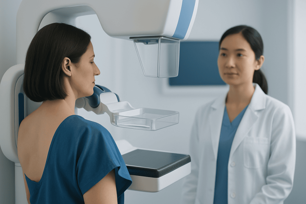
Ductal hyperplasia itself often causes no symptoms. It is frequently discovered after a mammogram shows calcifications, an area of distortion or another change that needs closer assessment. It may also be found incidentally in tissue removed for a different benign breast condition.
Assessment commonly begins with diagnostic mammography and may include breast ultrasound. If an abnormal area requires sampling, a radiologist can perform a core needle biopsy, often using mammographic guidance for calcifications. The tissue is examined under a microscope by a pathologist, who distinguishes UDH from ADH and from other conditions that may need different management.
A key part of decision-making is radiology-pathology concordance. This means confirming that the biopsy result adequately explains the imaging finding. If the imaging looks more concerning than the tissue result, if too little tissue was obtained, or if another high-risk lesion is present, surgical removal is more likely to be recommended.
Conditions such as breast cancer and DCIS are different from ductal hyperplasia. A specialist can explain what the pathology wording means, how it relates to the imaging results and whether additional testing is needed.
Ductal hyperplasia treatment: how it works and who may need it
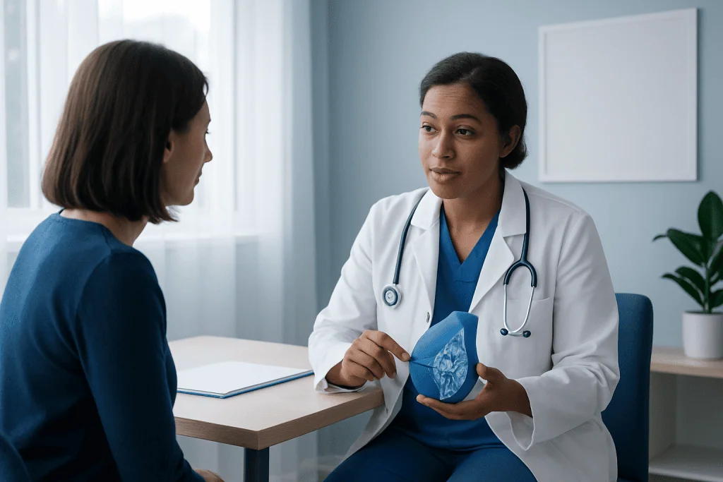
For UDH, treatment is generally reassurance and continuation of age- and risk-appropriate screening. UDH may slightly influence overall risk in some contexts, but it is not considered a precancerous lesion and does not routinely require surgery, medication or intensive follow-up.
For ADH, the next step after a needle biopsy depends on the details. Excisional biopsy is often considered when ADH is extensive, associated with a mass or suspicious calcifications, found alongside another high-risk lesion, or when imaging and pathology do not match. The procedure removes the biopsy area so that a pathologist can evaluate more tissue and exclude an overlooked cancer.
In selected cases, a multidisciplinary breast team may recommend close imaging surveillance instead of surgery. This may be considered when ADH is very limited, the abnormality appears to have been adequately sampled or removed by vacuum-assisted biopsy, and imaging-pathology concordance is strong. This decision should be individualised, because recommendations can vary according to local practice and the person’s overall risk profile.
If ADH is confirmed, clinicians may also discuss lifestyle measures, enhanced screening and risk-reducing medicines for people who may benefit. These medicines are not necessary for everyone; their potential benefits and side effects need a careful personal discussion.
What happens during an excisional biopsy and recovery
An excisional biopsy is usually an outpatient breast procedure. Before surgery, the area may be marked using imaging guidance, particularly when it cannot be felt as a lump. This localisation helps the surgeon remove the correct tissue while preserving as much normal breast tissue as possible.
On the day of surgery, the patient meets the surgical and anaesthesia teams. The procedure is commonly performed under general anaesthesia, although the anaesthesia plan depends on the individual and the planned operation. The surgeon makes a small incision, removes the targeted tissue and sends it to the laboratory for detailed examination. The incision is then closed and covered with a dressing.
Most people go home the same day. Tenderness, bruising, mild swelling and temporary numbness around the incision can occur. Many people can return to light daily activities within a few days, while strenuous exercise and heavy lifting may need to wait until the surgical team confirms healing is progressing well.
Pathology results often take several days to be finalised because the tissue must be processed and thoroughly examined. At the follow-up appointment, the team explains whether only ADH or another benign change was found, whether additional care is needed and what future screening plan is appropriate.
Benefits, limitations and possible risks
The main benefit of excisional biopsy for ADH is diagnostic certainty. It allows a larger tissue sample to be examined and can identify the small number of cases in which a more significant abnormality is present nearby. It may also remove the targeted imaging abnormality, although removal alone does not erase the longer-term risk association of ADH.
Surveillance avoids an operation for people whose findings are assessed as low risk. However, it requires reliable follow-up and may involve repeat imaging or another biopsy if an area changes. The most suitable approach depends on the quality of sampling, pathology findings, imaging features, personal history and patient preferences.
Possible risks of excisional biopsy include bleeding, infection, fluid collection, delayed wound healing, scarring and changes in breast shape or sensation. Anaesthesia also carries risks, although serious complications are uncommon. The care team reviews relevant medical conditions, medicines and allergies beforehand to reduce avoidable risks.
Whether managed with surgery or surveillance, ADH should prompt a conversation about long-term breast health. A clinician may calculate overall risk using family history, prior breast findings, reproductive history and, when relevant, genetic information.
Can ductal hyperplasia turn into cancer?
Usual ductal hyperplasia does not turn into cancer in the way a cancer precursor might. It is a benign overgrowth of otherwise typical duct cells. Its presence may be considered alongside other risk factors, but it does not mean that cancer is expected to develop.
Atypical ductal hyperplasia is different. It is not cancer, and many people with ADH never develop breast cancer. However, ADH is a marker of increased future risk in either breast and may sometimes be found near DCIS or invasive cancer that was not captured by a small needle biopsy sample. This is why careful review and, in some situations, excision are recommended.
Risk is best understood over years rather than as a certainty for any one person. Regular screening and an individualised risk-reduction plan can help detect breast changes early and support informed decisions.
How long does it take for atypical ductal hyperplasia to turn into cancer?
There is no predictable timetable because ADH does not inevitably become cancer. It is better described as a risk marker and, in some cases, a lesion that may be found alongside an unsampled cancer rather than a condition that follows a fixed progression path.
When cancer develops in a person with a history of ADH, it may occur years later, and it can occur in either breast. The reason for excision after some needle-biopsy diagnoses is not that ADH is known to transform within a certain number of months, but that a needle sample may not represent every part of the original imaging abnormality.
Ongoing care focuses on appropriate breast imaging, breast awareness and review of modifiable risks. The breast team can explain the person’s estimated level of risk and the intervals recommended for follow-up.
Is usual ductal hyperplasia serious, and should ductal hyperplasia be removed?
Usual ductal hyperplasia is not generally considered serious and does not usually need to be removed. If it was found after a biopsy of an imaging abnormality, the clinician will still confirm that the result explains what was seen on the mammogram or ultrasound. Once this is confirmed, routine screening is often appropriate.
Whether ductal hyperplasia should be removed depends mainly on whether it is atypical. ADH found on a needle biopsy is often removed surgically when there is concern that the sample may have missed a nearby more significant lesion. In carefully selected low-risk cases, close surveillance may be a reasonable alternative after expert multidisciplinary review.
People should not assume that surgery is always necessary or that observation is always sufficient. A clear discussion with a breast specialist should cover the imaging findings, pathology report, completeness of sampling, family history and personal priorities. Acibadem International’s multidisciplinary specialists at JCI-accredited hospitals evaluate breast conditions and treatment options for international patients.
When to seek medical care
A person should arrange medical assessment for a new breast lump, persistent localised breast pain, skin dimpling, nipple inversion that is new, a breast shape change, bloody or spontaneous nipple discharge, or swelling in the underarm area. These signs are often caused by benign conditions, but prompt assessment helps establish the cause.
Anyone who has received a biopsy report mentioning ADH, UDH, DCIS, atypia or a high-risk breast lesion should discuss the result with the clinician who arranged the biopsy or a breast specialist. It is helpful to ask whether the imaging and pathology are concordant and whether surgery, additional imaging or surveillance is advised.
After an excisional biopsy, urgent medical advice is appropriate for fever, worsening redness or drainage at the wound, rapidly increasing swelling, uncontrolled pain, shortness of breath or other concerning symptoms. Post-procedure instructions from the surgical team should always take priority.
Frequently asked questions
What is the difference between usual and atypical ductal hyperplasia?
Usual ductal hyperplasia is a benign increase in otherwise typical cells lining breast ducts. Atypical ductal hyperplasia has cells with more unusual growth patterns and is associated with a higher future breast cancer risk, although it is not cancer itself.
Does atypical ductal hyperplasia always require surgery?
No. Surgical excision is often recommended after ADH is diagnosed on a needle biopsy, but some carefully selected cases can be followed with imaging. The decision depends on the biopsy method, amount of atypia, imaging findings and whether imaging and pathology agree.
Can atypical ductal hyperplasia return after removal?
The excised area may not return, but ADH is associated with a broader, long-term increased risk of breast cancer in either breast. Follow-up screening and risk assessment remain important even after surgery.
Will an excisional biopsy change the appearance of the breast?
Most excisional biopsies remove a small amount of tissue and cause only a limited scar. Bruising, swelling or a temporary contour change can occur, and the final appearance depends on the location and volume of tissue removed.
What follow-up is needed after atypical ductal hyperplasia?
Follow-up commonly includes regular clinical breast reviews and mammography based on individual risk and local guidance. Some people may be offered additional imaging, genetic counselling or discussion of risk-reducing medicines when their overall risk is higher.
Can lifestyle changes lower breast cancer risk after ADH?
Healthy weight management, regular physical activity, limiting alcohol and avoiding smoking can support overall breast health and may help reduce breast cancer risk. These steps do not replace recommended screening or specialist follow-up.
References
- American Cancer Society
- National Cancer Institute
- American Society of Breast Surgeons
- National Comprehensive Cancer Network
- Mayo Clinic
This article is for general information only and is not a substitute for professional medical advice. Please consult a qualified doctor about your individual situation.
Oncology care in Turkey — second opinion and treatment plan
JCI-accredited · board-certified surgeons · reply within 24h
Add us as a Preferred Source to see more of our trusted health content across Google Search, AI Overviews and Discover.