

Cancer Spots on Spine MRI: Preparation, Procedure and Results

An MRI can detect suspicious spine lesions, but it cannot always identify their cause on its own. Spinal metastases commonly involve the vertebral bone marrow and may affect more than one spinal level.
Key Takeaways
- An MRI can detect suspicious spine lesions, but it cannot always identify their cause on its own.
- Spinal metastases commonly involve the vertebral bone marrow and may affect more than one spinal level.
- Urgent assessment is important for new weakness, trouble walking, numbness around the groin, or bladder or bowel changes.
- Further evaluation may include contrast MRI, CT, blood tests, PET imaging, or a biopsy.
- Treatment depends on the underlying diagnosis, spinal stability, nerve involvement, symptoms, and overall health.
Cancer spots on spine MRI are areas that look abnormal in the bones, spinal canal, or nearby tissues. They may represent cancer that has spread to the spine, but infection, benign bone changes, fractures, and inflammatory conditions can look similar, so MRI alone does not usually confirm a diagnosis.
Cancer spots on spine MRI: what the scan can and cannot show
Cancer spots on spine MRI are abnormal areas seen within the vertebrae, bone marrow, spinal canal, or tissues around the spine. In a person with a known cancer, these findings may raise concern that cancer has spread to the spine, also called spinal metastasis. However, an abnormal-looking area is not automatically cancer.
MRI is particularly useful because it shows bone marrow, nerves, the spinal cord, discs, and soft tissues in detail. Radiologists interpret the pattern, location, number, and appearance of lesions alongside symptoms, previous scans, and medical history. If cancer is suspected, the result is usually followed by further testing rather than treated as a final diagnosis by itself.
The clinical priority is to identify whether a lesion is causing pressure on the spinal cord or nerve roots, weakening a vertebra, or creating a risk of collapse. These concerns can be addressed promptly while the healthcare team works to establish the cause.
What does cancer look like on the MRI spine?
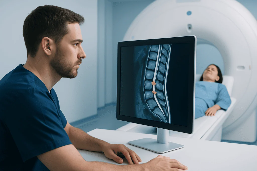
On MRI, cancer in the spine often appears as an area where normal fatty bone marrow has been replaced by tissue with a different signal. Many metastatic lesions look darker than normal marrow on T1-weighted images and brighter on fluid-sensitive sequences such as T2 or STIR images. After contrast is given, suspicious tissue may enhance, meaning it becomes more visible.
These patterns are not exclusive to cancer. Bone marrow swelling from infection, recent fracture, degenerative change, benign vascular lesions such as hemangiomas, and some blood-related conditions can also alter MRI signals. For this reason, the radiology report may use terms such as “indeterminate,” “suspicious,” or “recommend further evaluation” when the cause is not certain.
The appearance also depends on the cancer type and the lesion’s effect on bone. Some lesions mainly replace marrow, while others can erode bone or make it denser. A specialist may compare the MRI with CT, PET imaging, prior scans, and laboratory results to clarify what the finding means.
What are the typical MRI findings of spine metastasis?
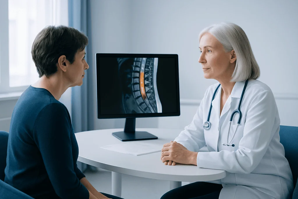
Spine metastases most often affect the vertebral bodies, the large weight-bearing bones at the front of each spinal segment. MRI may show one lesion or multiple lesions at different levels of the spine. The thoracic spine is a frequent location, although the neck and lower back can also be involved.
Typical findings can include replacement of normal marrow signal, enhancement after contrast, a fracture caused by weakened bone, or spread into the epidural space around the spinal cord. Epidural disease may narrow the spinal canal or compress the spinal cord or nerve roots. The scan can also show swelling, spinal alignment changes, and whether the spinal column may be unstable.
These features help clinicians assess urgency, but they do not replace tissue diagnosis when the primary cancer is unknown or when imaging is unclear. A biopsy may be recommended when it can safely provide information that will change treatment planning.
- Single or multiple marrow lesions in vertebrae
- Abnormal contrast enhancement within bone or soft tissue
- Compression fracture or loss of vertebral height
- Extension toward the spinal canal or nerve openings
- Spinal cord or nerve root compression in more advanced cases
How the spine MRI works and who may need it
MRI uses a strong magnetic field and radio waves to create detailed images; it does not use ionizing radiation. A spine MRI may be requested for persistent or unexplained back pain, pain that is worse at night, known cancer with new spinal symptoms, abnormal findings on another scan, or symptoms suggesting nerve or spinal cord involvement.
Contrast material, usually gadolinium-based, may be injected into a vein when clinicians need clearer detail about a lesion, inflammation, blood supply, or possible spread into surrounding tissues. The ordering clinician considers kidney function, prior contrast reactions, pregnancy, and the reason for the scan before deciding whether contrast is appropriate.
Not everyone can undergo a standard MRI without planning. People with certain implanted devices, metal fragments, severe claustrophobia, or difficulty lying flat should tell the imaging team in advance. Many modern implants are MRI-conditional, meaning scanning may still be possible under specified conditions.
Spine MRI procedure: preparation, step-by-step and recovery
Preparation is usually simple. The imaging team may ask the patient to complete a safety questionnaire, remove jewelry and other metal items, and change into a gown. They should report any implanted device, prior surgery, kidney disease, possible pregnancy, allergies, or previous reaction to contrast. Eating and regular medicines are often allowed unless the imaging center gives different instructions.
During the scan, the patient lies on a padded table that moves into the MRI machine. A receiving coil may be placed around the relevant body area. The scanner makes loud tapping or knocking sounds, so ear protection is provided. Remaining still is important because movement can blur the images. If contrast is needed, it is usually administered partway through the examination.
A spine MRI often takes about 30 to 60 minutes, depending on the number of spinal areas scanned and whether contrast is used. It is painless, although lying still may be uncomfortable for people with back pain. After the scan, most people can return to usual activities immediately. If sedating medicine was used for anxiety or claustrophobia, someone else may need to drive them home.
The benefits of MRI include detailed assessment without radiation and sensitive detection of marrow and nerve involvement. Risks are low but include discomfort, anxiety in enclosed spaces, and uncommon contrast reactions. The team uses screening procedures to reduce risks related to magnetic fields and implanted devices.
How long for MRI results if cancer?
The timing of MRI results varies by hospital, the urgency of the request, and whether a specialist review is needed. In an urgent situation, such as suspected spinal cord compression, images may be reviewed the same day and the treating team may contact the patient quickly. For non-urgent examinations, the formal radiology report may take several days.
If the MRI suggests cancer, additional tests can extend the time needed to reach a definite diagnosis. The healthcare team may arrange CT, PET imaging, blood tests, a scan of another part of the body, or a biopsy. This careful process helps distinguish cancer from other causes and identifies the cancer type when needed.
Patients can ask who will explain the report, when this will happen, and what symptoms should prompt earlier contact. A report containing uncertain language does not necessarily mean cancer; it often means that comparison or further assessment is needed.
How treatable is cancer in the spine?
How treatable cancer in the spine is depends on whether the cancer began in the spine or spread there from another organ, its type and biological features, the extent of disease, and whether the spinal cord or stability of the spine is affected. Many spinal metastases can be treated effectively to relieve pain, protect nerve function, strengthen the spine, and control local tumor growth.
Treatment may involve a multidisciplinary team including medical oncology, radiation oncology, radiology, spine surgery, pain specialists, and rehabilitation professionals. Options can include systemic anticancer medicines, radiation therapy, surgery to stabilize the spine or relieve pressure on nerves, and medicines that support bone health. The choice is individualized and may involve more than one approach.
When a lesion is pressing on the spinal cord, treatment may need to begin urgently. Spinal tumor surgery can be considered in selected patients to obtain tissue, decompress neural structures, or stabilize the spine. Acibadem International’s multidisciplinary specialists and JCI-accredited hospitals diagnose and treat spinal conditions for international patients.
When to seek medical care
A person should contact a doctor promptly for new or persistent back or neck pain, especially if there is a history of cancer, unexplained weight loss, fever, pain at night, or pain that does not improve as expected. New pain after a minor injury may also need assessment in people at risk of bone weakness.
Emergency medical care is needed for symptoms that may indicate spinal cord or cauda equina compression. These include new weakness in an arm or leg, difficulty walking, rapidly worsening numbness, numbness in the groin or inner thighs, or new loss of bladder or bowel control. Early assessment can help protect nerve function.
For people with an unexpected MRI finding, the next appropriate step is usually a timely appointment with the clinician who ordered the scan or a relevant specialist. They can explain whether the finding is likely benign, requires follow-up imaging, or needs further cancer evaluation.
Frequently asked questions
Can an MRI confirm cancer in the spine?
MRI can strongly suggest a spinal tumor or metastasis, especially when the pattern is typical and there is a known history of cancer. However, it may not confirm the diagnosis or identify the cancer type. Additional imaging, blood tests, and sometimes a biopsy may be needed.
Are all bright spots on a spine MRI cancer?
No. Bright areas on fluid-sensitive MRI sequences can result from many non-cancerous conditions, including inflammation, injury, infection, degenerative changes, and benign bone lesions. The complete scan pattern and clinical history are needed for interpretation.
Does a spine MRI require contrast when cancer is suspected?
Contrast can help show the boundaries and activity of suspicious tissue, involvement around nerves, and possible spread into the spinal canal. It is not needed in every case, and the radiologist or ordering clinician decides based on the clinical question and patient safety factors.
What happens after an MRI shows suspicious spinal lesions?
The doctor reviews the images and report together with symptoms, medical history, and previous tests. Further evaluation may include CT, PET imaging, laboratory studies, consultation with oncology or spine specialists, or biopsy. Urgent treatment may be arranged if there is spinal cord compression or an unstable fracture.
Can spinal metastases cause no symptoms?
Yes. Some spinal metastases are found incidentally before they cause pain or neurologic symptoms. Others cause localized pain, pain that travels into an arm or leg, numbness, weakness, or difficulty walking, depending on their location and effect on nearby structures.
Should back pain in a person with cancer always lead to an MRI?
Not always, but new, persistent, severe, or progressive back pain should be discussed promptly with the treating clinician. MRI is commonly used when there is concern for spinal involvement, nerve compression, or another serious cause that needs detailed evaluation.
References
- National Cancer Institute
- American Cancer Society
- Radiological Society of North America
- National Institute for Health and Care Excellence
- American College of Radiology
This article is for general information only and is not a substitute for professional medical advice. Please consult a qualified doctor about your individual situation.
Joint and spine care in Turkey — expert assessment & treatment
JCI-accredited · board-certified surgeons · reply within 24h
Add us as a Preferred Source to see more of our trusted health content across Google Search, AI Overviews and Discover.

More from the Health Library



Related Specialists

Dr. Mahir Bülent Özgen
Urology
Dr. Nihat Cem Çelebi
Pediatrics
Prof. Dr. Çiğdem Kalaycık Ertuğay
Otorhinolaryngology
Asst. Prof. Dr. Berkhan Yilmaz
Aesthetic Plastic & Reconstructive Surgery