

Mammogram Radiation Dose: How It Works, Results and What to Expect
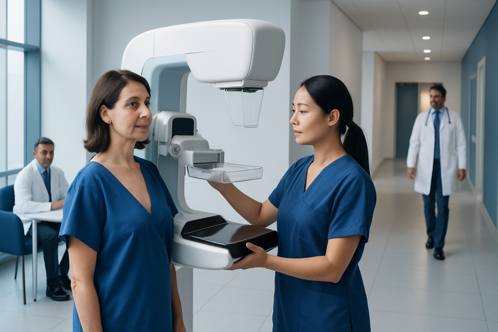
A standard two-view screening mammogram delivers a low radiation dose, similar to the amount of natural background radiation received over a short period of everyday life. Digital mammography is designed to use the lowest radiation dose needed to produce diagnostically useful breast images.
Key Takeaways
- A standard two-view screening mammogram delivers a low radiation dose, similar to the amount of natural background radiation received over a short period of everyday life.
- Digital mammography is designed to use the lowest radiation dose needed to produce diagnostically useful breast images.
- Compression is brief but important because it spreads breast tissue, improves image clarity, and can reduce the dose needed.
- Mammograms may be used for routine screening or to investigate symptoms such as a new lump, nipple discharge, or breast changes.
- Pregnancy, breast implants, prior breast surgery, and previous imaging should be discussed with the imaging team before the appointment.
- A mammogram result may be normal, may recommend follow-up imaging, or may identify an area that needs further assessment; an abnormal result does not automatically mean cancer.
A mammogram uses a low dose of X-ray radiation to create detailed images of breast tissue. For most people who are advised to have screening or diagnostic mammography, the benefit of finding breast cancer early outweighs the very small radiation-related risk.
Overview: What Is the Mammogram Radiation Dose?
The mammogram radiation dose is low. A typical screening mammogram takes X-ray images of each breast from two angles and uses only the amount of radiation needed to obtain clear images. Modern digital systems closely control exposure, and the potential benefit of detecting breast cancer at an earlier, more treatable stage generally outweighs the very small radiation risk for people who are recommended to have mammography.
Radiation dose is often measured in milligray (mGy), a unit describing energy absorbed by tissue. The exact dose varies with breast thickness and density, equipment, image views, and whether extra images are needed. Rather than focusing on one fixed number, it is helpful to know that mammography is a regulated, low-dose imaging examination and that facilities monitor image quality and radiation exposure.
Mammography is used in two main ways. Screening mammography is performed before symptoms develop, while diagnostic mammography evaluates a concern such as a lump, focal pain, skin change, nipple discharge, or an unusual screening result. Both uses can be part of breast cancer care when further evaluation or treatment is needed.
How Mammography Works and Why Compression Matters
Mammography uses low-energy X-rays to show differences in breast tissue. Fatty tissue, glandular tissue, calcifications, and some masses absorb X-rays differently, creating an image that a radiologist can interpret. Digital mammography records the image electronically, allowing careful review and, when appropriate, adjustment of contrast or magnification without repeating the original exposure.
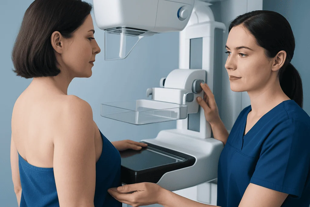
During the examination, the breast is positioned on a detector and gently but firmly compressed for a few seconds. Compression may feel uncomfortable, but it should not be harmful. It reduces breast thickness, limits movement, separates overlapping tissues, and helps produce a sharper image. These effects can also allow the imaging system to use less radiation than would otherwise be required.
Standard screening images usually include a top-to-bottom view and an angled side view of each breast. Additional views may be taken if the radiologist needs to clarify an area. A diagnostic mammogram can therefore take longer and may involve a slightly higher total dose than routine screening, but it remains a low-dose examination designed around the clinical question.
Who May Need a Mammogram

People are offered mammography based on age, personal health history, family history, breast density, inherited cancer risk, and national or local screening guidance. Recommendations differ between countries and organizations, so the most suitable screening schedule should be discussed with a doctor or breast specialist. People with average risk may begin regular screening at a different age from those with a strong family history or a known genetic risk.
Diagnostic mammography may be recommended at any appropriate age when there is a breast concern. Symptoms that may lead to imaging include a new breast or underarm lump, persistent localized breast pain, nipple inversion, bloody or spontaneous nipple discharge, skin dimpling, swelling, or a change in breast shape. Depending on age and the concern, an ultrasound examination may be used alongside mammography or may be the first test.
Individuals with breast implants, previous breast surgery, a history of breast cancer, or prior chest radiation should tell the imaging center before their appointment. Extra positioning or specialized views may be needed. Those at increased inherited risk may also need a personalized surveillance plan that includes MRI; this can be discussed in the context of breast cancer risk and evaluation.
What Happens During the Procedure
Before the appointment, the person may be asked to avoid deodorant, talcum powder, lotion, or perfume on the breasts or underarms, because some products can leave particles that resemble calcifications on images. Wearing a two-piece outfit can make changing easier. Previous mammograms should be made available whenever possible, as comparison with earlier images can reduce unnecessary callbacks.
At the imaging center, a radiographer will explain the process and position one breast at a time. The breast is placed on the detector, compression is applied briefly, and the X-ray is taken. The process is repeated for the usual views of both breasts. The image-taking part is short, and a routine screening appointment commonly takes around 20 to 30 minutes in total, although timing varies by center.
It is important to tell the radiographer if there is any possibility of pregnancy, if breastfeeding, if there are implants, or if a particular position is painful. The team can adapt positioning where possible and advise whether the examination should proceed. Mammography is often possible during breastfeeding, although expressing or feeding beforehand may improve comfort and image quality.
If additional images are requested, this does not mean cancer has been found. It may simply mean that an area is not fully visible, breast tissue overlaps on one image, or the radiologist needs a closer look. Further assessment can include targeted mammographic views, breast ultrasound, MRI, or a biopsy when clinically indicated.
Results, Recovery Timeline, Benefits and Limitations
There is no physical recovery period after a mammogram. Mild tenderness or temporary redness from compression can occur, especially in people with sensitive breasts, but this usually settles quickly. Normal daily activities can be resumed immediately. Results may be communicated directly by the imaging center, through a referring doctor, or at a follow-up breast clinic appointment.
A report may describe a normal result, a benign finding such as a cyst or stable calcification, an area needing comparison with prior images, or a recommendation for additional tests. A callback after screening is relatively common and is not a diagnosis. Most recalled findings are not cancer, but completing the recommended follow-up promptly is important.
The key benefit of mammography is its ability to detect some breast cancers before they can be felt. It can also identify microcalcifications, which may be an early sign of certain breast changes. However, no screening test is perfect. Dense breast tissue can make some abnormalities harder to see, and mammography can sometimes miss a cancer or identify a finding that ultimately proves harmless.
When an imaging result is suspicious, a doctor may recommend a tissue sample to establish a diagnosis. This can include breast biopsy procedures guided by mammography, ultrasound, or MRI, depending on where the area is seen most clearly.
Risks and How Radiation Safety Is Managed
The main concern people raise is radiation exposure. Mammography does use ionizing radiation, but the dose is low and carefully managed. Digital machines are subject to quality-control standards, and radiographers use positioning, compression, and exposure settings tailored to the breast being imaged. The goal is always to obtain sufficient diagnostic detail while keeping radiation as low as reasonably achievable.
Other possible drawbacks include brief discomfort from compression, anxiety while waiting for results, false-positive findings that lead to extra tests, and false-negative results. These limitations are why mammogram results are interpreted together with symptoms, examination findings, personal risk factors, and previous imaging rather than in isolation.
Pregnancy should always be mentioned before an X-ray examination. If breast imaging is necessary during pregnancy, the medical team will choose the most appropriate approach based on the symptoms and stage of pregnancy. For people who are not pregnant, routine mammography radiation does not remain in the body and does not make a person radioactive.
Questions about individual risk are appropriate, particularly for people who have had repeated imaging, previous radiation treatment, or a genetic cancer predisposition. A radiologist or breast specialist can explain why a particular test is recommended and whether another imaging method would add useful information.
Preparing for Screening and Supporting Breast Health
Scheduling a mammogram when the breasts are least tender may improve comfort. For people who menstruate, this is often after the menstrual period rather than in the days just before it. Patients should bring relevant medical information and should tell the team about breast symptoms, surgeries, implants, hormone use, prior cancer, and family history.
Regular screening is only one aspect of breast health. Knowing what is normal for the breasts and reporting new or persistent changes supports earlier assessment. Lifestyle measures such as maintaining a healthy weight where possible, being physically active, limiting alcohol, and avoiding tobacco can support overall health, although they do not eliminate breast cancer risk.
People should follow the screening plan agreed with their clinician rather than delaying recommended mammography because of radiation concerns. The dose from a medically appropriate mammogram is low, and the examination is performed because it can provide information that is not available through self-examination alone.
Acibadem International’s multidisciplinary specialists and JCI-accredited hospitals can assess breast symptoms, coordinate imaging, and provide care for international patients when further investigation is required.
When to Seek Medical Care
A person should arrange medical assessment promptly for a new breast or underarm lump, a persistent focal area of pain, unexplained swelling, skin dimpling, redness that does not settle, a new nipple change, or spontaneous nipple discharge, particularly if it is bloody. These symptoms are often caused by non-cancerous conditions, but they should not be ignored or assessed only through routine screening.
Medical advice is also appropriate after a mammogram if a person has not received expected results, does not understand a recommendation for further testing, or notices a new breast change despite a recent normal mammogram. A normal result is reassuring but does not replace evaluation of a new symptom.
Urgent assessment may be needed for severe breast redness, fever, significant swelling, or rapidly worsening pain, especially during breastfeeding, because infection or another acute condition may need treatment. For non-urgent questions about screening timing, radiation exposure, or breast density, a primary care doctor, gynecologist, or breast clinic can provide individualized guidance.
Frequently asked questions
How much radiation is in a mammogram?
A mammogram uses a low dose of X-ray radiation. The exact amount varies according to breast size and density, the equipment used, and the number of images required. Modern digital systems are designed to keep exposure as low as possible while still producing clear images.
Is mammogram radiation dangerous?
For most people, the radiation-related risk from recommended mammography is very small. The potential benefit of finding breast cancer early generally outweighs this risk. Imaging teams use controlled techniques and quality standards to limit radiation exposure.
Does a diagnostic mammogram use more radiation than a screening mammogram?
A diagnostic mammogram may involve more images than a standard screening mammogram, so the total dose can be somewhat higher. It is still a low-dose examination. The additional views are taken only when they can help clarify a symptom or an area seen on prior imaging.
Can mammography be performed during pregnancy?
Anyone who may be pregnant should tell the imaging team before a mammogram. If breast imaging is needed, the clinician will weigh the reason for the test and select the safest, most informative approach. Ultrasound may be used in some situations, particularly for evaluating a new breast symptom.
Does breast compression increase radiation exposure?
No. Compression is used to spread the breast tissue and reduce motion, improving image quality. Because the breast becomes thinner during imaging, compression can help the system obtain a useful image with less radiation than might otherwise be needed.
Can mammogram radiation cause breast cancer?
Ionizing radiation can carry a theoretical risk, but the dose used in mammography is low. For people who are advised to have mammograms, this very small potential risk is balanced against the proven value of detecting some breast cancers earlier. A doctor can discuss individual screening needs and concerns.
References
- World Health Organization
- International Atomic Energy Agency
- U.S. Food and Drug Administration
- American College of Radiology
- National Cancer Institute
This article is for general information only and is not a substitute for professional medical advice. Please consult a qualified doctor about your individual situation.
Oncology care in Turkey — second opinion and treatment plan
JCI-accredited · board-certified surgeons · reply within 24h
Add us as a Preferred Source to see more of our trusted health content across Google Search, AI Overviews and Discover.

More from the Health Library



Related Specialists

Dr. Semra Demirli Atıcı
General Surgery
Assoc. Prof. Dr. Emine Acar Akkaya
Nuclear Medicine
Dr. Cem Tangay
Orthopedic Surgery & Traumatology
Dt. Oğuzhan Hakan Özdemir
Oral & Dental Health