Ultrasound on Eyeball: Preparation, Procedure and Results
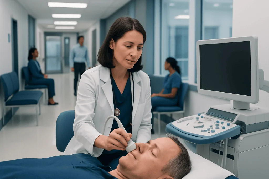
Eye ultrasound is a noninvasive imaging test that does not use radiation. It can help assess the retina, vitreous gel, optic nerve area, eye muscles, and orbit.
Key Takeaways
- Eye ultrasound is a noninvasive imaging test that does not use radiation.
- It can help assess the retina, vitreous gel, optic nerve area, eye muscles, and orbit.
- The examination usually takes only a few minutes and is generally comfortable.
- Most people can return to usual activities immediately unless dilating drops or another treatment was used.
- Results must be interpreted alongside symptoms and a complete eye examination.
An ultrasound on the eyeball, also called ocular ultrasound or eye ultrasound, uses high-frequency sound waves to create images of the eye and the tissues behind it. It is commonly used when a doctor needs a clearer view through the eye, including when cataract, bleeding, swelling, or injury makes a standard examination difficult.
Overview: What Is an Ultrasound on the Eyeball?
An ultrasound on eyeball is an imaging examination that uses sound waves rather than X-rays or radiation. A small handheld device, called an ultrasound eye probe, sends sound waves into or around the closed eyelid. A computer converts the returning echoes into images that help an eye specialist assess structures inside the eye and in the eye socket.
The test is often called an ocular ultrasound, eye scan, B-scan ultrasound, or ultrasound eye procedure. It can be particularly useful when the doctor cannot see the back of the eye clearly during a routine examination. For example, clouding of the cornea or lens, bleeding within the eye, or significant swelling may limit the view of the retina.
There are different ultrasound techniques. B-scan ultrasound produces a two-dimensional image of the inside of the eye and orbit, while A-scan measurements may be used to calculate the length of the eye before cataract surgery or to evaluate certain eye findings. The appropriate technique depends on the clinical question.
Why Would a Doctor Order an Eye Ultrasound?
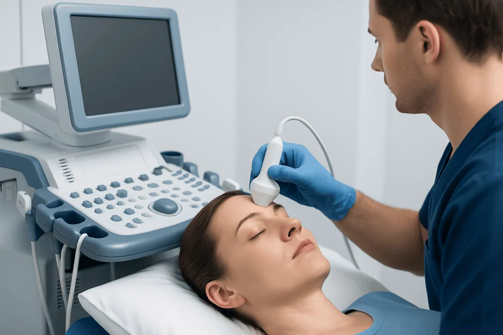
A doctor may order an eye ultrasound to investigate new visual symptoms, assess an eye injury, or clarify an abnormal finding seen during an eye examination. The scan can provide important information about the vitreous, the clear gel that fills the eye; the retina; the choroid; and the tissues surrounding the eye.
Common reasons for imaging include suspected retinal detachment, vitreous hemorrhage, an unexplained loss of vision, a possible mass in or behind the eye, eye pain with swelling, or trauma. It may also be used before surgery when the retina cannot be examined directly. An ultrasound does not replace all eye tests, but it can be a valuable complement to dilated examination and other imaging.
An eye specialist may use the examination to distinguish among conditions with similar symptoms. For example, flashes, floaters, or a shadow in vision can be associated with several causes, including retinal detachment, which needs prompt ophthalmic assessment. The scan helps guide the next steps, but a diagnosis is based on the full clinical picture.
Who May Need the Ultrasound Eye Procedure?

Eye ultrasound is suitable for many adults and children when an ophthalmologist considers it clinically helpful. It is especially useful in people with an opaque cornea, a dense cataract, or bleeding that prevents direct visualization of the back of the eye. It may also be requested after an injury or when there is concern about changes in the tissues around the eye.
Before the examination, it is important to tell the care team about recent eye surgery, a known or suspected eye injury, active infection, severe pain, allergies to anesthetic drops, and any medications being taken. These details help determine the safest scanning technique. In particular, a direct-contact technique is usually avoided if there is any concern for an open-globe injury, where the wall of the eye may be penetrated.
In many situations, the probe is placed gently over the closed eyelid with gel, so it does not touch the eyeball itself. In other situations, numbing drops may be used and the probe may be placed on the eye surface through a protective fluid-filled cup. The clinician selects the method that is appropriate for the individual situation.
How Do I Prepare for an Eye Ultrasound?
Most people need little preparation for an eye ultrasound. Eating and drinking are usually allowed as normal unless the appointment is combined with another procedure that has separate instructions. Patients should bring their glasses, contact lens case if relevant, a medication list, and details of previous eye operations or eye conditions.
Contact lenses are generally removed before the test. Eye makeup may also need to be removed if the probe will be placed over the eyelid, because gel is used to obtain clear images. If the clinician plans to use dilating drops, vision may become blurred and light sensitivity can occur for several hours afterward.
Patients should inform the clinician immediately if they have had a penetrating eye injury, sudden severe eye pain, or surgery very recently. They should not press on an injured eye before medical assessment. Preparation instructions can vary slightly by clinic, so following the guidance from the ophthalmology team is the best approach.
Ultrasound Eye Procedure: How It Works Step by Step
The ultrasound eye procedure is usually performed by an ophthalmologist, an eye imaging specialist, or a trained technician. The patient sits or lies comfortably and is asked to look in certain directions so the examiner can obtain images from different angles. The examination room may be dimmed to improve comfort and allow other parts of the eye assessment to be completed.
For a common B-scan, the patient closes the eyelid and a small amount of ultrasound gel is placed on the lid. The ultrasound eye probe is moved gently over the gel without pressing firmly on the eye. The probe sends sound waves through the eyelid, and images appear on a monitor in real time. The examiner may ask the patient to look up, down, left, or right.
When a more precise measurement is needed, anesthetic eye drops may be given first. The eye is numbed to minimize discomfort, and the specialist uses the selected measurement technique. The patient may notice gentle contact or cool gel, but the test should not be painful. If pain occurs, the patient should tell the examiner.
Images are reviewed by the ophthalmologist along with the person’s symptoms, medical history, vision test results, and findings from a slit-lamp or dilated eye examination. Depending on the findings, the clinician may recommend monitoring, further imaging, medication, or a procedure such as retinal detachment surgery.
How Long Does an Ultrasound of the Eye Take?
The scan itself commonly takes about 5 to 15 minutes. The total appointment may take longer if it includes vision testing, pupil dilation, a detailed eye examination, discussion of results, or additional imaging. A complex assessment after trauma or with limited visibility inside the eye may also require extra time.
Patients can usually expect the procedure to be completed during one outpatient visit. The clinician may be able to discuss preliminary findings immediately, particularly when the test is performed during an urgent eye assessment. In some cases, images need formal review before a final interpretation is provided.
Timing should not be the main concern when urgent symptoms are present. Sudden vision changes, a curtain-like shadow, severe eye pain, or an eye injury require timely medical assessment rather than waiting for a routine imaging appointment.
Results, Benefits, Risks and Recovery
Eye ultrasound may show whether the retina is in its normal position, whether blood or debris is present in the vitreous, or whether there are changes in the tissues behind the eye. It can also help characterize a suspected lesion or assess the orbit after injury. A normal result can be reassuring, while an abnormal result helps the ophthalmologist determine whether further testing or treatment is needed.
The main benefits are that the test is quick, does not use ionizing radiation, and can image through certain visual obstructions. It is generally well tolerated. The main minor effects are temporary coolness from the gel, brief discomfort from probe pressure, or mild irritation if anesthetic drops were used. Serious complications are uncommon when the examination is performed appropriately.
Recovery is usually immediate. Gel can be wiped away, and there is typically no wound care or activity restriction. If dilating or numbing drops were used, vision may be temporarily blurred. Patients should avoid rubbing a numb eye, since it may be less sensitive to irritation or accidental injury until the drops wear off.
Can You Drive After an Eye Ultrasound?
Many people can drive after an eye ultrasound if only gel was applied to the closed eyelid and their vision is unchanged. However, driving is not advisable if dilating drops, numbing drops, sedating medication, or another procedure has affected vision or comfort. After dilation, glare and blurred near vision may make driving unsafe for several hours.
People should arrange a companion or alternative transport if their appointment is likely to include dilation, especially if they are unfamiliar with how their eyes respond to dilating drops. The clinic team can advise based on the planned tests and the person’s vision on the day.
Even when driving is technically possible, it is sensible to avoid driving if there is eye pain, reduced vision, double vision, light sensitivity, or any concern after injury. Safety should take priority over convenience.
When to Seek Medical Care
Prompt medical care is important for sudden loss or marked reduction of vision, new flashing lights, a rapid increase in floaters, a dark curtain or shadow across vision, severe eye pain, significant redness with nausea, or an eye injury. These symptoms do not always indicate a serious condition, but they require timely assessment because some eye problems benefit from early treatment.
After trauma, a person should avoid pressing on the eye, attempting to remove an embedded object, or using eye drops unless advised by a clinician. A protective shield may be placed loosely over the eye while urgent care is arranged. Emergency services may be appropriate for a major injury or sudden severe symptoms.
For planned assessment, ophthalmology teams may use eye ultrasound alongside other investigations and treatments, including cataract surgery planning when the view into the eye is limited. Acibadem International’s multidisciplinary specialists and JCI-accredited hospitals assess and treat eye conditions for international patients, with care tailored to the person’s clinical needs.
Frequently asked questions
Is an ultrasound on eyeball painful?
An eye ultrasound is usually not painful. Many scans are performed over a closed eyelid, where the patient may only feel cool gel and gentle movement of the probe. If direct eye contact is needed, numbing drops are commonly used to improve comfort.
Does an eye ultrasound use radiation?
No. Eye ultrasound uses sound waves and does not expose the patient to ionizing radiation. This differs from imaging tests such as CT scans, which use X-rays.
Can an eye ultrasound detect retinal detachment?
Yes, ocular ultrasound can help identify retinal detachment, particularly when the retina cannot be seen clearly because of blood, cataract, or other clouding inside the eye. The result is interpreted with the eye examination and the person’s symptoms. Sudden flashes, floaters, or a curtain-like shadow need urgent eye assessment.
Do I need to remove contact lenses for an eye ultrasound?
Contact lenses are usually removed before an eye ultrasound. This helps keep the examination comfortable and avoids interfering with a scan performed near or on the eye surface. Patients can ask the clinic whether to bring a lens case and glasses for use afterward.
When will I get the results of my eye ultrasound?
In urgent settings, an ophthalmologist may discuss preliminary results shortly after the scan. Some images require detailed review, so the final interpretation may be provided later in the appointment or at a follow-up visit. The timing depends on why the test was ordered and whether more examinations are needed.
Can eye ultrasound be done after an eye injury?
It can be useful after some eye injuries, but the method must be selected carefully. If there is a suspected open-globe injury, pressure on the eye should be avoided and urgent ophthalmic evaluation is needed. The care team will decide whether ultrasound is safe and necessary.
References
- American Academy of Ophthalmology
- Royal College of Ophthalmologists
- National Eye Institute
- American Society of Retina Specialists
This article is for general information only and is not a substitute for professional medical advice. Please consult a qualified doctor about your individual situation.
Ultrasound in Turkey — costs, top hospitals & a free quote
JCI-accredited · board-certified surgeons · reply within 24h
Add us as a Preferred Source to see more of our trusted health content across Google Search, AI Overviews and Discover.

Check-up & Preventive Medicine
Comprehensive health screenings designed for early detection and prevention.
3 specialists in this unit