

3D Imaging and Navigation System for Prostate Biopsy
Diagnosis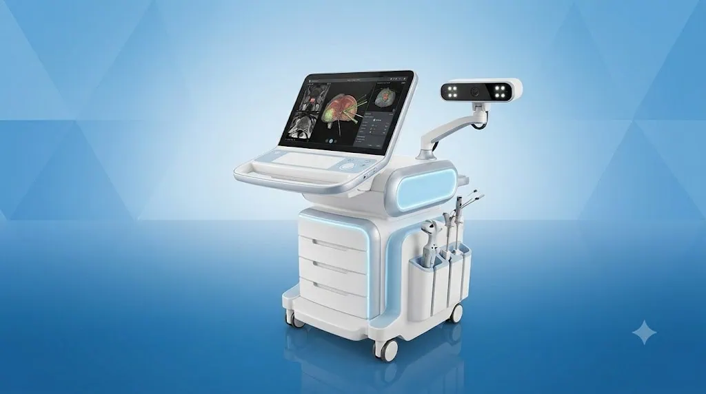
Quick answer
A 3D imaging and navigation system for prostate biopsy fuses multiparametric MRI with real-time ultrasound to guide tissue sampling from suspicious areas of the prostate. At Acibadem in Turkey, urologists use this technology to support targeted biopsy planning and systematic sampling when clinically appropriate.
3D Imaging and Navigation System for Prostate Biopsy
A 3D Imaging and Navigation System for Prostate Biopsy is an advanced diagnostic technology that helps urologists take more precise tissue samples from the prostate. It combines detailed magnetic resonance imaging (MRI) performed before the biopsy with real-time ultrasound images taken during the procedure.
This approach is often called MRI-ultrasound fusion biopsy or MRI-fusion guided prostate biopsy. It allows the specialist to identify areas that looked suspicious on multiparametric MRI, create a three-dimensional map of the prostate and guide the biopsy needle toward these areas with millimetric accuracy.
At Acibadem International, this technology supports a more informed approach to prostate cancer diagnosis. By improving the ability to target areas of concern, it can help specialists make clearer decisions about further investigations, active surveillance, treatment planning or reassurance when results are benign.
What Is MRI-Fusion Guided Prostate Biopsy?
A prostate biopsy is a procedure in which very small tissue samples are collected from the prostate and examined by a pathologist. It may be recommended when there is concern about prostate cancer, such as an elevated prostate-specific antigen (PSA) level, an abnormal rectal examination or a suspicious area seen on MRI.
Traditional ultrasound-guided biopsy uses ultrasound images to guide sampling. While ultrasound is valuable, some suspicious prostate areas can be difficult to distinguish clearly on ultrasound alone. Multiparametric MRI provides more detailed information about the prostate and may reveal changes that require closer evaluation.
The 3D imaging and navigation system brings these two sources of information together. MRI images are reviewed before the procedure, and suspicious regions are marked by the medical team. During the biopsy, live ultrasound images are matched with the MRI data to build a detailed three-dimensional navigation model. This helps the urologist track the biopsy needle and target the planned areas accurately.
The system can also support systematic sampling, where samples are taken from standard prostate regions. Whether targeted samples, systematic samples or both are appropriate depends on your MRI findings, PSA level, previous biopsy history and overall clinical assessment.
How the 3D Imaging and Navigation System Works
The process begins with a high-quality multiparametric MRI scan of the prostate. This examination provides detailed images that can help identify areas with features requiring further investigation. The radiology and urology teams review the images carefully and use them to plan the biopsy.
On the day of the procedure, ultrasound is used to create real-time images of the prostate. The navigation platform combines the MRI information with these ultrasound images. This is known as image fusion.
Using the fused images, the urologist can view the prostate in three dimensions and identify the MRI-marked targets during the biopsy. The system records sample locations, which can be useful for pathology correlation, follow-up imaging and future treatment planning if needed.
Key Steps in MRI-Ultrasound Fusion
- MRI review: Multiparametric MRI images are assessed to identify and score areas that may need targeted sampling.
- Target planning: Suspicious regions are outlined on the MRI images before the biopsy.
- Real-time ultrasound: Ultrasound creates live images of the prostate during the procedure.
- 3D reconstruction: The system builds a three-dimensional model of the prostate anatomy.
- Image fusion: MRI targets are aligned with live ultrasound images to guide sampling.
- Navigation and documentation: The specialist tracks sampling locations and records them for the clinical file.
Image fusion is an advanced guidance method, but it does not replace clinical judgement. Your urologist interprets the MRI findings, reviews your personal risk factors and determines the most suitable biopsy strategy for your case.
Clinical Applications
MRI-fusion guidance is used primarily in urology for the assessment of suspected prostate cancer. It can be particularly helpful when MRI has identified a lesion that requires targeted investigation or when previous tests have not provided a clear explanation for ongoing clinical concern.
| Clinical application | How MRI-fusion guidance may help | When it may be considered |
|---|---|---|
| Suspicious prostate MRI finding | Targets the specific area identified on multiparametric MRI for tissue sampling. | When MRI shows a lesion that requires pathological assessment. |
| Elevated or rising PSA level | Supports a more focused biopsy plan when PSA results and imaging suggest further investigation is needed. | When PSA trends, prostate size and clinical assessment raise concern. |
| Previous negative biopsy with ongoing concern | May help evaluate areas that could have been difficult to sample with conventional ultrasound guidance alone. | When PSA remains elevated, MRI is suspicious or symptoms and examination findings need clarification. |
| Active surveillance assessment | Can support targeted sampling of known or newly suspicious areas during monitoring of selected prostate cancers. | For patients whose urologist recommends repeat assessment as part of an individual surveillance plan. |
| Biopsy mapping for treatment planning | Documents where samples were obtained and helps correlate pathology findings with MRI anatomy. | When detailed information is needed to discuss treatment options or follow-up. |
This technology may assist in diagnosing prostate cancer, but it is not a diagnosis on its own. Final conclusions depend on the pathology results, MRI interpretation, PSA values, examination findings and discussion within the appropriate clinical team.
Conditions and Concerns It Can Help Assess
The 3D Imaging and Navigation System for Prostate Biopsy is designed to support investigation of prostate abnormalities. It is most commonly used when there is a need to rule out, confirm or better characterise possible prostate cancer.
- Elevated PSA levels or a PSA level that is increasing over time
- Abnormal findings during a digital rectal examination
- Suspicious lesions or areas seen on multiparametric prostate MRI
- Persistent concern after a previous biopsy that did not detect cancer
- Known prostate cancer requiring reassessment during active surveillance
- Need for more detailed biopsy information before considering treatment options
Not every patient with an elevated PSA level needs a biopsy, and not every MRI finding represents cancer. Your specialist will consider your age, medical history, PSA density, MRI report, family history, medications and previous test results before recommending the next step.
Benefits and Advantages for Patients
The main advantage of MRI-fusion biopsy technology is precision. It helps the specialist direct biopsy samples toward areas that have already been identified as potentially significant on MRI, rather than relying only on standard sampling patterns.
For many patients, this means a more personalised diagnostic pathway. Targeted information may help reduce uncertainty and provide a stronger foundation for decisions about monitoring, additional tests or treatment.
| Patient benefit | What it means in practice |
|---|---|
| More targeted sampling | MRI-identified areas of concern can be sampled with image-guided navigation. |
| Detailed 3D visualisation | The prostate, planned targets and biopsy locations can be viewed within a three-dimensional model. |
| Personalised biopsy planning | Your biopsy approach can be based on MRI findings, PSA results, previous tests and clinical risk factors. |
| Support for clearer clinical decisions | Targeted pathology results can help guide discussions about surveillance, further investigation or treatment. |
| Useful follow-up documentation | Recorded sample locations may assist with future comparison, repeat biopsy planning or treatment assessment. |
| Coordinated specialist care | Radiology, urology, pathology and international patient teams can work from a structured diagnostic plan. |
While this technology offers important advantages, no biopsy technique can guarantee detection of every prostate cancer. The value of MRI-fusion guidance is that it adds detailed imaging information and navigational support to the expertise of the clinical team.
What to Expect During Your Procedure
Your experience begins before you travel or arrive at the hospital. Our international patient team can help coordinate the review of your available MRI images, PSA results, previous biopsy reports and medical history. If additional imaging or tests are needed, your care coordinator will explain the recommended sequence of appointments.
Before the Biopsy
A urology specialist reviews whether MRI-fusion guided biopsy is appropriate for you. You may be asked about allergies, current medications, blood thinners, diabetes, previous infections and any history of urinary difficulties. It is important to tell your team about all medicines, supplements and medical conditions.
Your doctor will explain how to prepare. Instructions may include guidance about eating and drinking, medication adjustments and infection prevention. Do not stop prescribed medicines, especially blood-thinning medication, unless your treating doctor gives you clear instructions.
You will have an opportunity to discuss the procedure, possible alternatives, expected recovery and potential risks. Informed consent is obtained before the biopsy.
During the Biopsy
The biopsy may be performed through the rectum or through the skin between the scrotum and anus, known as the transperineal route. The most suitable approach depends on your anatomy, MRI findings, infection-risk assessment and the urologist’s clinical recommendation.
Comfort measures and anaesthesia are planned individually. Depending on the procedure and your needs, this may involve local anaesthesia, sedation or another appropriate anaesthetic approach. Your team will explain what is recommended and how it may affect your recovery and travel plans.
During the procedure, the urologist uses ultrasound to visualise the prostate and activates the MRI-fusion navigation system. The MRI targets are aligned with the real-time ultrasound images, allowing planned samples to be taken from the areas of concern. Additional systematic samples may be collected when clinically appropriate.
The procedure itself is usually completed within a relatively short period, although your total time at the hospital may be longer because of preparation, anaesthesia, observation and discharge instructions.
After the Biopsy
After the procedure, you will be monitored until the team is satisfied that you are comfortable and ready for discharge or onward care. Mild discomfort, temporary blood in the urine, stool or semen, and short-term urinary symptoms can occur after a prostate biopsy. Your doctor will explain what is expected for your chosen biopsy approach.
You should seek urgent medical advice if you develop fever, chills, worsening pain, inability to pass urine, heavy bleeding or symptoms that concern you. These symptoms can indicate a complication that needs prompt assessment.
The tissue samples are examined by an experienced pathology team. Results are usually discussed during a follow-up consultation, where your specialist explains the findings in clear language and recommends any next steps.
Safety, Quality and Accreditation
At Acibadem International, MRI-fusion prostate biopsy is performed by trained urology, radiology, anaesthesia and nursing professionals working within structured clinical pathways. Patient safety begins with careful selection, review of MRI quality, infection prevention measures, appropriate anaesthesia planning and clear post-procedure guidance.
Our JCI-accredited hospitals follow internationally recognised quality and patient-safety standards. These standards support reliable clinical processes, multidisciplinary communication, infection-control practices and a strong focus on informed consent and patient experience.
Every biopsy involves potential risks, including bleeding, infection, urinary retention, temporary discomfort and anaesthesia-related effects. The level of risk varies according to the biopsy route, your general health, medications and individual medical history. Your urologist will explain the risks and benefits relevant to you before proceeding.
For patients travelling from abroad, safety planning also includes practical considerations. If sedation or anaesthesia is used, you may need a companion and may be advised not to travel immediately. Your international patient coordinator can help arrange the timing of consultations, procedure day and follow-up around your medical needs.
Why This Technology Matters for International Patients
Travelling for prostate assessment can feel complex, particularly when you are already concerned about a possible diagnosis. A coordinated MRI-fusion biopsy pathway can help make the process more organised by bringing imaging review, urology consultation, biopsy planning, pathology and follow-up into one carefully managed care journey.
Before your visit, our international patient team can guide you on which documents are useful to share, such as MRI reports and images, PSA records, previous pathology results, medication lists and referral letters. Where possible, having these records reviewed in advance helps the clinical team prepare for your consultation.
Language support and personalised coordination are available to help you understand each stage of care. Your coordinator can assist with appointment planning, communication between departments, documentation for your insurer where applicable, and guidance about expected recovery before you return home.
If further treatment is required, the biopsy findings can be reviewed alongside MRI results and other clinical information by the appropriate specialists. This supports a personalised discussion of options, which may include monitoring, surgery, radiotherapy, focal approaches or other treatments depending on the diagnosis and your individual circumstances.
Frequently Asked Questions
Is MRI-fusion prostate biopsy safe?
MRI-fusion biopsy is routinely used by trained specialists to improve guidance during prostate sampling. As with any biopsy, there are possible risks, including infection, bleeding, discomfort and urinary symptoms. Your doctor will assess your individual risk factors and explain the safety measures used for your procedure.
Does MRI-fusion guidance mean I will only need targeted samples?
Not always. In some cases, the urologist may recommend targeted samples from MRI-visible lesions together with systematic samples from other prostate regions. This decision is based on your MRI findings, PSA level, prior biopsy history and overall clinical assessment.
Can this technology diagnose prostate cancer without a biopsy?
No. MRI can identify areas that may need further investigation, but a prostate cancer diagnosis usually requires pathology assessment of tissue samples. MRI-fusion navigation helps guide the biopsy to areas of concern.
Will my insurance cover the procedure?
Insurance coverage depends on your policy, provider requirements and the planned medical services. You can share your insurance details with our international patient team, who can help clarify available options and provide information before your visit.
How do I get started?
You can submit your medical records for an initial review. Please include recent PSA results, prostate MRI reports and images if available, previous biopsy reports, your medical history and a list of current medications. A specialist can then advise whether 3D imaging and navigation for prostate biopsy may be appropriate for your individual care plan.
Related Medical Units
Available at these Hospitals

















Related Treatments
Speak with our medical team
Find out how this technology could support your diagnosis or treatment.